|
Theodor Boveri
Theodor Heinrich Boveri (12 October 1862 – 15 October 1915) was a German zoologist, comparative anatomist and co-founder of modern cytology. He was notable for the first hypothesis regarding cellular processes that cause cancer, and for describing ''chromatin diminution in nematodes''. Boveri was married to the American biologist Marcella O'Grady (1863–1950). Their daughter Margret Boveri (1900–1975) became one of the best-known journalists in post-World War II Germany. Work Using an optical microscope, Boveri examined the processes involved in the fertilization of the animal egg cell; his favorite research objects were the nematode ''Parascaris'' and sea urchins. Boveri's work with sea urchins showed that it was necessary to have all chromosomes present in order for proper embryonic development to take place. This discovery was an important part of the Boveri–Sutton chromosome theory. He also discovered, in 1888, the importance of the centrosome for the formation of th ... [...More Info...] [...Related Items...] OR: [Wikipedia] [Google] [Baidu] |
Bamberg
Bamberg (, , ; East Franconian: ''Bambärch'') is a town in Upper Franconia, Germany, on the river Regnitz close to its confluence with the river Main. The town dates back to the 9th century, when its name was derived from the nearby ' castle. Cited as one of Germany's most beautiful towns, with medieval streets and Europe's largest intact old city wall, the old town of Bamberg has been a UNESCO World Heritage Site since 1993. From the 10th century onwards, Bamberg became a key link with the Slav peoples, notably those of Poland and Pomerania. It experienced a period of great prosperity from the 12th century onwards, during which time it was briefly the centre of the Holy Roman Empire. Emperor Henry II was also buried in the old town, alongside his wife Kunigunde. The town's architecture from this period strongly influenced that in Northern Germany and Hungary. From the middle of the 13th century onwards, the bishops were princes of the Empire and ruled Bamberg, overseeing the c ... [...More Info...] [...Related Items...] OR: [Wikipedia] [Google] [Baidu] |
Chromatin
Chromatin is a complex of DNA and protein found in eukaryotic cells. The primary function is to package long DNA molecules into more compact, denser structures. This prevents the strands from becoming tangled and also plays important roles in reinforcing the DNA during cell division, preventing DNA damage, and regulating gene expression and DNA replication. During mitosis and meiosis, chromatin facilitates proper segregation of the chromosomes in anaphase; the characteristic shapes of chromosomes visible during this stage are the result of DNA being coiled into highly condensed chromatin. The primary protein components of chromatin are histones. An octamer of two sets of four histone cores (Histone H2A, Histone H2B, Histone H3, and Histone H4) bind to DNA and function as "anchors" around which the strands are wound.Maeshima, K., Ide, S., & Babokhov, M. (2019). Dynamic chromatin organization without the 30-nm fiber. ''Current opinion in cell biology, 58,'' 95–104. https://doi.o ... [...More Info...] [...Related Items...] OR: [Wikipedia] [Google] [Baidu] |
Mitosis
In cell biology, mitosis () is a part of the cell cycle in which replicated chromosomes are separated into two new nuclei. Cell division by mitosis gives rise to genetically identical cells in which the total number of chromosomes is maintained. Therefore, mitosis is also known as equational division. In general, mitosis is preceded by S phase of interphase (during which DNA replication occurs) and is often followed by telophase and cytokinesis; which divides the cytoplasm, organelles and cell membrane of one cell into two new cells containing roughly equal shares of these cellular components. The different stages of mitosis altogether define the mitotic (M) phase of an animal cell cycle—the division of the mother cell into two daughter cells genetically identical to each other. The process of mitosis is divided into stages corresponding to the completion of one set of activities and the start of the next. These stages are preprophase (specific to plant cells), prophase ... [...More Info...] [...Related Items...] OR: [Wikipedia] [Google] [Baidu] |
Mitotic Spindle
In cell biology, the spindle apparatus refers to the cytoskeletal structure of eukaryotic cells that forms during cell division to separate sister chromatids between daughter cells. It is referred to as the mitotic spindle during mitosis, a process that produces genetically identical daughter cells, or the meiotic spindle during meiosis, a process that produces gametes with half the number of chromosomes of the parent cell. Besides chromosomes, the spindle apparatus is composed of hundreds of proteins. Microtubules comprise the most abundant components of the machinery. Spindle structure Attachment of microtubules to chromosomes is mediated by kinetochores, which actively monitor spindle formation and prevent premature anaphase onset. Microtubule polymerization and depolymerization dynamic drive chromosome congression. Depolymerization of microtubules generates tension at kinetochores; bipolar attachment of sister kinetochores to microtubules emanating from opposite cell pol ... [...More Info...] [...Related Items...] OR: [Wikipedia] [Google] [Baidu] |
Centrosome
In cell biology, the centrosome (Latin centrum 'center' + Greek sōma 'body') (archaically cytocentre) is an organelle that serves as the main microtubule organizing center (MTOC) of the animal cell, as well as a regulator of cell-cycle progression. The centrosome provides structure for the cell. The centrosome is thought to have evolved only in the metazoan lineage of eukaryotic cells. Fungi and plants lack centrosomes and therefore use other structures to organize their microtubules. Although the centrosome has a key role in efficient mitosis in animal cells, it is not essential in certain fly and flatworm species. Centrosomes are composed of two centrioles arranged at right angles to each other, and surrounded by a dense, highly structured mass of protein termed the pericentriolar material (PCM). The PCM contains proteins responsible for microtubule nucleation and anchoring — including γ-tubulin, pericentrin and ninein. In general, each centriole of the centrosome is based ... [...More Info...] [...Related Items...] OR: [Wikipedia] [Google] [Baidu] |
Boveri–Sutton Chromosome Theory
The Boveri–Sutton chromosome theory (also known as the chromosome theory of inheritance or the Sutton–Boveri theory) is a fundamental unifying theory of genetics which identifies chromosomes as the carriers of genetic material.1902: Theodor Boveri (1862-1915) and Walter Sutton (1877-1916) propose that chromosomes bear hereditary factors in accordance with Mendelian laws Genetics and Genomics Timeline. Genome News Network an online publication of the J. Craig Venter Institute. Holmgren Lab Northwestern Uni ... [...More Info...] [...Related Items...] OR: [Wikipedia] [Google] [Baidu] |
Embryonic Development
An embryo is an initial stage of development of a multicellular organism. In organisms that reproduce sexually, embryonic development is the part of the life cycle that begins just after fertilization of the female egg cell by the male sperm cell. The resulting fusion of these two cells produces a single-celled zygote that undergoes many cell divisions that produce cells known as blastomeres. The blastomeres are arranged as a solid ball that when reaching a certain size, called a morula, takes in fluid to create a cavity called a blastocoel. The structure is then termed a blastula, or a blastocyst in mammals. The mammalian blastocyst hatches before implantating into the endometrial lining of the womb. Once implanted the embryo will continue its development through the next stages of gastrulation, neurulation, and organogenesis. Gastrulation is the formation of the three germ layers that will form all of the different parts of the body. Neurulation forms the nervous syst ... [...More Info...] [...Related Items...] OR: [Wikipedia] [Google] [Baidu] |
Chromosome
A chromosome is a long DNA molecule with part or all of the genetic material of an organism. In most chromosomes the very long thin DNA fibers are coated with packaging proteins; in eukaryotic cells the most important of these proteins are the histones. These proteins, aided by chaperone proteins, bind to and condense the DNA molecule to maintain its integrity. These chromosomes display a complex three-dimensional structure, which plays a significant role in transcriptional regulation. Chromosomes are normally visible under a light microscope only during the metaphase of cell division (where all chromosomes are aligned in the center of the cell in their condensed form). Before this happens, each chromosome is duplicated ( S phase), and both copies are joined by a centromere, resulting either in an X-shaped structure (pictured above), if the centromere is located equatorially, or a two-arm structure, if the centromere is located distally. The joined copies are now called si ... [...More Info...] [...Related Items...] OR: [Wikipedia] [Google] [Baidu] |
Sea Urchin
Sea urchins () are spiny, globular echinoderms in the class Echinoidea. About 950 species of sea urchin live on the seabed of every ocean and inhabit every depth zone from the intertidal seashore down to . The spherical, hard shells (tests) of sea urchins are round and spiny, ranging in diameter from . Sea urchins move slowly, crawling with tube feet, and also propel themselves with their spines. Although algae are the primary diet, sea urchins also eat slow-moving (sessile) animals. Predators that eat sea urchins include a wide variety of fish, starfish, crabs, marine mammals. Sea urchins are also used as food especially in Japan. Adult sea urchins have fivefold symmetry, but their pluteus larvae feature bilateral (mirror) symmetry, indicating that the sea urchin belongs to the Bilateria group of animal phyla, which also comprises the chordates and the arthropods, the annelids and the molluscs, and are found in every ocean and in every climate, from the tropics to the pol ... [...More Info...] [...Related Items...] OR: [Wikipedia] [Google] [Baidu] |
Parascaris
''Parascaris'' is a genus of nematodes in the family Ascarididae. It contains two species, ''Parascaris equorum ''Parascaris equorum'' is a species of ascarid that is the equine roundworm. Amongst horse owners, the parasites are colloquially called "Ascarids". This is a host-specific helminth intestinal parasite that can infect horses, donkeys, and zeb ...'' and '' Parascaris univalens'', which are morphologically identical, but can be distinguished by chromosome number. Both species parasitize horses. References Ascaridida Secernentea genera {{nematode-stub ... [...More Info...] [...Related Items...] OR: [Wikipedia] [Google] [Baidu] |
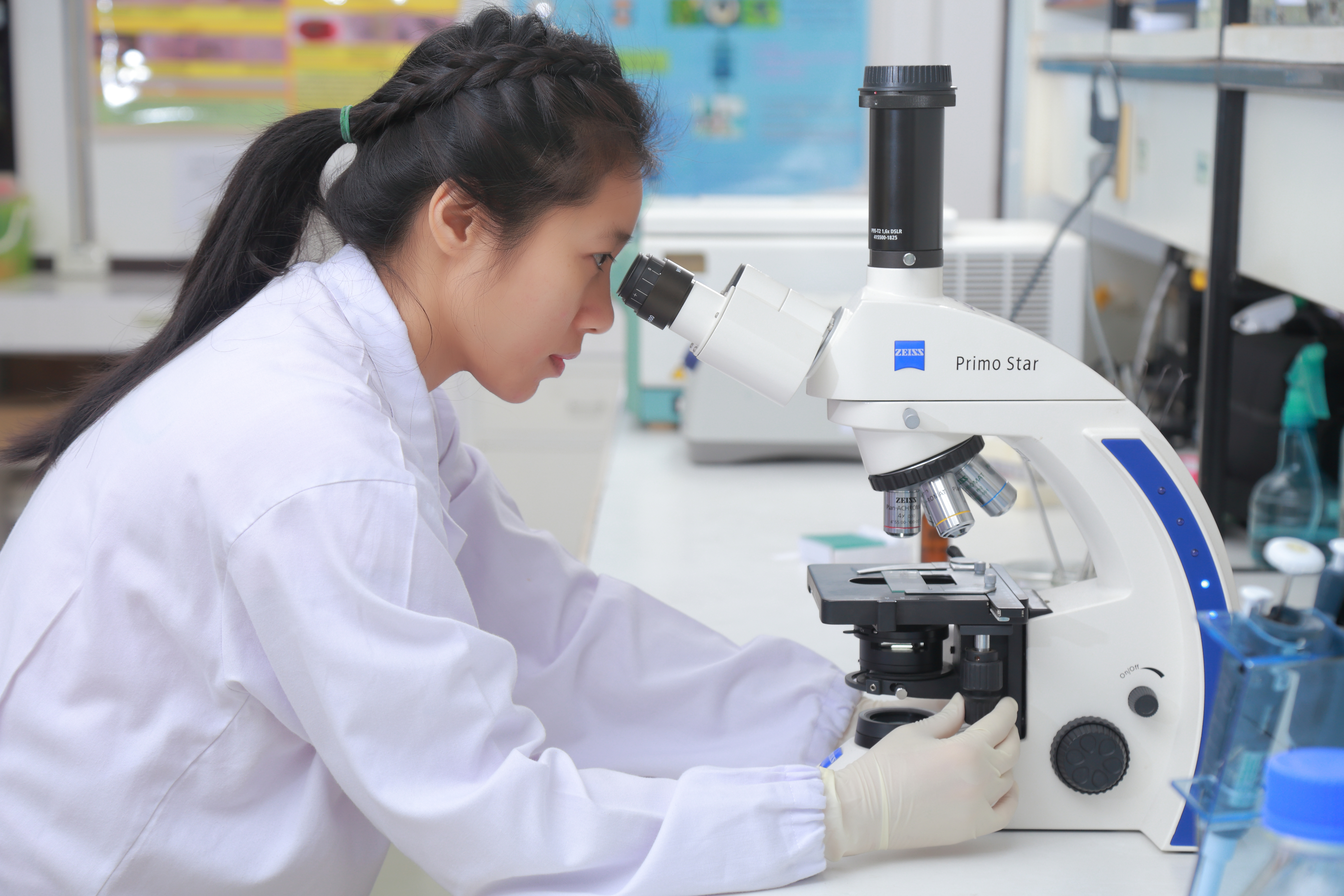
Optical Microscope
The optical microscope, also referred to as a light microscope, is a type of microscope that commonly uses visible light and a system of lenses to generate magnified images of small objects. Optical microscopes are the oldest design of microscope and were possibly invented in their present compound form in the 17th century. Basic optical microscopes can be very simple, although many complex designs aim to improve resolution and sample contrast. The object is placed on a stage and may be directly viewed through one or two eyepieces on the microscope. In high-power microscopes, both eyepieces typically show the same image, but with a stereo microscope, slightly different images are used to create a 3-D effect. A camera is typically used to capture the image (micrograph). The sample can be lit in a variety of ways. Transparent objects can be lit from below and solid objects can be lit with light coming through ( bright field) or around (dark field) the objective lens. Polarised ... [...More Info...] [...Related Items...] OR: [Wikipedia] [Google] [Baidu] |


.jpg)
