|
Midclavicular Line
{{short description, None Anatomical lines, or "reference lines," are theoretical lines drawn through anatomical structures and are used to describe anatomical location. The following reference lines are identified in '' Terminologia Anatomica'': * Anterior median line * Lateral sternal line: A vertical line corresponding to the lateral margin of the sternum. * Parasternal line: A vertical line equidistant from the sternal and mid-clavicular lines. * Mid-clavicular line: A vertical line passing through the midpoint of the clavicle. * Mammillary line * Anterior axillary line: A vertical line on the anterior torso marked by the anterior axillary fold. * Midaxillary line: A vertical line passing through the apex of the axilla. * Posterior axillary line: A vertical line passing through the posterior axillary fold. * Scapular line: A vertical line passing through the inferior angle of the scapula. * Paravertebral line: A vertical line corresponding to the tips of the transverse proc ... [...More Info...] [...Related Items...] OR: [Wikipedia] [Google] [Baidu] |
Axillary Lines
The axillary lines are the anterior axillary line, midaxillary line and the posterior axillary line. The anterior axillary line is a coronal line on the anterior torso marked by the anterior axillary fold. It's the imaginary line that runs down from the point midway between the middle of the clavicle and the lateral end of the clavicle. The V5 ECG lead is placed on the left anterior axillary line, horizontally even with V4. The midaxillary line is a coronal line on the torso between the anterior and posterior axillary lines. It is a landmark used in thoracentesis, and the V6 electrode of the 10 electrode ECG. The posterior axillary line is a coronal line on the posterior torso marked by the posterior axillary fold. Additional images File:Axillary lines.png, The left side of the thorax with lines labeled See also * List of anatomical lines {{short description, None Anatomical lines, or "reference lines," are theoretical lines drawn through anatomical structures an ... [...More Info...] [...Related Items...] OR: [Wikipedia] [Google] [Baidu] |
Scapular Line
The scapular line, also known as the linea scapularis, is a vertical line passing through the inferior angle of the scapula. It has been used in the evaluation of brachial plexus birth palsy. References External links * http://www.meddean.luc.edu/Lumen/MedEd/MEDICINE/PULMONAR/apd/lines.htm Anatomy {{Anatomy-stub ... [...More Info...] [...Related Items...] OR: [Wikipedia] [Google] [Baidu] |
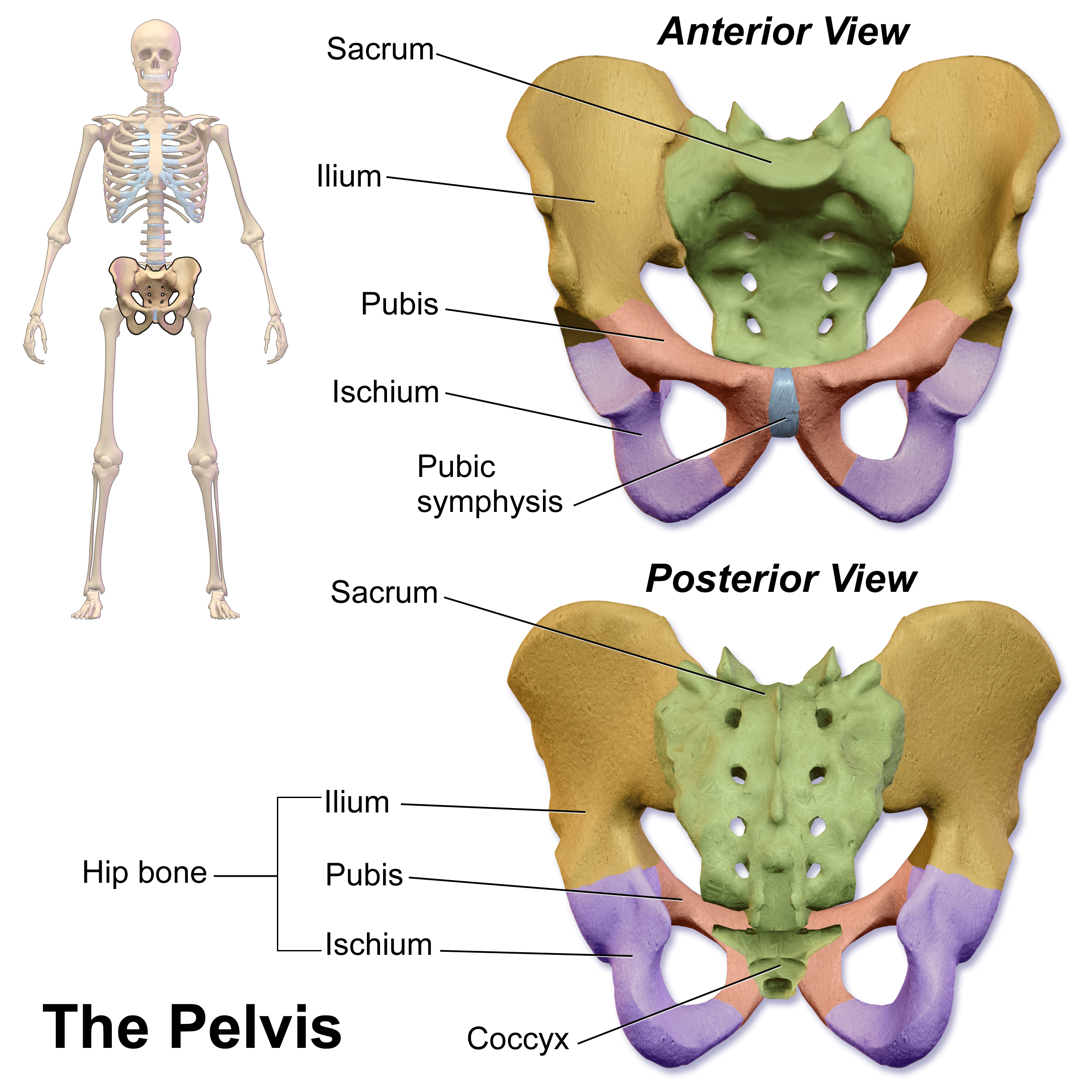
Pubic Symphysis
The pubic symphysis (: symphyses) is a secondary cartilaginous joint between the left and right superior rami of the pubis of the hip bones. It is in front of and below the urinary bladder. In males, the suspensory ligament of the penis attaches to the pubic symphysis. In females, the pubic symphysis is attached to the suspensory ligament of the clitoris. In most adults, it can be moved roughly 2 mm and with 1 degree rotation. This increases for women at the time of childbirth. The name comes from the Greek word ''symphysis'', meaning 'growing together'. Structure The pubic symphysis is a nonsynovial amphiarthrodial joint. The width of the pubic symphysis at the front is 3–5 mm greater than its width at the back. This joint is connected by fibrocartilage and may contain a fluid-filled cavity; the center is avascular, possibly due to the nature of the compressive forces passing through this joint, which may lead to harmful vascular disease. The ends of both pubi ... [...More Info...] [...Related Items...] OR: [Wikipedia] [Google] [Baidu] |
Ilium (bone)
The ilium () (: ilia) is the uppermost and largest region of the coxal bone, and appears in most vertebrates including mammals and birds, but not bony fish. All reptiles have an ilium except snakes, with the exception of some snake species which have a tiny bone considered to be an ilium. The ilium of the human is divisible into two parts, the body and the wing; the separation is indicated on the top surface by a curved line, the arcuate line, and on the external surface by the margin of the acetabulum. The name comes from the Latin ('' ile'', ''ilis''), meaning "groin" or "flank". Structure The ilium consists of the body and wing. Together with the ischium and pubis, to which the ilium is connected, these form the pelvic bone, with only a faint line indicating the place of union. The body () forms less than two-fifths of the acetabulum; and also forms part of the acetabular fossa. The internal surface of the body is part of the wall of the lesser pelvis and gives o ... [...More Info...] [...Related Items...] OR: [Wikipedia] [Google] [Baidu] |
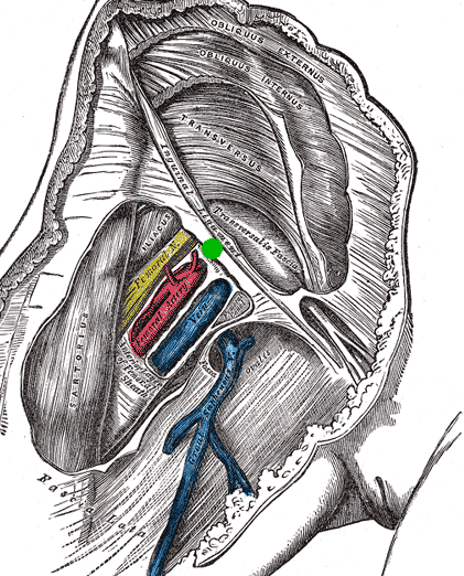
Mid-inguinal Point
The mid-inguinal point (MIP) is located at the middle point of an imaginary line drawn between the anterior superior iliac spine (ASIS) and the pubic symphysis. The mid-inguinal point is superior to (above) the inguinal ligament, and should not to be confused with the midpoint of the inguinal ligament itself, which is located halfway between the anterior superior iliac spine and the pubic tubercle. Significance The external iliac artery becomes the femoral artery when it passes deep to the inguinal ligament, at the mid-inguinal point. As such, the point is along the superior boundary of the femoral triangle. References {{Authority control Abdomen Ligaments ... [...More Info...] [...Related Items...] OR: [Wikipedia] [Google] [Baidu] |
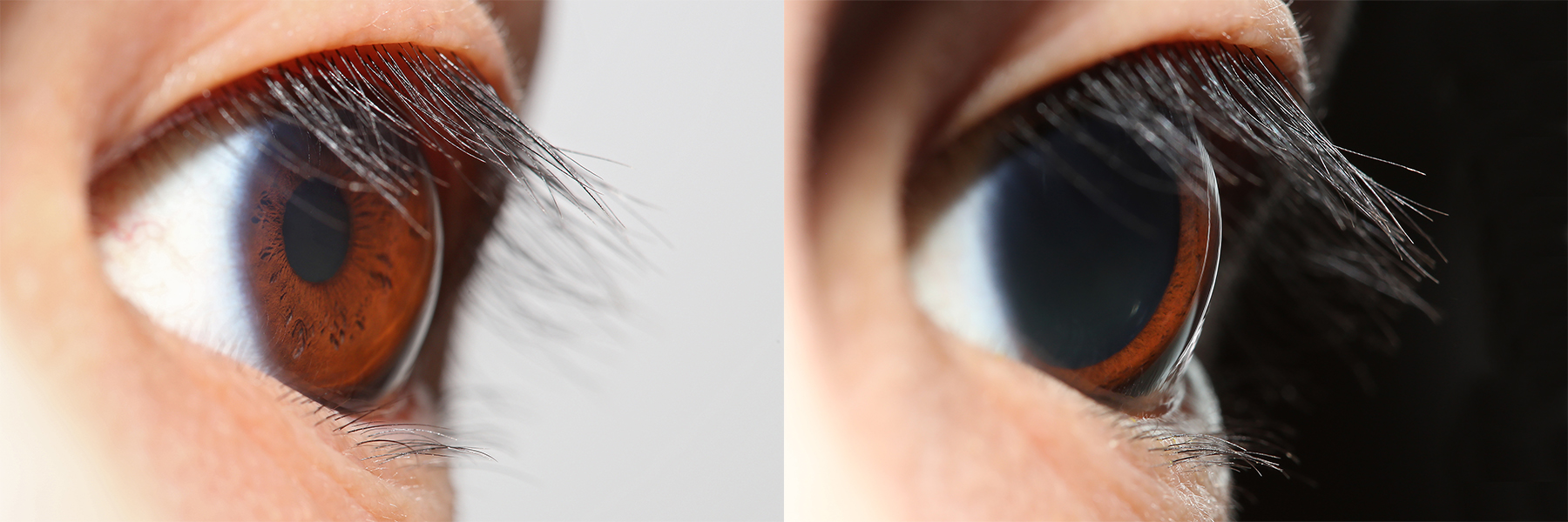
Pupil
The pupil is a hole located in the center of the iris of the eye that allows light to strike the retina.Cassin, B. and Solomon, S. (1990) ''Dictionary of Eye Terminology''. Gainesville, Florida: Triad Publishing Company. It appears black because light rays entering the pupil are either absorbed by the tissues inside the eye directly, or absorbed after diffuse reflections within the eye that mostly miss exiting the narrow pupil. The size of the pupil is controlled by the iris, and varies depending on many factors, the most significant being the amount of light in the environment. The term "pupil" was coined by Gerard of Cremona. In humans, the pupil is circular, but its shape varies between species; some cats, reptiles, and foxes have vertical slit pupils, goats and sheep have horizontally oriented pupils, and some catfish have annular types. In optical terms, the anatomical pupil is the eye's aperture and the iris is the aperture stop. The image of the pupil as seen from o ... [...More Info...] [...Related Items...] OR: [Wikipedia] [Google] [Baidu] |
Vertebra
Each vertebra (: vertebrae) is an irregular bone with a complex structure composed of bone and some hyaline cartilage, that make up the vertebral column or spine, of vertebrates. The proportions of the vertebrae differ according to their spinal segment and the particular species. The basic configuration of a vertebra varies; the vertebral body (also ''centrum'') is of bone and bears the load of the vertebral column. The upper and lower surfaces of the vertebra body give attachment to the intervertebral discs. The posterior part of a vertebra forms a vertebral arch, in eleven parts, consisting of two pedicles (pedicle of vertebral arch), two laminae, and seven processes. The laminae give attachment to the ligamenta flava (ligaments of the spine). There are vertebral notches formed from the shape of the pedicles, which form the intervertebral foramina when the vertebrae articulate. These foramina are the entry and exit conduits for the spinal nerves. The body of the vertebr ... [...More Info...] [...Related Items...] OR: [Wikipedia] [Google] [Baidu] |
Scapula
The scapula (: scapulae or scapulas), also known as the shoulder blade, is the bone that connects the humerus (upper arm bone) with the clavicle (collar bone). Like their connected bones, the scapulae are paired, with each scapula on either side of the body being roughly a mirror image of the other. The name derives from the Classical Latin word for trowel or small shovel, which it was thought to resemble. In compound terms, the prefix omo- is used for the shoulder blade in medical terminology. This prefix is derived from ὦμος (ōmos), the Ancient Greek word for shoulder, and is cognate with the Latin , which in Latin signifies either the shoulder or the upper arm bone. The scapula forms the back of the shoulder girdle. In humans, it is a flat bone, roughly triangular in shape, placed on a posterolateral aspect of the thoracic cage. Structure The scapula is a thick, flat bone lying on the thoracic wall that provides an attachment for three groups of muscles: intrinsic, e ... [...More Info...] [...Related Items...] OR: [Wikipedia] [Google] [Baidu] |
Posterior Axillary Fold
The axilla (: axillae or axillas; also known as the armpit, underarm or oxter) is the area on the human body directly under the shoulder joint. It includes the axillary space, an anatomical space within the shoulder girdle between the arm and the thoracic cage, bounded superiorly by the imaginary plane between the superior borders of the first rib, clavicle and scapula (above which are considered part of the neck), medially by the serratus anterior muscle and thoracolumbar fascia, anteriorly by the pectoral muscles and posteriorly by the subscapularis, teres major and latissimus dorsi muscle. The soft skin covering the lateral axilla contains many hair and sweat glands. In humans, the formation of body odor happens mostly in the axilla. These odorant substances have been suggested by some to serve as pheromones, which play a role related to mate selection, although this is a controversial topic within the scientific community. The underarms seem more important than the pubic ar ... [...More Info...] [...Related Items...] OR: [Wikipedia] [Google] [Baidu] |
Axilla
The axilla (: axillae or axillas; also known as the armpit, underarm or oxter) is the area on the human body directly under the shoulder joint. It includes the axillary space, an anatomical space within the shoulder girdle between the arm and the thoracic cage, bounded superiorly by the imaginary plane between the superior borders of the first rib, clavicle and scapula (above which are considered part of the neck), medially by the serratus anterior muscle and thoracolumbar fascia, anteriorly by the pectoral muscles and posteriorly by the subscapularis, teres major and latissimus dorsi muscle. The soft skin covering the lateral axilla contains many hair and sweat glands. In humans, the formation of body odor happens mostly in the axilla. These odorant substances have been suggested by some to serve as pheromones, which play a role related to mate selection, although this is a controversial topic within the scientific community. The underarms seem more importa ... [...More Info...] [...Related Items...] OR: [Wikipedia] [Google] [Baidu] |
Anterior Axillary Fold
The axilla (: axillae or axillas; also known as the armpit, underarm or oxter) is the area on the human body directly under the shoulder joint. It includes the axillary space, an anatomical space within the shoulder girdle between the arm and the thoracic cage, bounded superiorly by the imaginary plane between the superior borders of the first rib, clavicle and scapula (above which are considered part of the neck), medially by the serratus anterior muscle and thoracolumbar fascia, anteriorly by the pectoral muscles and posteriorly by the subscapularis, teres major and latissimus dorsi muscle. The soft skin covering the lateral axilla contains many hair and sweat glands. In humans, the formation of body odor happens mostly in the axilla. These odorant substances have been suggested by some to serve as pheromones, which play a role related to mate selection, although this is a controversial topic within the scientific community. The underarms seem more important than the pubic ar ... [...More Info...] [...Related Items...] OR: [Wikipedia] [Google] [Baidu] |


