|
Pupil
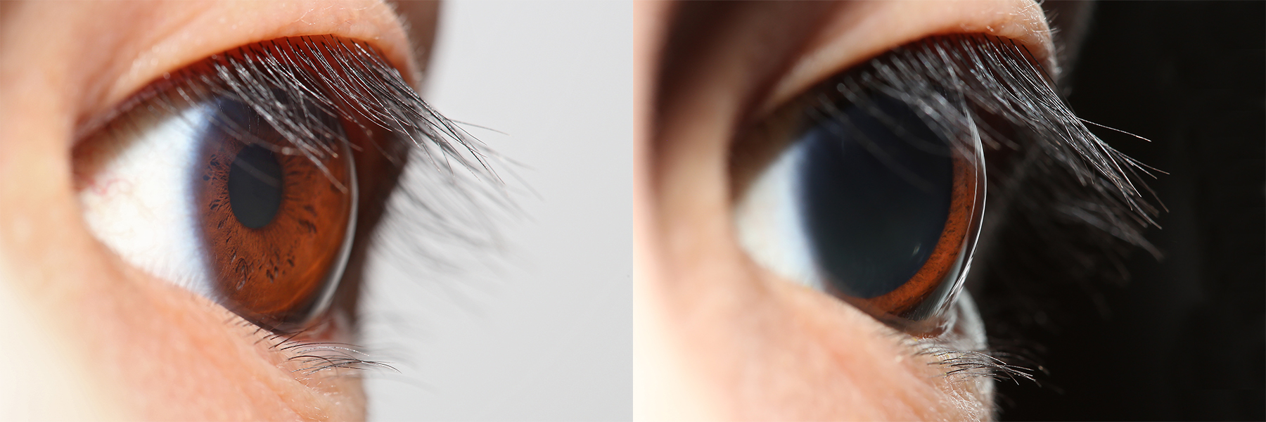
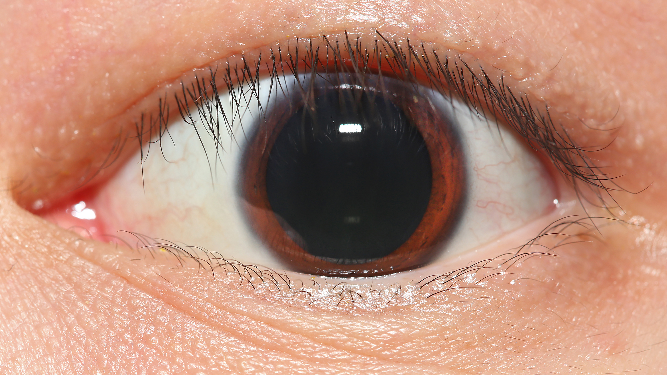
The pupil is a hole located in the center of the iris of the eye that allows light to strike the retina.Cassin, B. and Solomon, S. (1990) ''Dictionary of Eye Terminology''. Gainesville, Florida: Triad Publishing Company. It appears black because light rays entering the pupil are either absorbed by the tissues inside the eye directly, or absorbed after diffuse reflections within the eye that mostly miss exiting the narrow pupil. The size of the pupil is controlled by the iris, and varies depending on many factors, the most significant being the amount of light in the environment. The term "pupil" was coined by Gerard of Cremona. In humans, the pupil is circular, but its shape varies between species; some cats, reptiles, and foxes have vertical slit pupils, goats and sheep have horizontally oriented pupils, and some catfish have annular types. In optical terms, the anatomical pupil is the eye's aperture and the iris is the aperture stop. The image of the pupil as seen from o ... [...More Info...] [...Related Items...] OR: [Wikipedia] [Google] [Baidu] |
Pupillary Light Reflex
The pupillary light reflex (PLR) or photopupillary reflex is a reflex that controls the diameter of the pupil, in response to the intensity ( luminance) of light that falls on the retinal ganglion cells of the retina in the back of the eye, thereby assisting in adaptation of vision to various levels of lightness/darkness. A greater intensity of light causes the pupil to constrict ( miosis/myosis; thereby allowing less light in), whereas a lower intensity of light causes the pupil to dilate ( mydriasis, expansion; thereby allowing more light in). Thus, the pupillary light reflex regulates the intensity of light entering the eye. Light shone into one eye will cause both pupils to constrict. Terminology The pupil is the dark circular opening in the center of the iris and is where light enters the eye. By analogy with a camera, the pupil is equivalent to aperture, whereas the iris is equivalent to the diaphragm. It may be helpful to consider the ''Pupillary reflex'' as an Iris ... [...More Info...] [...Related Items...] OR: [Wikipedia] [Google] [Baidu] |
Collarette (iris)
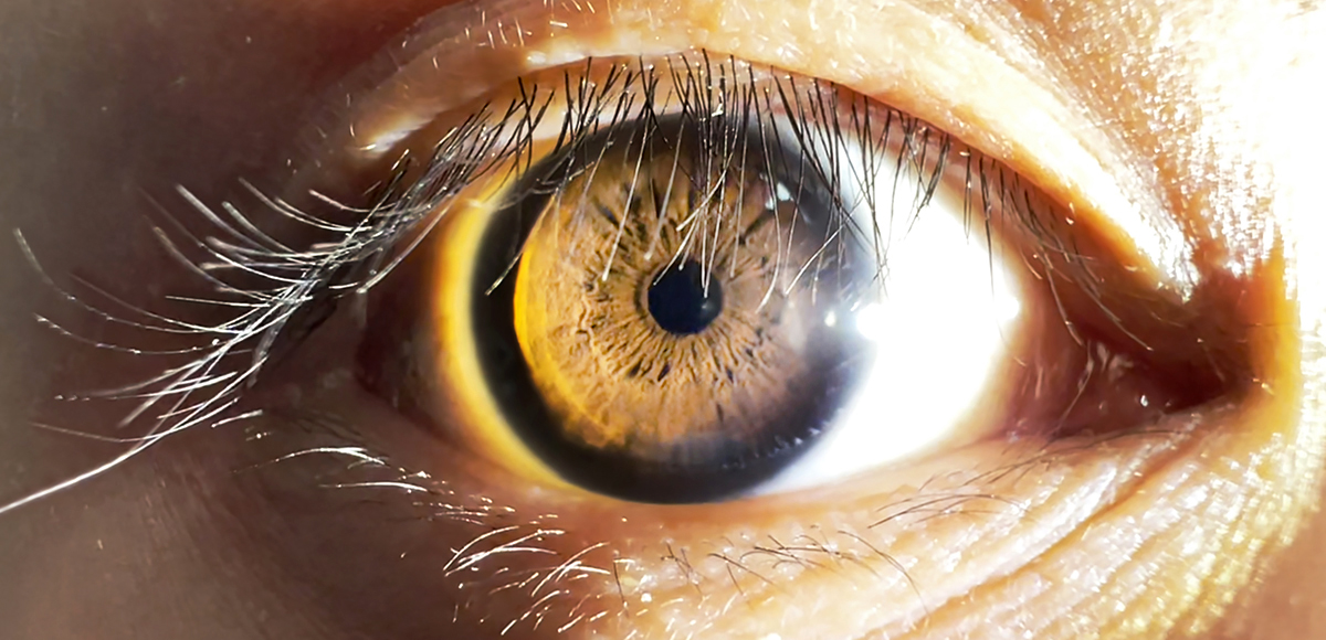
The iris (: irides or irises) is a thin, annular structure in the eye in most mammals and birds that is responsible for controlling the diameter and size of the pupil, and thus the amount of light reaching the retina. In optical terms, the pupil is the eye's aperture, while the iris is the diaphragm. Eye color is defined by the iris. Etymology The word "iris" is derived from the Greek word for "rainbow", also its goddess plus messenger of the gods in the ''Iliad'', because of the many colours of this eye part. Structure The iris consists of two layers: the front pigmented fibrovascular layer known as a stroma and, behind the stroma, pigmented epithelial cells. The stroma is connected to a sphincter muscle ( sphincter pupillae), which contracts the pupil in a circular motion, and a set of dilator muscles (dilator pupillae), which pull the iris radially to enlarge the pupil, pulling it in folds. The sphincter pupillae is the opposing muscle of the dilator pupillae. The pupil ... [...More Info...] [...Related Items...] OR: [Wikipedia] [Google] [Baidu] |
Entrance Pupil
In an optical system, the entrance pupil is the optical image of the physical aperture stop, as 'seen' through the optical elements in front of the stop. The corresponding image of the aperture stop as seen through the optical elements behind it is called the ''exit pupil''. The entrance pupil defines the cone of rays that can enter and pass through the optical system. Rays that fall outside of the entrance pupil will not pass through the system. If there is no lens in front of the aperture (as in a pinhole camera), the entrance pupil's location and size are identical to those of the aperture. Optical elements in front of the aperture will produce a magnified or diminished image of the aperture that is displaced from the aperture location. The entrance pupil is usually a virtual image: it lies behind the first optical surface of the system. The entrance pupil is a useful concept for determining the size of the cone of rays that an optical system will accept. Once the size ... [...More Info...] [...Related Items...] OR: [Wikipedia] [Google] [Baidu] |
Aperture Stop
In optics, the aperture of an optical system (including a system consisting of a single lens) is the hole or opening that primarily limits light propagated through the system. More specifically, the entrance pupil as the front side image of the aperture and focal length of an optical system determine the cone angle of a bundle of ray (optics), rays that comes to a focus (optics), focus in the image plane. An optical system typically has many structures that limit ray bundles (ray bundles are also known as ''pencils'' of light). These structures may be the edge of a lens (optics), lens or mirror, or a ring or other fixture that holds an optical element in place or may be a special element such as a diaphragm (optics), diaphragm placed in the optical path to limit the light admitted by the system. In general, these structures are called stops, and the aperture stop is the stop that primarily determines the cone of rays that an optical system accepts (see entrance pupil). As a ... [...More Info...] [...Related Items...] OR: [Wikipedia] [Google] [Baidu] |
Human Eye
The human eye is a sensory organ in the visual system that reacts to light, visible light allowing eyesight. Other functions include maintaining the circadian rhythm, and Balance (ability), keeping balance. The eye can be considered as a living optics, optical device. It is approximately spherical in shape, with its outer layers, such as the outermost, white part of the eye (the sclera) and one of its inner layers (the pigmented choroid) keeping the eye essentially stray light, light tight except on the eye's optic axis. In order, along the optic axis, the optical components consist of a first lens (the cornea, cornea—the clear part of the eye) that accounts for most of the optical power of the eye and accomplishes most of the Focus (optics), focusing of light from the outside world; then an aperture (the pupil) in a Diaphragm (optics), diaphragm (the Iris (anatomy), iris—the coloured part of the eye) that controls the amount of light entering the interior of the eye; then an ... [...More Info...] [...Related Items...] OR: [Wikipedia] [Google] [Baidu] |
Pupillary Response
Pupillary response is a physiological response that varies the size of the pupil between 1.5 mm and 8 mm, via the optic and oculomotor cranial nerve. A constriction response (miosis), is the narrowing of the pupil, which may be caused by scleral buckles or drugs such as opiates/ opioids or anti-hypertension medications. Constriction of the pupil occurs when the circular muscle, controlled by the parasympathetic nervous system (PSNS), contracts, and also to an extent when the radial muscle relaxes. A dilation response (mydriasis), is the widening of the pupil and may be caused by adrenaline; anticholinergic agents; stimulant drugs such as MDMA, cocaine, and amphetamines; and some hallucinogenics (e.g. LSD). Dilation of the pupil occurs when the smooth cells of the radial muscle, controlled by the sympathetic nervous system (SNS), contract, and also when the cells of the iris sphincter muscle relax. The responses can have a variety of causes, from an involuntary reflex reac ... [...More Info...] [...Related Items...] OR: [Wikipedia] [Google] [Baidu] |
Dilator Pupillae
The iris dilator muscle (pupil dilator muscle, pupillary dilator, radial muscle of iris, radiating fibers), is a smooth muscle of the eye, running radially in the iris and therefore fit as a dilator. The pupillary dilator consists of a spokelike arrangement of modified contractile cells called myoepithelial cells. These cells are stimulated by the sympathetic nervous system. When stimulated, the cells contract, widening the pupil and allowing more light to enter the eye. The ciliary muscle, pupillary sphincter muscle and pupillary dilator muscle sometimes are called intrinsic ocular muscles or intraocular muscles. Structure Innervation It is innervated by the sympathetic system, which acts by releasing noradrenaline, which acts on α1-receptors. Thus, when presented with a threatening stimulus that activates the fight-or-flight response, this innervation contracts the muscle and dilates the pupil, thus temporarily letting more light reach the retina. The dilator muscle is ... [...More Info...] [...Related Items...] OR: [Wikipedia] [Google] [Baidu] |
Iris Sphincter Muscle
The iris sphincter muscle (pupillary sphincter, pupillary constrictor, circular muscle of iris, circular fibers) is a muscle in the part of the eye called the iris. It encircles the pupil of the iris, appropriate to its function as a constrictor of the pupil. The ciliary muscle, pupillary sphincter muscle and pupillary dilator muscle sometimes are called intrinsic ocular muscles or intraocular muscles. Comparative anatomy This structure is found in vertebrates and in some cephalopods. General structure All the myocytes are of the smooth muscle type. Its dimensions are about 0.75 mm wide by 0.15 mm thick. Mode of action In humans, it functions to constrict the pupil in bright light (pupillary light reflex) or during accommodation. In , the muscle cells themselves are photosensitive causing iris action without brain input. Innervation It is controlled by parasympathetic postganglionic fibers releasing acetylcholine acting primarily on the muscarinic acetylc ... [...More Info...] [...Related Items...] OR: [Wikipedia] [Google] [Baidu] |


