|
Collarette (iris)
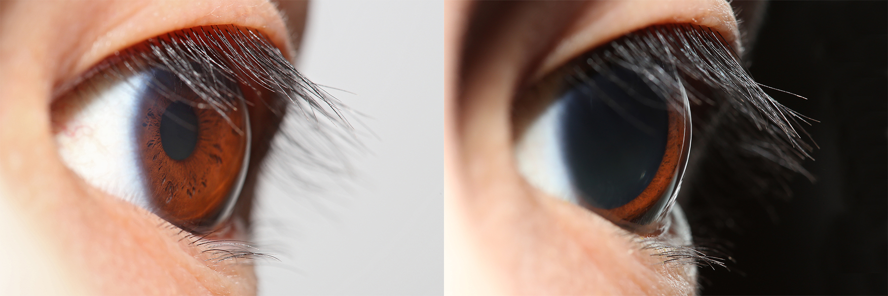
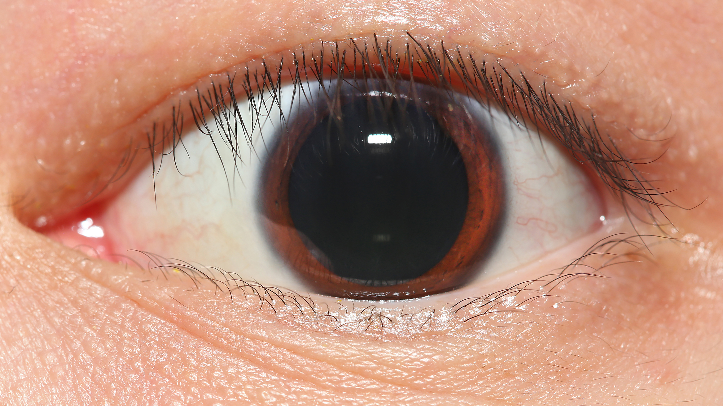
The iris (: irides or irises) is a thin, annular structure in the eye in most mammals and birds that is responsible for controlling the diameter and size of the pupil, and thus the amount of light reaching the retina. In optical terms, the pupil is the eye's aperture, while the iris is the diaphragm. Eye color is defined by the iris. Etymology The word "iris" is derived from the Greek word for "rainbow", also its goddess plus messenger of the gods in the ''Iliad'', because of the many colours of this eye part. Structure The iris consists of two layers: the front pigmented fibrovascular layer known as a stroma and, behind the stroma, pigmented epithelial cells. The stroma is connected to a sphincter muscle ( sphincter pupillae), which contracts the pupil in a circular motion, and a set of dilator muscles (dilator pupillae), which pull the iris radially to enlarge the pupil, pulling it in folds. The sphincter pupillae is the opposing muscle of the dilator pupillae. The pupil ... [...More Info...] [...Related Items...] OR: [Wikipedia] [Google] [Baidu] |
Eye Color
Eye color is a polygene, polygenic phenotypic trait determined by two factors: the pigmentation of the eye's Iris (anatomy), iris and the frequency-dependence of the scattering of light by the Turbidity, turbid medium in the Stroma of iris, stroma of the iris. In humans, the pigmentation of the Iris (anatomy), iris varies from light brown to black, depending on the concentration of melanin in the iris pigment epithelium (located on the back of the iris), the melanin content within the iris stroma (located at the front of the iris), and the cellular density of the stroma. The appearance of blue, green, and hazel eyes results from the Tyndall effect, Tyndall scattering of light in the stroma, a phenomenon similar to Rayleigh scattering which accounts for the blue sky. Neither blue nor green pigments are present in the human iris or vitreous humour. This is an example of structural color, which depends on the lighting conditions, especially for lighter-colored eyes. The brightly c ... [...More Info...] [...Related Items...] OR: [Wikipedia] [Google] [Baidu] |
Iris Regions
Iris most often refers to: *Iris (anatomy), part of the eye * Iris (color), an ambiguous color term *Iris (mythology), a Greek goddess * ''Iris'' (plant), a genus of flowering plants * Iris (given name), a feminine given name, and a list of people so named Iris or IRIS may also refer to: Arts and media Fictional entities * Iris (''American Horror Story''), an ''American Horror Story: Hotel'' character * Iris (''Fire Force''), a character in the manga series ''Fire Force'' * Iris (''Mega Man''), a ''Mega Man X4'' character ** Iris, a ''Mega Man Battle Network'' character * Iris (''Pokémon'') ** Iris (''Pokémon'' anime) * Sorceress Iris, a ''Magicians of Xanth'' character * Iris, a kaiju character in '' Gamera 3: The Revenge of Iris'' * Iris, a '' LoliRock'' character * Iris, a '' Lufia II: Rise of the Sinistrals'' (1995) character * Iris, a '' Phoenix Wright: Ace Attorney − Trials and Tribulations'' character * Iris, a ''Ruby Gloom'' character * Iris, a ''Taxi Driver ... [...More Info...] [...Related Items...] OR: [Wikipedia] [Google] [Baidu] |
Intraocular Pressure
Intraocular pressure (IOP) is the fluid pressure inside the eye. Tonometry is the method eye care professionals use to determine this. IOP is an important aspect in the evaluation of patients at risk of glaucoma. Most tonometers are calibrated to measure pressure in millimeters of mercury (mmHg). Physiology Intraocular pressure is determined by the production and drainage of aqueous humour by the ciliary body and its drainage via the trabecular meshwork and uveoscleral outflow. The reason for this is because the vitreous humour in the posterior segment has a relatively fixed volume and thus does not affect intraocular pressure regulation. An important quantitative relationship (Goldmann's equation) is as follows: :P_o = \frac + P_v Where: * P_o is the IOP in millimeters of mercury (mmHg) * F the rate of aqueous humour formation in microliters per minute (μL/min) * U the resorption of aqueous humour through the uveoscleral route (μL/min) * C is the facility of outflow in mic ... [...More Info...] [...Related Items...] OR: [Wikipedia] [Google] [Baidu] |
Aqueous Humour
The aqueous humour is a transparent water-like fluid similar to blood plasma, but containing low protein concentrations. It is secreted from the ciliary body, a structure supporting the lens of the eyeball. It fills both the anterior and the posterior chambers of the eye, and is not to be confused with the vitreous humour, which is located in the space between the lens and the retina, also known as the posterior cavity or vitreous chamber. Blood cannot normally enter the eyeball. Structure Composition * Amino acids: transported by ciliary muscles * 98% water * Electrolytes ( pH = 7.4 -one source gives 7.1) ** Sodium = 142.09 ** Potassium = 2.2 - 4.0 ** Calcium = 1.8 ** Magnesium = 1.1 ** Chloride = 131.6 ** HCO3− = 20.15 ** Phosphate = 0.62 ** Osm = 304 * Ascorbic acid * Glutathione * Immunoglobulins Function * Maintains the intraocular pressure and inflates the globe of the eye. It is this hydrostatic pressure that keeps the eyeball in a roughly spherical shape and keeps ... [...More Info...] [...Related Items...] OR: [Wikipedia] [Google] [Baidu] |
Trabecular Meshwork
The trabecular meshwork is an area of tissue in the eye located around the base of the cornea, near the ciliary body, and is responsible for draining the aqueous humor from the eye via the anterior chamber (the chamber on the front of the eye covered by the cornea). The tissue is spongy and lined by trabeculocytes; it allows fluid to drain into a set of tubes called Schlemm's canal which is lined by endothelium with blood and lymphatic properties that allow aqueous humor to flow into the blood system. Structure The meshwork is divided up into three parts, with characteristically different ultrastructures: #''Inner uveal meshwork'' - Closest to the anterior chamber angle, contains thin cord-like trabeculae, orientated predominantly in a radial fashion, enclosing trabeculae spaces larger than the corneoscleral meshwork. #''Corneoscleral meshwork'' - Contains a large amount of elastin, arranged as a series of thin, flat, perforated sheets arranged in a laminar pattern; c ... [...More Info...] [...Related Items...] OR: [Wikipedia] [Google] [Baidu] |
Uvea
The uvea (; derived from meaning "grape"), also called the uveal layer, uveal coat, uveal tract, vascular tunic or vascular layer, is the pigmented middle layer of the three concentric layers that make up an eye, precisely between the inner retina and the outer fibrous layer composed of the sclera and cornea The cornea is the transparency (optics), transparent front part of the eyeball which covers the Iris (anatomy), iris, pupil, and Anterior chamber of eyeball, anterior chamber. Along with the anterior chamber and Lens (anatomy), lens, the cornea .... History and etymology The originally medieval Latin term comes from the Latin word ''uva'' ("grape") and is a reference to its grape-like appearance (reddish-blue or almost black colour, wrinkled appearance and grape-like size and shape when stripped intact from a cadaveric eye). In fact, it is a partial loan translation of the Ancient Greek term for the choroid, which literally means “covering resembling a grape ... [...More Info...] [...Related Items...] OR: [Wikipedia] [Google] [Baidu] |
Ciliary Body
The ciliary body is a part of the eye that includes the ciliary muscle, which controls the shape of the lens, and the ciliary epithelium, which produces the aqueous humor. The aqueous humor is produced in the non-pigmented portion of the ciliary body. The ciliary body is part of the uvea, the layer of tissue that delivers oxygen and nutrients to the eye tissues. The ciliary body joins the ora serrata of the choroid to the root of the iris.Cassin, B. and Solomon, S. ''Dictionary of Eye Terminology''. Gainesville, Florida: Triad Publishing Company, 1990. Structure The ciliary body is a ring-shaped thickening of tissue inside the eye that divides the posterior chamber from the vitreous body. It contains the ciliary muscle, vessels, and fibrous connective tissue. Folds on the inner ciliary epithelium are called ciliary processes, and these secrete aqueous humor into the posterior chamber. The aqueous humor then flows through the iris into the anterior chamber. The ciliary bo ... [...More Info...] [...Related Items...] OR: [Wikipedia] [Google] [Baidu] |
Encyclopædia Britannica 2006 Ultimate Reference Suite DVD
An encyclopedia is a reference work or compendium providing summaries of knowledge, either general or special, in a particular field or discipline. Encyclopedias are divided into articles or entries that are arranged alphabetically by article name or by thematic categories, or else are hyperlinked and searchable. Encyclopedia entries are longer and more detailed than those in most dictionaries. Generally speaking, encyclopedia articles focus on ''factual information'' concerning the subject named in the article's title; this is unlike dictionary entries, which focus on linguistic information about words, such as their etymology, meaning, pronunciation, use, and grammatical forms.Béjoint, Henri (2000)''Modern Lexicography'', pp. 30–31. Oxford University Press. Encyclopedias have existed for around 2,000 years and have evolved considerably during that time as regards language (written in a major international or a vernacular language), size (few or many volumes), intent ( ... [...More Info...] [...Related Items...] OR: [Wikipedia] [Google] [Baidu] |
Epithelium
Epithelium or epithelial tissue is a thin, continuous, protective layer of cells with little extracellular matrix. An example is the epidermis, the outermost layer of the skin. Epithelial ( mesothelial) tissues line the outer surfaces of many internal organs, the corresponding inner surfaces of body cavities, and the inner surfaces of blood vessels. Epithelial tissue is one of the four basic types of animal tissue, along with connective tissue, muscle tissue and nervous tissue. These tissues also lack blood or lymph supply. The tissue is supplied by nerves. There are three principal shapes of epithelial cell: squamous (scaly), columnar, and cuboidal. These can be arranged in a singular layer of cells as simple epithelium, either simple squamous, simple columnar, or simple cuboidal, or in layers of two or more cells deep as stratified (layered), or ''compound'', either squamous, columnar or cuboidal. In some tissues, a layer of columnar cells may appear to be stratified d ... [...More Info...] [...Related Items...] OR: [Wikipedia] [Google] [Baidu] |
Pupillary Light Reflex
The pupillary light reflex (PLR) or photopupillary reflex is a reflex that controls the diameter of the pupil, in response to the intensity ( luminance) of light that falls on the retinal ganglion cells of the retina in the back of the eye, thereby assisting in adaptation of vision to various levels of lightness/darkness. A greater intensity of light causes the pupil to constrict ( miosis/myosis; thereby allowing less light in), whereas a lower intensity of light causes the pupil to dilate ( mydriasis, expansion; thereby allowing more light in). Thus, the pupillary light reflex regulates the intensity of light entering the eye. Light shone into one eye will cause both pupils to constrict. Terminology The pupil is the dark circular opening in the center of the iris and is where light enters the eye. By analogy with a camera, the pupil is equivalent to aperture, whereas the iris is equivalent to the diaphragm. It may be helpful to consider the ''Pupillary reflex'' as an Iris ... [...More Info...] [...Related Items...] OR: [Wikipedia] [Google] [Baidu] |
Dilator Pupillae
The iris dilator muscle (pupil dilator muscle, pupillary dilator, radial muscle of iris, radiating fibers), is a smooth muscle of the eye, running radially in the iris and therefore fit as a dilator. The pupillary dilator consists of a spokelike arrangement of modified contractile cells called myoepithelial cells. These cells are stimulated by the sympathetic nervous system. When stimulated, the cells contract, widening the pupil and allowing more light to enter the eye. The ciliary muscle, pupillary sphincter muscle and pupillary dilator muscle sometimes are called intrinsic ocular muscles or intraocular muscles. Structure Innervation It is innervated by the sympathetic system, which acts by releasing noradrenaline, which acts on α1-receptors. Thus, when presented with a threatening stimulus that activates the fight-or-flight response, this innervation contracts the muscle and dilates the pupil, thus temporarily letting more light reach the retina. The dilator muscle is ... [...More Info...] [...Related Items...] OR: [Wikipedia] [Google] [Baidu] |


