|
Intraocular Muscle
Intrinsic ocular muscles or intraocular muscles{{cite book , last1=Ludwig , first1=Parker E. , last2=Aslam , first2=Sanah , last3=Czyz , first3=Craig N. , title=StatPearls , date=2024 , publisher=StatPearls Publishing , chapter-url=https://www.ncbi.nlm.nih.gov/books/NBK470534/ , chapter=Anatomy, Head and Neck: Eye Muscles , pmid=29262013 are muscles of the inside of the eye structure. The intraocular muscles are responsible for adjusting the shape of the lens and the size of the pupil. They're different from the extraocular muscles that are outside of the eye and control the external movement of the eye. There are three intrisic ocular muscles: the ciliary muscle, pupillary sphincter muscle ( sphincter pupillae) and pupillary dilator muscle (dilator pupillae). All of them are smooth muscles. The ciliary muscle is attached to the zonular fibers and the zonular fibers are the suspensory ligaments of the lens. The ciliary muscle controls accommodation by altering the shape of ... [...More Info...] [...Related Items...] OR: [Wikipedia] [Google] [Baidu] |
Zonular Fiber
The zonule of Zinn () (Zinn's membrane, ciliary zonule) (after Johann Gottfried Zinn) is a ring of fibrous strands forming a zonule (little band) that connects the ciliary body with the crystalline lens of the eye. The Zonular fibers are viscoelastic cables, although their component microfibrils are stiff structures. These fibers are sometimes collectively referred to as the suspensory ligaments of the lens, as they act like suspensory ligaments. Development The non-pigmented ciliary epithelial cells of the eye synthesize portions of the zonules. Anatomy The zonule of Zinn is split into two layers: a thin layer, which lies near the hyaloid fossa, and a thicker layer, which is a collection of zonular fibers. Together, the fibers are known as the suspensory ligament of the lens. The zonules are about 1–2 μm in diameter. The zonules attach to the lens capsule 2 mm anterior and 1 mm posterior to the equator, and arise of the ciliary epithelium from the pars plana region ... [...More Info...] [...Related Items...] OR: [Wikipedia] [Google] [Baidu] |
Extraocular Muscle
The extraocular muscles, or extrinsic ocular muscles, are the seven extrinsic muscles of the eye in humans and other animals. Six of the extraocular muscles, the four recti muscles, and the superior and inferior oblique muscles, control movement of the eye. The other muscle, the levator palpebrae superioris, controls eyelid elevation. The actions of the six muscles responsible for eye movement depend on the position of the eye at the time of muscle contraction. The ciliary muscle, pupillary sphincter muscle and pupillary dilator muscle sometimes are called intrinsic ocular muscles or intraocular muscles. Structure Since only a small part of the eye called the fovea provides sharp vision, the eye must move to follow a target. Eye movements must be precise and fast. This is seen in scenarios like reading, where the reader must shift gaze constantly. Although under voluntary control, most eye movement is accomplished without conscious effort. Precisely how the integration betwe ... [...More Info...] [...Related Items...] OR: [Wikipedia] [Google] [Baidu] |
Miosis
Miosis, or myosis (), is excessive constriction of the pupil. citing: Mosby's Medical Dictionary, 8th ed. The opposite condition, mydriasis, is the dilation of the pupil. Anisocoria is the condition of one pupil being more dilated than the other. Causes Age * Senile miosis (a reduction in the size of a person's pupil in old age)Diseases *[...More Info...] [...Related Items...] OR: [Wikipedia] [Google] [Baidu] |
Mydriasis
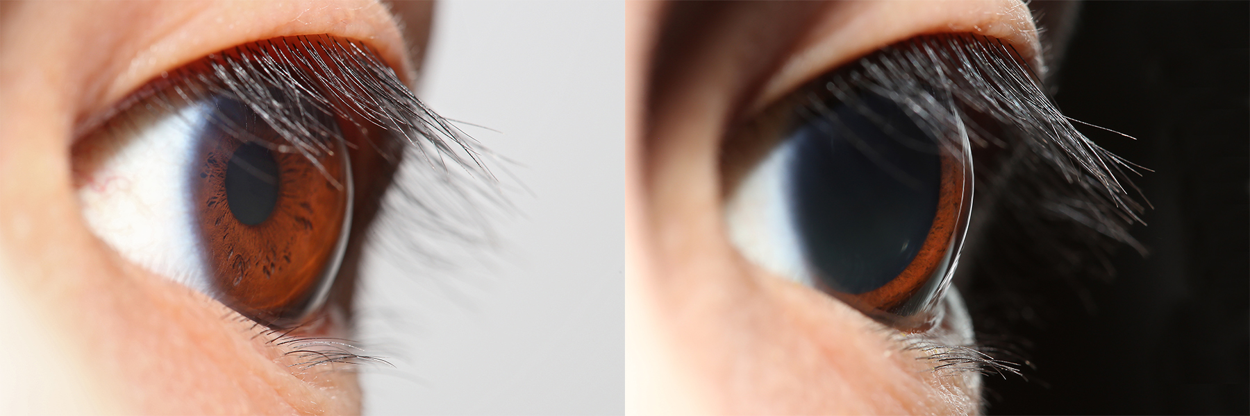
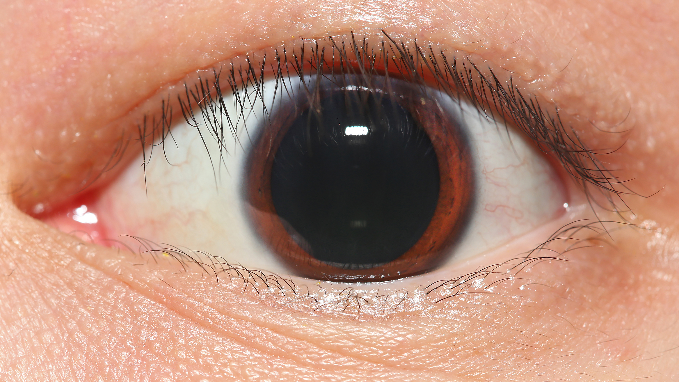
Mydriasis is the Pupillary dilation, dilation of the pupil, usually having a non-physiological cause, or sometimes a physiological pupillary response. Non-physiological causes of mydriasis include disease, Physical trauma, trauma, or the use of certain types of drug, drugs. It may also be of unknown cause. Normally, as part of the pupillary light reflex, the pupil dilates in the dark and miosis, constricts in the light to respectively improve vividity at night and to protect the retina from sunlight damage during the day. A ''mydriatic'' pupil will remain excessively large even in a bright environment. The excitation of the radial fibres of the iris which increases the pupillary aperture is referred to as a mydriasis. More generally, mydriasis also refers to the natural dilation of pupils, for instance in low light conditions or under sympathetic stimulation. Mydriasis is frequently induced by drugs for certain Ophthalmology, ophthalmic examinations and procedures, particularly th ... [...More Info...] [...Related Items...] OR: [Wikipedia] [Google] [Baidu] |
Pupil
The pupil is a hole located in the center of the iris of the eye that allows light to strike the retina.Cassin, B. and Solomon, S. (1990) ''Dictionary of Eye Terminology''. Gainesville, Florida: Triad Publishing Company. It appears black because light rays entering the pupil are either absorbed by the tissues inside the eye directly, or absorbed after diffuse reflections within the eye that mostly miss exiting the narrow pupil. The size of the pupil is controlled by the iris, and varies depending on many factors, the most significant being the amount of light in the environment. The term "pupil" was coined by Gerard of Cremona. In humans, the pupil is circular, but its shape varies between species; some cats, reptiles, and foxes have vertical slit pupils, goats and sheep have horizontally oriented pupils, and some catfish have annular types. In optical terms, the anatomical pupil is the eye's aperture and the iris is the aperture stop. The image of the pupil as seen from o ... [...More Info...] [...Related Items...] OR: [Wikipedia] [Google] [Baidu] |
Iris (anatomy)
The iris (: irides or irises) is a thin, annular structure in the eye in most mammals and birds that is responsible for controlling the diameter and size of the pupil, and thus the amount of light reaching the retina. In optical terms, the pupil is the eye's aperture, while the iris is the diaphragm (optics), diaphragm. Eye color is defined by the iris. Etymology The word "iris" is derived from the Greek word for "rainbow", also Iris (mythology), its goddess plus messenger of the gods in the ''Iliad'', because of the many eye color, colours of this eye part. Structure The iris consists of two layers: the front pigmented Wikt:fibrovascular, fibrovascular layer known as a stroma of iris, stroma and, behind the stroma, pigmented epithelial cells. The stroma is connected to a sphincter muscle (sphincter pupillae), which contracts the pupil in a circular motion, and a set of dilator muscles (dilator pupillae), which pull the iris radially to enlarge the pupil, pulling it in folds. ... [...More Info...] [...Related Items...] OR: [Wikipedia] [Google] [Baidu] |
Accommodation (vertebrate Eye)
Accommodation is the process by which the vertebrate eye changes optical power to maintain a clear image or focus on an object as its distance varies. In this, distances vary for individuals from the far point—the maximum distance from the eye for which a clear image of an object can be seen, to the near point—the minimum distance for a clear image. Accommodation usually acts like a reflex, including part of the accommodation-convergence reflex, but it can also be consciously controlled. The main ways animals may change focus are: * Changing the shape of the lens. * Changing the position of the lens relative to the retina. * Changing the axial length of the eyeball. * Changing the shape of the cornea. Focusing mechanisms Focusing the light scattered by objects in a three dimensional environment into a two dimensional collection of individual bright points of light requires the light to be bent. To get a good image of these points of light on a defined area requires a p ... [...More Info...] [...Related Items...] OR: [Wikipedia] [Google] [Baidu] |
Smooth Muscle
Smooth muscle is one of the three major types of vertebrate muscle tissue, the others being skeletal and cardiac muscle. It can also be found in invertebrates and is controlled by the autonomic nervous system. It is non- striated, so-called because it has no sarcomeres and therefore no striations (''bands'' or ''stripes''). It can be divided into two subgroups, ''single-unit'' and ''multi-unit'' smooth muscle. Within single-unit muscle, the whole bundle or sheet of smooth muscle cells contracts as a syncytium. Smooth muscle is found in the walls of hollow organs, including the stomach, intestines, bladder and uterus. In the walls of blood vessels, and lymph vessels, (excluding blood and lymph capillaries) it is known as vascular smooth muscle. There is smooth muscle in the tracts of the respiratory, urinary, and reproductive systems. In the eyes, the ciliary muscles, iris dilator muscle, and iris sphincter muscle are types of smooth muscles. The iris dilator and s ... [...More Info...] [...Related Items...] OR: [Wikipedia] [Google] [Baidu] |
Intraocular Muscle
Intrinsic ocular muscles or intraocular muscles{{cite book , last1=Ludwig , first1=Parker E. , last2=Aslam , first2=Sanah , last3=Czyz , first3=Craig N. , title=StatPearls , date=2024 , publisher=StatPearls Publishing , chapter-url=https://www.ncbi.nlm.nih.gov/books/NBK470534/ , chapter=Anatomy, Head and Neck: Eye Muscles , pmid=29262013 are muscles of the inside of the eye structure. The intraocular muscles are responsible for adjusting the shape of the lens and the size of the pupil. They're different from the extraocular muscles that are outside of the eye and control the external movement of the eye. There are three intrisic ocular muscles: the ciliary muscle, pupillary sphincter muscle ( sphincter pupillae) and pupillary dilator muscle (dilator pupillae). All of them are smooth muscles. The ciliary muscle is attached to the zonular fibers and the zonular fibers are the suspensory ligaments of the lens. The ciliary muscle controls accommodation by altering the shape of ... [...More Info...] [...Related Items...] OR: [Wikipedia] [Google] [Baidu] |
Dilator Pupillae
The iris dilator muscle (pupil dilator muscle, pupillary dilator, radial muscle of iris, radiating fibers), is a smooth muscle of the eye, running radially in the iris and therefore fit as a dilator. The pupillary dilator consists of a spokelike arrangement of modified contractile cells called myoepithelial cells. These cells are stimulated by the sympathetic nervous system. When stimulated, the cells contract, widening the pupil and allowing more light to enter the eye. The ciliary muscle, pupillary sphincter muscle and pupillary dilator muscle sometimes are called intrinsic ocular muscles or intraocular muscles. Structure Innervation It is innervated by the sympathetic system, which acts by releasing noradrenaline, which acts on α1-receptors. Thus, when presented with a threatening stimulus that activates the fight-or-flight response, this innervation contracts the muscle and dilates the pupil, thus temporarily letting more light reach the retina. The dilator muscle is ... [...More Info...] [...Related Items...] OR: [Wikipedia] [Google] [Baidu] |
Pupillary Dilator Muscle
The iris dilator muscle (pupil dilator muscle, pupillary dilator, radial muscle of iris, radiating fibers), is a smooth muscle of the eye, running radially in the iris and therefore fit as a dilator. The pupillary dilator consists of a spokelike arrangement of modified contractile cells called myoepithelial cells. These cells are stimulated by the sympathetic nervous system. When stimulated, the cells contract, widening the pupil and allowing more light to enter the eye. The ciliary muscle, pupillary sphincter muscle and pupillary dilator muscle sometimes are called intrinsic ocular muscles or intraocular muscles. Structure Innervation It is innervated by the sympathetic system, which acts by releasing noradrenaline, which acts on α1-receptors. Thus, when presented with a threatening stimulus that activates the fight-or-flight response, this innervation contracts the muscle and dilates the pupil, thus temporarily letting more light reach the retina. The dilator muscle is inne ... [...More Info...] [...Related Items...] OR: [Wikipedia] [Google] [Baidu] |


