Canine Space on:
[Wikipedia]
[Google]
[Amazon]
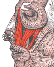
The canine space (also termed the infra-orbital space), is a fascial space of the head and neck (sometimes also termed fascial spaces or tissue spaces). It is a thin 

potential space
In anatomy, a potential space is a space between two adjacent structures that are normally pressed together (directly apposed). Many anatomic spaces are potential spaces, which means that they are potential rather than realized (with their realiz ...
on the face, and is paired on either side. It is located between the levator anguli oris muscle inferiorly and the levator labii superioris muscle
The levator labii superioris (pl. ''levatores labii superioris'', also called quadratus labii superioris, pl. ''quadrati labii superioris'') is a muscle of the human body used in facial expression. It is a broad sheet, the origin of which exten ...
superiorly. The term is derived from the fact that the space is in the region of the canine fossa
In the musculoskeletal anatomy of the human head, lateral to the incisive fossa of the maxilla is a depression called the canine fossa. It is larger and deeper than the comparable incisive fossa, and is separated from it by a vertical ridge, the ...
, and that infections originating from the maxillary canine tooth may spread to involve the space. ''Infra-orbital'' is derived from '' infra-'' meaning below and orbit
In celestial mechanics, an orbit is the curved trajectory of an object such as the trajectory of a planet around a star, or of a natural satellite around a planet, or of an artificial satellite around an object or position in space such as ...
which refers to the eye socket.


Structure
Boundaries
The boundaries of the canine space are: * the nasal cartilages anteriorly * the buccal space posteriorly * the quadratus labii superioris muscle (levator labii superioris) superiorly * theoral mucosa
The oral mucosa is the mucous membrane lining the inside of the mouth. It comprises stratified squamous epithelium, termed "oral epithelium", and an underlying connective tissue termed '' lamina propria''. The oral cavity has sometimes been des ...
of the maxillary labial sulcus inferiorly
* the quadratus labii superioris muscle superficially
* and the deep border is created by the levator anguli oris muscle.
Communications
The canine space communicates with the buccal space posteriorly.Function
Contents
The contents of the canine space are: * the angular artery and angular vein * the infra-orbital nerve (a branch of the maxillary division of thetrigeminal nerve
In neuroanatomy, the trigeminal nerve ( lit. ''triplet'' nerve), also known as the fifth cranial nerve, cranial nerve V, or simply CN V, is a cranial nerve responsible for sensation in the face and motor functions such as biting and chew ...
)
Clinical significance
Canine space infections may occur by spread of infection from the buccal space. Signs and symptoms of a canine space abscess might include swelling that obliterates thenasolabial fold
The nasolabial folds, commonly known as "smile lines" or "laugh lines", are facial features. They are the two skin folds that run from each side of the nose to the corners of the mouth. They are defined by facial structures that support the bucca ...
. If left untreated, infections of this space will eventually spontaneously drain via the medial or lateral canthus of the eye, as this is the path of least resistance. Treatment is usually by surgical incision and drainage
Incision and drainage (I&D), also known as clinical lancing, are minor surgical procedures to release pus or pressure built up under the skin, such as from an abscess, boil, or infected paranasal sinus. It is performed by treating the area with a ...
, and the incision is placed inside the mouth to avoid a facial scar.
Rarely, when infections of the canine space erode into the infra-orbital vein or the inferior ophthalmic vein
The inferior ophthalmic vein is a vein of the orbit that - together with the superior ophthalmic vein - represents the principal drainage system of the orbit. It begins from a venous network in the front of the orbit, then passes backwards throu ...
(via the sinuses
Paranasal sinuses are a group of four paired air-filled spaces that surround the nasal cavity. The maxillary sinuses are located under the eyes; the frontal sinuses are above the eyes; the ethmoidal sinuses are between the eyes and the sphenoid ...
), there can be spread via the common ophthalmic vein through the superior orbital fissure and into the cavernous sinus
The cavernous sinus within the human head is one of the dural venous sinuses creating a cavity called the lateral sellar compartment bordered by the temporal bone of the skull and the sphenoid bone, lateral to the sella turcica.
Structure
The cave ...
. This can result in septic cavernous sinus thrombosis
The cavernous sinus within the human head is one of the dural venous sinuses creating a cavity called the lateral sellar compartment bordered by the temporal bone of the skull and the sphenoid bone, lateral to the sella turcica.
Structure
The ca ...
, which is a rare, but life-threatening condition.
Odontogenic infection
Odontogenic infection
An odontogenic infection is an infection that originates within a tooth or in the closely surrounding tissues. The term is derived from '' odonto-'' (Ancient Greek: , – 'tooth') and '' -genic'' (Ancient Greek: , ; – 'birth'). The most common ...
s may spread to involve the canine space. The most likely causative tooth is the maxillary canine or maxillary first premolar. This occurs when pus (e.g. from a periapical abscess
A dental abscess is a localized collection of pus associated with a tooth. The most common type of dental abscess is a periapical abscess, and the second most common is a periodontal abscess. In a periapical abscess, usually the origin is a ba ...
), perforates the buccal cortical plate of the maxilla above the level of attachment of the levator anguli oris muscle. This is more likely if the tooth root is long (the maxillary canine has the longest root of all the teeth), and its apex lies at a level above the muscle attachment.
References
{{Digestive tract Mouth Otorhinolaryngology Oral and maxillofacial surgery Fascial spaces of the head and neck