|
Potential Space
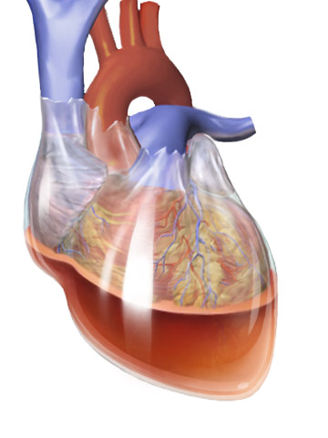
In anatomy, a potential space is a space between two adjacent structures that are normally pressed together (directly apposed). Many anatomic spaces are potential spaces, which means that they are potential rather than realized (with their realization being dynamic according to physiologic or pathophysiologic events). In other words, they are like an empty plastic bag that has not been opened (two walls collapsed against each other; no interior volume until opened) or a balloon that has not been inflated. The pleural space, between the visceral and parietal pleura of the lung, is a potential space. Though it only contains a small amount of fluid normally, it can sometimes accumulate fluid or air that widens the space. The pericardial space is another potential space that may fill with fluid (effusion) in certain disease states (e.g. pericarditis; a large pericardial effusion may result in cardiac tamponade. Examples * costodiaphragmatic recess * pericardial cavity *epidural space ... [...More Info...] [...Related Items...] OR: [Wikipedia] [Google] [Baidu] |
Anatomy
Anatomy () is the branch of biology concerned with the study of the structure of organisms and their parts. Anatomy is a branch of natural science that deals with the structural organization of living things. It is an old science, having its beginnings in prehistoric times. Anatomy is inherently tied to developmental biology, embryology, comparative anatomy, evolutionary biology, and phylogeny, as these are the processes by which anatomy is generated, both over immediate and long-term timescales. Anatomy and physiology, which study the structure and function (biology), function of organisms and their parts respectively, make a natural pair of related disciplines, and are often studied together. Human anatomy is one of the essential basic research, basic sciences that are applied in medicine. The discipline of anatomy is divided into macroscopic scale, macroscopic and microscopic scale, microscopic. Gross anatomy, Macroscopic anatomy, or gross anatomy, is the examination of an ... [...More Info...] [...Related Items...] OR: [Wikipedia] [Google] [Baidu] |
Pleural Cavity
The pleural cavity, pleural space, or interpleural space is the potential space between the pleurae of the pleural sac that surrounds each lung. A small amount of serous pleural fluid is maintained in the pleural cavity to enable lubrication between the membranes, and also to create a pressure gradient. The serous membrane that covers the surface of the lung is the visceral pleura and is separated from the outer membrane the parietal pleura by just the film of pleural fluid in the pleural cavity. The visceral pleura follows the fissures of the lung and the root of the lung structures. The parietal pleura is attached to the mediastinum, the upper surface of the diaphragm, and to the inside of the ribcage. Structure In humans, the left and right lungs are completely separated by the mediastinum, and there is no communication between their pleural cavities. Therefore, in cases of a unilateral pneumothorax, the contralateral lung will remain functioning normally unless there is ... [...More Info...] [...Related Items...] OR: [Wikipedia] [Google] [Baidu] |
Lung
The lungs are the primary organs of the respiratory system in humans and most other animals, including some snails and a small number of fish. In mammals and most other vertebrates, two lungs are located near the backbone on either side of the heart. Their function in the respiratory system is to extract oxygen from the air and transfer it into the bloodstream, and to release carbon dioxide from the bloodstream into the atmosphere, in a process of gas exchange. Respiration is driven by different muscular systems in different species. Mammals, reptiles and birds use their different muscles to support and foster breathing. In earlier tetrapods, air was driven into the lungs by the pharyngeal muscles via buccal pumping, a mechanism still seen in amphibians. In humans, the main muscle of respiration that drives breathing is the diaphragm. The lungs also provide airflow that makes vocal sounds including human speech possible. Humans have two lungs, one on the left and on ... [...More Info...] [...Related Items...] OR: [Wikipedia] [Google] [Baidu] |
Pericarditis
Pericarditis is inflammation of the pericardium, the fibrous sac surrounding the heart. Symptoms typically include sudden onset of sharp chest pain, which may also be felt in the shoulders, neck, or back. The pain is typically less severe when sitting up and more severe when lying down or breathing deeply. Other symptoms of pericarditis can include fever, weakness, palpitations, and shortness of breath. The onset of symptoms can occasionally be gradual rather than sudden. The cause of pericarditis often remains unknown but is believed to be most often due to a viral infection. Other causes include bacterial infections such as tuberculosis, uremic pericarditis, heart attack, cancer, autoimmune disorders, and chest trauma. Diagnosis is based on the presence of chest pain, a pericardial rub, specific electrocardiogram (ECG) changes, and fluid around the heart. A heart attack may produce similar symptoms to pericarditis. Treatment in most cases is with NSAIDs and possibly the ant ... [...More Info...] [...Related Items...] OR: [Wikipedia] [Google] [Baidu] |
Cardiac Tamponade
Cardiac tamponade, also known as pericardial tamponade (), is the buildup of fluid in the pericardium (the sac around the heart), resulting in compression of the heart. Onset may be rapid or gradual. Symptoms typically include those of obstructive shock including shortness of breath, weakness, lightheadedness, and cough. Other symptoms may relate to the underlying cause. Common causes of cardiac tamponade include cancer, kidney failure, chest trauma, myocardial infarction, and pericarditis. Other causes include connective tissues diseases, hypothyroidism, aortic rupture, autoimmune disease, and complications of cardiac surgery. In Africa, tuberculosis is a relatively common cause. Diagnosis may be suspected based on low blood pressure, jugular venous distension, or quiet heart sounds (together known as Beck's triad). A pericardial rub may be present in cases due to inflammation. The diagnosis may be further supported by specific electrocardiogram (ECG) changes, chest X-ra ... [...More Info...] [...Related Items...] OR: [Wikipedia] [Google] [Baidu] |
Costodiaphragmatic Recess
The costodiaphragmatic recess, also called the costophrenic recess or phrenicocostal sinus, costodiaphragmatic-recess Retrieved May 2011 Imaging In anatomy, the costophrenic angles are the places where the diaphragm (''-phrenic'') meets the ribs (''costo-''). Each costophrenic angle can normally be seen as on chest x-ray as a sharply-pointed, downward indentation (dark) between each hemi-diaphragm (white) and the adjacent chest wall (white). A small portion of each lung normally reaches into the costophrenic angle. The normal angle usually measures thirty degrees. Pleural effusion With pleural effusion, fluid often builds up in the costophrenic angle (due to gravity). This can push the lung upwards, resulting in "blunting" of the costophrenic angle. The posterior angle is the deepest. Obtuse angulation is sign of disease. Chest x-ray is the first test done to confirm the presence of pleural fluid. The lateral upright chest x-ray should be examined when a pleural effusion is s ... [...More Info...] [...Related Items...] OR: [Wikipedia] [Google] [Baidu] |
Pericardial Cavity
The pericardium, also called pericardial sac, is a double-walled sac containing the heart and the roots of the great vessels. It has two layers, an outer layer made of strong connective tissue (fibrous pericardium), and an inner layer made of serous membrane (serous pericardium). It encloses the pericardial cavity, which contains pericardial fluid, and defines the middle mediastinum. It separates the heart from interference of other structures, protects it against infection and blunt trauma, and lubricates the heart's movements. The English name originates from the Ancient Greek prefix "''peri-''" (περί; "around") and the suffix "''-cardion''" (κάρδιον; "heart"). Anatomy The pericardium is a tough fibroelastic sac which covers the heart from all sides except at the cardiac root (where the great vessels join the heart) and the bottom (where only the serous pericardium exists to cover the upper surface of the central tendon of diaphragm, central tendon of thoracic d ... [...More Info...] [...Related Items...] OR: [Wikipedia] [Google] [Baidu] |
Epidural Space
In anatomy, the epidural space is the potential space between the dura mater and vertebrae ( spine). The anatomy term "epidural space" has its origin in the Ancient Greek language; , "on, upon" + dura mater also known as "epidural cavity", "extradural space" or "peridural space". In humans the epidural space contains lymphatics, spinal nerve roots, loose connective tissue, adipose tissue, small arteries, dural venous sinuses and a network of internal vertebral venous plexuses. Cranial epidural space In the skull, the periosteal layer of the dura mater adheres to the inner surface of the skull bones while the meningeal layer lays over the arachnoid mater. Between them is the epidural space. The two layers of the dura mater separate at several places, with the meningeal layer projecting deeper into the brain parenchyma forming fibrous septa that compartmentalize the brain tissue. At these sites, the epidural space is wide enough to house the epidural venous sinuses. There are fo ... [...More Info...] [...Related Items...] OR: [Wikipedia] [Google] [Baidu] |
Subdural Space
The subdural space (or subdural cavity) is a potential space that can be opened by the separation of the arachnoid mater from the dura mater as the result of trauma, pathologic process, or the absence of cerebrospinal fluid as seen in a cadaver. In the cadaver, due to the absence of cerebrospinal fluid in the subarachnoid space, the arachnoid mater falls away from the dura mater. It may also be the site of trauma, such as a subdural hematoma, causing abnormal separation of dura and arachnoid mater. Hence, the subdural space is referred to as " potential" or "artificial" space. See also * Epidural space * Subarachnoid space * Meninges * Subdural hematoma A subdural hematoma (SDH) is a type of bleeding in which a collection of blood—usually but not always associated with a traumatic brain injury—gathers between the inner layer of the dura mater and the arachnoid mater of the meninges surround ... References External links * * Meninges {{Neuroanatomy-stub ... [...More Info...] [...Related Items...] OR: [Wikipedia] [Google] [Baidu] |
Peritoneal Cavity
The peritoneal cavity is a potential space between the parietal peritoneum (the peritoneum that surrounds the abdominal wall) and visceral peritoneum (the peritoneum that surrounds the internal organs). The parietal and visceral peritonea are layers of the peritoneum named depending on their function/location. It is one of the spaces derived from the coelomic cavity of the embryo, the others being the pleural cavities around the lungs and the pericardial cavity around the heart. It is the largest serosal sac, and the largest fluid-filled cavity, in the body and secretes approximately 50 ml of fluid per day. This fluid acts as a lubricant and has anti-inflammatory properties. The peritoneal cavity is divided into two compartments – one above, and one below the transverse colon. Compartments The peritoneal cavity is divided by the transverse colon (and its mesocolon) into an upper supracolic compartment, and a lower infracolic compartment. The liver, spleen, stomach, and lesser ... [...More Info...] [...Related Items...] OR: [Wikipedia] [Google] [Baidu] |
Buccal Space
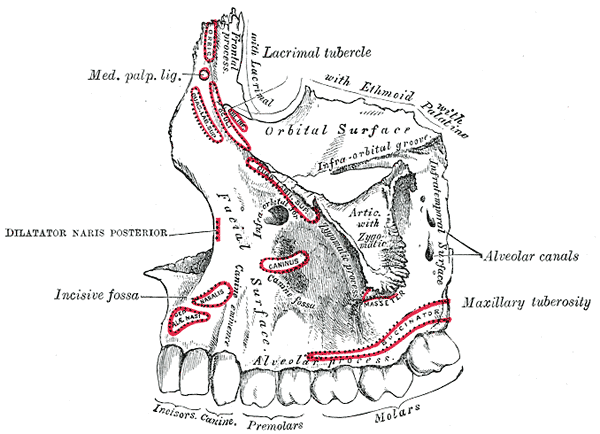
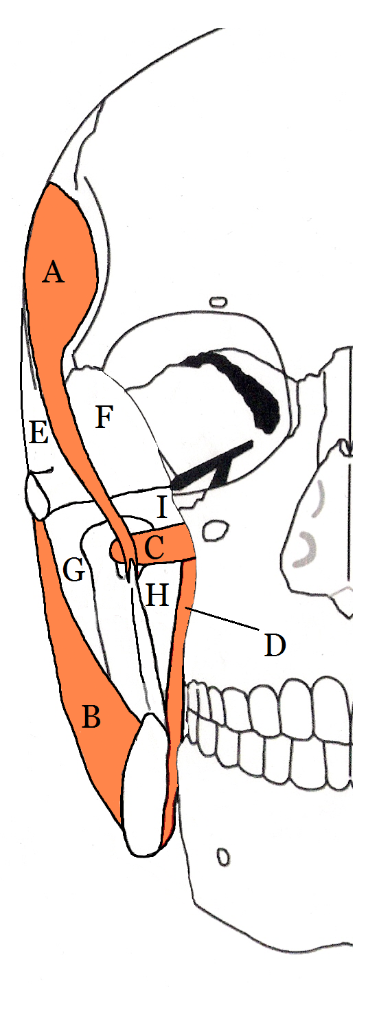
The buccal space (also termed the buccinator space) is a fascial space of the head and neck (sometimes also termed fascial tissue spaces or tissue spaces). It is a potential space in the cheek, and is paired on each side. The buccal space is superficial to the buccinator muscle and deep to the platysma muscle and the skin. The buccal space is part of the subcutaneous space, which is continuous from head to toe. Structure Boundaries The boundaries of each buccal space are: * the angle of the mouth anteriorly, * the masseter muscle posteriorly, * the zygomatic process of the maxilla and the zygomaticus muscles superiorly, * the depressor anguli oris muscle and the attachment of the deep fascia to the mandible inferiorly, * the buccinator muscle medially (the buccal space is superficial to the buccinator), * the platysma muscle, subcutaneous tissue and skin laterally (the space is deep to platysma). Communications * to the pterygomandibular space, infratemporal space, submassete ... [...More Info...] [...Related Items...] OR: [Wikipedia] [Google] [Baidu] |
Fascial Spaces Of The Head And Neck
Fascial spaces (also termed fascial tissue spaces or tissue spaces) are potential spaces that exist between the fasciae and underlying organs and other tissues. In health, these spaces do not exist; they are only created by pathology, e.g. the spread of pus or cellulitis in an infection. The fascial spaces can also be opened during the dissection of a cadaver. The fascial spaces are different from the fasciae themselves, which are bands of connective tissue that surround structures, e.g. muscles. The opening of fascial spaces may be facilitated by pathogenic bacterial release of enzymes which cause tissue lysis (e.g. hyaluronidase and collagenase). The spaces filled with loose areolar connective tissue may also be termed clefts. Other contents such as salivary glands, blood vessels, nerves and lymph nodes are dependent upon the location of the space. Those containing neurovascular tissue (nerves and blood vessels) may also be termed compartments. Generally, the spread of infection ... [...More Info...] [...Related Items...] OR: [Wikipedia] [Google] [Baidu] |


