|
Quadratus Labii Superioris Muscles
The levator labii superioris (pl. ''levatores labii superioris'', also called quadratus labii superioris, pl. ''quadrati labii superioris'') is a muscle of the human body The human body is the structure of a Human, human being. It is composed of many different types of Cell (biology), cells that together create Tissue (biology), tissues and subsequently organ systems. They ensure homeostasis and the life, viabi ... used in facial expression. It is a broad sheet, the origin of which extends from the side of the nose to the zygomatic bone. Structure Its medial fibers form the ''angular head'' (also known as the levator labii superioris alaeque nasi muscle,) which arises by a pointed extremity from the upper part of the frontal process of the maxilla and passing obliquely downward and lateralward divides into two slips. One of these is inserted into the greater alar cartilage and skin of the nose; the other is prolonged into the lateral part of the upper lip, blending ... [...More Info...] [...Related Items...] OR: [Wikipedia] [Google] [Baidu] |
Medial Infra-orbital Margin
The infraorbital margin is the lower margin of the eye socket. Structure It consists of the zygomatic bone and the maxilla, on which it separates the anterior and the orbital surface of the body of the maxilla. Function It is an attachment for the levator labii superioris muscle The levator labii superioris (pl. ''levatores labii superioris'', also called quadratus labii superioris, pl. ''quadrati labii superioris'') is a muscle of the human body used in facial expression. It is a broad sheet, the origin of which exten .... Bones of the head and neck {{musculoskeletal-stub ... [...More Info...] [...Related Items...] OR: [Wikipedia] [Google] [Baidu] |
Levator Labii Superioris Alaeque Nasi Muscle
The levator labii superioris alaeque nasi muscle is, translated from Latin, the "lifter of both the upper lip and of the wing of the nose". It has the longest name of any muscle in an animal. The muscle is attached to the upper frontal process of the maxilla and inserts into the skin of the lateral part of the nostril and upper lip. Overview Historically known as Otto's muscle, it dilates the nostril and elevates the upper lip, enabling one to snarl. Elvis Presley is famous for his use of this expression, earning the muscle's nickname "The Elvis muscle". A mnemonic to remember its name is, "Little Ladies Snore All Night." Snore- because it is the labial elevator closest to the nose. The levator labii superioris alaeque nasi is sometimes referred to as the "angular head" of the levator labii superioris muscle. See also * Levator labii superioris * Frontalis muscle The frontalis muscle () is a muscle which covers parts of the forehead of the skull. Some sources consider the fron ... [...More Info...] [...Related Items...] OR: [Wikipedia] [Google] [Baidu] |
Zygomaticus Minor Muscle
The zygomaticus minor muscle is a muscle of facial expression. It originates from the zygomatic bone, lateral to the rest of the levator labii superioris muscle, and inserts into the outer part of the upper lip. It draws the upper lip backward, upward, and outward and is used in smiling. It is innervated by the facial nerve (VII). Structure The zygomaticus minor muscle originates from the zygomatic bone. It inserts into the tissue around the upper lip, particularly blending its fibres with orbicularis oris muscle. It lies lateral to the rest of levator labii superioris muscle, and medial to its stronger synergist zygomaticus major muscle. It travels at an angle of approximately 30°. It has a mean width of around 0.5 cm. Nerve supply The zygomaticus minor muscle is supplied by the buccal branch of the facial nerve (VII). Variation The zygomaticus minor muscle may have either a straight or a curved course along its length. It may attach to both the upper lip and the latera ... [...More Info...] [...Related Items...] OR: [Wikipedia] [Google] [Baidu] |
Levator Anguli Oris
The levator anguli oris (caninus) is a facial muscle of the mouth arising from the canine fossa, immediately below the infraorbital foramen. It elevates angle of mouth medially. Its fibers are inserted into the angle of the mouth, intermingling with those of the zygomaticus, triangularis, and orbicularis oris In human anatomy, the orbicularis oris muscle is a complex of muscles in the lips that encircles the mouth. It is a sphincter, or circular muscle, but it is actually composed of four independent quadrants that interlace and give only an appearance .... Specifically, the levator anguli oris is innervated by the buccal branches of the facial nerve. Additional images File:Sobo 1909 264.png File:Sobo 1909 263.png, Seen from the inside. References External links PTCentral Muscles of the head and neck {{muscle-stub ... [...More Info...] [...Related Items...] OR: [Wikipedia] [Google] [Baidu] |
Orbit (anatomy)
In anatomy, the orbit is the cavity or socket of the skull in which the eye and its appendages are situated. "Orbit" can refer to the bony socket, or it can also be used to imply the contents. In the adult human, the volume of the orbit is , of which the eye occupies . The orbital contents comprise the eye, the orbital and retrobulbar fascia, extraocular muscles, cranial nerves II, III, IV, V, and VI, blood vessels, fat, the lacrimal gland with its sac and duct, the eyelids, medial and lateral palpebral ligaments, cheek ligaments, the suspensory ligament, septum, ciliary ganglion and short ciliary nerves. Structure The orbits are conical or four-sided pyramidal cavities, which open into the midline of the face and point back into the head. Each consists of a base, an apex and four walls."eye, human."Encyclopædia Britannica from Encyclopædia Britannica 2006 Ultimate Reference Suite DVD 2009 Openings There are two important foramina, or windows, two important fissu ... [...More Info...] [...Related Items...] OR: [Wikipedia] [Google] [Baidu] |
Orbicularis Oris
In human anatomy, the orbicularis oris muscle is a complex of muscles in the lips that encircles the mouth. It is a sphincter, or circular muscle, but it is actually composed of four independent quadrants that interlace and give only an appearance of circularity.Saladin, "Anatomy & Physiology: The Unity of Form and Function". 5th edition. McGraw Hill. Page 330 It is also one of the muscles used in the playing of all brass instruments and some woodwind instruments. This muscle closes the mouth and puckers the lips when it contracts. Structure The orbicularis oris is not a simple sphincter muscle like the orbicularis oculi; it consists of numerous strata of muscular fibers surrounding the orifice of the mouth, but having different direction. It consists partly of fibers derived from the other facial muscles which are inserted into the lips, and partly of fibers proper to the lips. Of the former, a considerable number are derived from the buccinator and form the deeper stratum of th ... [...More Info...] [...Related Items...] OR: [Wikipedia] [Google] [Baidu] |
Greater Alar Cartilage
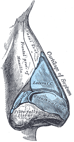
The major alar cartilage (greater alar cartilage) (lower lateral cartilage) is a thin, flexible plate, situated immediately below the lateral nasal cartilage, and bent upon itself in such a manner as to form the medial wall and lateral wall of the nostril of its own side. The portion which forms the medial wall (crus mediale) is loosely connected with the corresponding portion of the opposite cartilage, the two forming, together with the thickened integument and subjacent tissue, the nasal septum. The part which forms the lateral wall (crus laterale) is curved to correspond with the ala of the nose; it is oval and flattened, narrow behind, where it is connected with the frontal process of the maxilla by a tough fibrous membrane, in which are found three or four small cartilaginous plates, the lesser alar cartilages (cartilagines alares minores; sesamoid cartilages). Above, it is connected by fibrous tissue to the lateral cartilage and front part of the cartilage of the septum; ... [...More Info...] [...Related Items...] OR: [Wikipedia] [Google] [Baidu] |
Maxilla
The maxilla (plural: ''maxillae'' ) in vertebrates is the upper fixed (not fixed in Neopterygii) bone of the jaw formed from the fusion of two maxillary bones. In humans, the upper jaw includes the hard palate in the front of the mouth. The two maxillary bones are fused at the intermaxillary suture, forming the anterior nasal spine. This is similar to the mandible (lower jaw), which is also a fusion of two mandibular bones at the mandibular symphysis. The mandible is the movable part of the jaw. Structure In humans, the maxilla consists of: * The body of the maxilla * Four processes ** the zygomatic process ** the frontal process of maxilla ** the alveolar process ** the palatine process * three surfaces – anterior, posterior, medial * the Infraorbital foramen * the maxillary sinus * the incisive foramen Articulations Each maxilla articulates with nine bones: * two of the cranium: the frontal and ethmoid * seven of the face: the nasal, zygomatic, lacrimal, inferior n ... [...More Info...] [...Related Items...] OR: [Wikipedia] [Google] [Baidu] |
Frontal Process Of Maxilla
The frontal process of maxilla is a strong plate, which projects upward, medialward, and backward from the maxilla, forming part of the lateral boundary of the nose. Its ''lateral surface'' is smooth, continuous with the anterior surface of the body, and gives attachment to the quadratus labii superioris, the orbicularis oculi, and the medial palpebral ligament. Its ''medial surface'' forms part of the lateral wall of the nasal cavity; at its upper part is a rough, uneven area, which articulates with the ethmoid, closing in the anterior ethmoidal cells; below this is an oblique ridge, the ethmoidal crest, the posterior end of which articulates with the middle nasal concha, while the anterior part is termed the agger nasi; the crest forms the upper limit of the atrium of the middle meatus. The ''upper border'' articulates with the frontal bone and the ''anterior'' with the nasal; the ''posterior border'' is thick, and hollowed into a groove, which is continuous below with the lac ... [...More Info...] [...Related Items...] OR: [Wikipedia] [Google] [Baidu] |
Zygomatic Bone
In the human skull, the zygomatic bone (from grc, ζῠγόν, zugón, yoke), also called cheekbone or malar bone, is a paired irregular bone which articulates with the maxilla, the temporal bone, the sphenoid bone and the frontal bone. It is situated at the upper and lateral part of the face and forms the prominence of the cheek, part of the lateral wall and floor of the orbit, and parts of the temporal fossa and the infratemporal fossa. It presents a malar and a temporal surface; four processes (the frontosphenoidal, orbital, maxillary, and temporal), and four borders. Etymology The term ''zygomatic'' derives from the Ancient Greek , ''zygoma'', meaning "yoke". The zygomatic bone is occasionally referred to as the zygoma, but this term may also refer to the zygomatic arch. Structure Surfaces The ''malar surface'' is convex and perforated near its center by a small aperture, the zygomaticofacial foramen, for the passage of the zygomaticofacial nerve and vessels; below ... [...More Info...] [...Related Items...] OR: [Wikipedia] [Google] [Baidu] |
Upper Lip
The lips are the visible body part at the mouth of many animals, including humans. Lips are soft, movable, and serve as the opening for food intake and in the articulation of sound and speech. Human lips are a tactile sensory organ, and can be an erogenous zone when used in kissing and other acts of intimacy. Structure The upper and lower lips are referred to as the "Labium superius oris" and "Labium inferius oris", respectively. The juncture where the lips meet the surrounding skin of the mouth area is the vermilion border, and the typically reddish area within the borders is called the vermilion zone. The vermilion border of the upper lip is known as the cupid's bow. The fleshy protuberance located in the center of the upper lip is a tubercle known by various terms including the procheilon (also spelled ''prochilon''), the "tuberculum labii superioris", and the "labial tubercle". The vertical groove extending from the procheilon to the nasal septum is called the philtr ... [...More Info...] [...Related Items...] OR: [Wikipedia] [Google] [Baidu] |


