|
Hibernating Myocardium
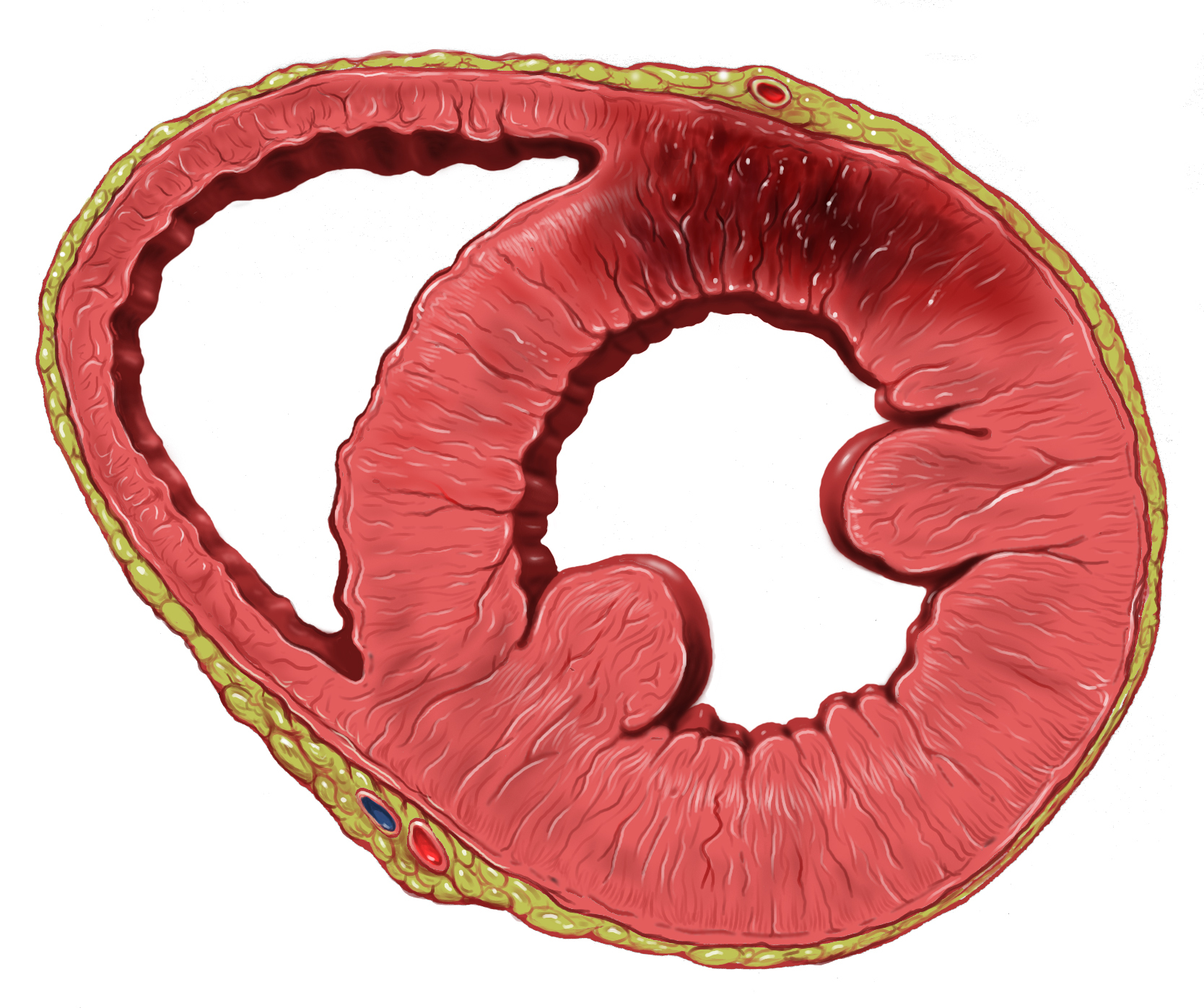
In cardiology, hibernating myocardium is a state when some segments of the myocardium exhibit abnormalities of contractile function. These abnormalities can be visualised with echocardiography, cardiac magnetic resonance imaging (CMR), nuclear medicine (PET) or ventriculography. Echocardiography: A wall motion abnormality at rest which improves during a low-dose dobutamine stress test is classified as "hibernating myocardium." Low dose dobutamine stimulates contractile function and thus helps to predict functional recovery after revascularization. Cardiac magnetic resonance: The most frequently used MR contrast agents based on Gd-chelates accumulate in the extracellular space which is increased in scarred myocardium. This leads to a signal increase which can be visualised with the "late gadolinium enhancement technique." This is probably the most accurate way to visualise scarred myocardium. An alternative (or additional) technique with CMR is the use of low dose dobutamine simila ... [...More Info...] [...Related Items...] OR: [Wikipedia] [Google] [Baidu] |
Cardiology
Cardiology () is a branch of medicine that deals with disorders of the heart and the cardiovascular system. The field includes medical diagnosis and treatment of congenital heart defects, coronary artery disease, heart failure, valvular heart disease and electrophysiology. Physicians who specialize in this field of medicine are called cardiologists, a specialty of internal medicine. Pediatric cardiologists are pediatricians who specialize in cardiology. Physicians who specialize in cardiac surgery are called cardiothoracic surgeons or cardiac surgeons, a specialty of general surgery. Specializations All cardiologists study the disorders of the heart, but the study of adult and child heart disorders each require different training pathways. Therefore, an adult cardiologist (often simply called "cardiologist") is inadequately trained to take care of children, and pediatric cardiologists are not trained to treat adult heart disease. Surgical aspects are not included in cardiology ... [...More Info...] [...Related Items...] OR: [Wikipedia] [Google] [Baidu] |
Myocardium
Cardiac muscle (also called heart muscle, myocardium, cardiomyocytes and cardiac myocytes) is one of three types of vertebrate muscle tissues, with the other two being skeletal muscle and smooth muscle. It is an involuntary, striated muscle that constitutes the main tissue of the wall of the heart. The cardiac muscle (myocardium) forms a thick middle layer between the outer layer of the heart wall (the pericardium) and the inner layer (the endocardium), with blood supplied via the coronary circulation. It is composed of individual cardiac muscle cells joined by intercalated discs, and encased by collagen fibers and other substances that form the extracellular matrix. Cardiac muscle contracts in a similar manner to skeletal muscle, although with some important differences. Electrical stimulation in the form of a cardiac action potential triggers the release of calcium from the cell's internal calcium store, the sarcoplasmic reticulum. The rise in calcium causes the cell's m ... [...More Info...] [...Related Items...] OR: [Wikipedia] [Google] [Baidu] |
Echocardiography
An echocardiography, echocardiogram, cardiac echo or simply an echo, is an ultrasound of the heart. It is a type of medical imaging of the heart, using standard ultrasound or Doppler ultrasound. Echocardiography has become routinely used in the diagnosis, management, and follow-up of patients with any suspected or known heart diseases. It is one of the most widely used diagnostic imaging modalities in cardiology. It can provide a wealth of helpful information, including the size and shape of the heart (internal chamber size quantification), pumping capacity, location and extent of any tissue damage, and assessment of valves. An echocardiogram can also give physicians other estimates of heart function, such as a calculation of the cardiac output, ejection fraction, and diastolic function (how well the heart relaxes). Echocardiography is an important tool in assessing wall motion abnormality in patients with suspected cardiac disease. It is a tool which helps in reaching an ear ... [...More Info...] [...Related Items...] OR: [Wikipedia] [Google] [Baidu] |
Cardiac Magnetic Resonance Imaging
Cardiac magnetic resonance imaging (cardiac MRI), also known as cardiovascular MRI, is a magnetic resonance imaging (MRI) technology used for non-invasive assessment of the function and structure of the cardiovascular system. Conditions in which it is performed include congenital heart disease, cardiomyopathies and valvular heart disease, diseases of the aorta such as dissection, aneurysm and coarctation, coronary heart disease and it can be used to look at pulmonary veins. It is contraindicated if there is a permanent pacemaker or defibrillator, intracerebral clips or claustrophobia. Conventional MRI sequences are adapted for cardiac imaging by using ECG gating and high temporal resolution protocols. The development of cardiac MRI is an active field of research and continues to see a rapid expansion of new and emerging techniques. Uses Cardiovascular MRI is complementary to other imaging techniques, such as echocardiography, cardiac CT, and nuclear medicine. The technique has a ... [...More Info...] [...Related Items...] OR: [Wikipedia] [Google] [Baidu] |
Cardiac Ventriculography
Cardiac ventriculography is a medical imaging test used to determine a person's heart function in the right, or left ventricle.Google books no page number Cardiac ventriculography involves injecting contrast media into the heart's ventricle(s) to measure the volume of blood pumped. Cardiac ventriculography can be performed with a radionuclide in ''radionuclide ventriculography'' or with an iodine-based contrast in cardiac chamber catheterization. The 3 major measurements obtained by cardiac ventriculography are: # Ejection Fraction # Stroke Volume # Cardiac Output These three measurements share a commonality of ratios between end systolic volume and end diastolic volume and all lend mathematical structure to the common medical term systole. Radionuclide ventriculography ''Radionuclide ventriculography'' is a form of nuclear imaging, where a gamma camera is used to create an image following injection of radioactive material, usually Technetium-99m Technetium-99m (99mTc) is a ... [...More Info...] [...Related Items...] OR: [Wikipedia] [Google] [Baidu] |
Ischemia
Ischemia or ischaemia is a restriction in blood supply to any tissue, muscle group, or organ of the body, causing a shortage of oxygen that is needed for cellular metabolism (to keep tissue alive). Ischemia is generally caused by problems with blood vessels, with resultant damage to or dysfunction of tissue i.e. hypoxia and microvascular dysfunction. It also implies local hypoxia in a part of a body resulting from constriction (such as vasoconstriction, thrombosis, or embolism). Ischemia causes not only insufficiency of oxygen, but also reduced availability of nutrients and inadequate removal of metabolic wastes. Ischemia can be partial (poor perfusion) or total blockage. The inadequate delivery of oxygenated blood to the organs must be resolved either by treating the cause of the inadequate delivery or reducing the oxygen demand of the system that needs it. For example, patients with myocardial ischemia have a decreased blood flow to the heart and are prescribed with medi ... [...More Info...] [...Related Items...] OR: [Wikipedia] [Google] [Baidu] |
Revascularization
In medical and surgical therapy, revascularization is the restoration of perfusion to a body part or organ that has had ischemia. It is typically accomplished by surgical means. Vascular bypass and angioplasty are the two primary means of revascularization. The term derives from the prefix re-, in this case meaning "restoration" and vasculature, which refers to the circulatory structures of an organ. It is often combined with "urgent" to form urgent vascularization. Revascularization involves a thorough analysis and diagnosis and treatment of the existing diseased vasculature of the affected organ, and can be aided by the use of different imaging modalities such as magnetic resonance imaging, PET scan, CT scan, and X-ray fluoroscopy. Applications For coronary artery disease (ischemic heart disease), coronary artery bypass surgery and percutaneous coronary intervention (coronary balloon angioplasty) are the two primary means of revascularization. When those cannot be done, ... [...More Info...] [...Related Items...] OR: [Wikipedia] [Google] [Baidu] |
Angina Pectoris
Angina, also known as angina pectoris, is chest pain or pressure, usually caused by insufficient blood flow to the heart muscle (myocardium). It is most commonly a symptom of coronary artery disease. Angina is typically the result of obstruction or spasm of the arteries that supply blood to the heart muscle. The main mechanism of coronary artery obstruction is atherosclerosis as part of coronary artery disease. Other causes of angina include abnormal heart rhythms, heart failure and, less commonly, anemia. The term derives from the Latin ''angere'' ("to strangle") and ''pectus'' ("chest"), and can therefore be translated as "a strangling feeling in the chest". There is a weak relationship between severity of angina and degree of oxygen deprivation in the heart muscle, however, the severity of angina does not always match the degree of oxygen deprivation to the heart or the risk of a myocardial infarction (heart attack). Some people may experience severe pain even though the ... [...More Info...] [...Related Items...] OR: [Wikipedia] [Google] [Baidu] |
Myocardial Infarction
A myocardial infarction (MI), commonly known as a heart attack, occurs when blood flow decreases or stops to the coronary artery of the heart, causing damage to the heart muscle. The most common symptom is chest pain or discomfort which may travel into the shoulder, arm, back, neck or jaw. Often it occurs in the center or left side of the chest and lasts for more than a few minutes. The discomfort may occasionally feel like heartburn. Other symptoms may include shortness of breath, nausea, feeling faint, a cold sweat or feeling tired. About 30% of people have atypical symptoms. Women more often present without chest pain and instead have neck pain, arm pain or feel tired. Among those over 75 years old, about 5% have had an MI with little or no history of symptoms. An MI may cause heart failure, an irregular heartbeat, cardiogenic shock or cardiac arrest. Most MIs occur due to coronary artery disease. Risk factors include high blood pressure, smoking, diabetes, ... [...More Info...] [...Related Items...] OR: [Wikipedia] [Google] [Baidu] |
Stunned Myocardium
Myocardial stunning or transient post-ischemic myocardial dysfunction is a state of mechanical cardiac dysfunction that can occur in a portion of myocardium without necrosis after a brief interruption in perfusion, despite the timely restoration of normal coronary blood flow. In this situation, even after ischemia has been relieved (by for instance angioplasty or coronary artery bypass surgery) and myocardial blood flow (MBF) returns to normal, myocardial function is still depressed for a variable period of time, usually days to weeks. This reversible reduction of function of heart contraction after reperfusion is not accounted for by tissue damage or reduced blood flow, but rather, its thought to represent a perfusion-contraction "mismatch". Myocardial stunning was first described in laboratory canine experiments in the 1970s where LV wall abnormalities were observed following coronary artery occlusion and subsequent reperfusion. Cause Clinical situations associated with myocard ... [...More Info...] [...Related Items...] OR: [Wikipedia] [Google] [Baidu] |
Myocardial Scarring
Myocardial scarring is the accumulation of fibrous tissue resulting after some form of trauma to the cardiac tissue. Fibrosis is the formation of excess tissue in replacement of necrotic or extensively damaged tissue. Fibrosis in the heart is often hard to detect because fibromas, scar tissue or small tumors formed in one cell line, are often formed. Because they are so small, they can be hard to detect by methods such as magnetic resonance imaging. A cell line is a path of fibrosis that follow only a line of cells. Causes Myocardial infarction A myocardial infarction, also known as a heart attack, often result in the formation of fibrosis. A myocardial infarction is an ischemic event, or a restriction of blood flow to body tissue, such as by atherothrombosis. Without blood flow to the myocardium, it is deprived of oxygen, causing tissue death and irreversible damage. The tissue destroyed by the infarction is replaced with non-functioning fibrosis, restoring some of the struct ... [...More Info...] [...Related Items...] OR: [Wikipedia] [Google] [Baidu] |


