|
Drug Reward
Brain stimulation reward (BSR) is a pleasurable phenomenon elicited via direct stimulation of specific brain regions, originally discovered by James Olds and Peter Milner. BSR can serve as a robust operant reinforcer. Targeted stimulation activates the reward system circuitry and establishes response habits similar to those established by natural rewards, such as food and sex. Experiments on BSR soon demonstrated that stimulation of the lateral hypothalamus, along with other regions of the brain associated with natural reward, was both rewarding as well as motivation-inducing. Electrical brain stimulation and intracranial drug injections produce robust reward sensation due to a relatively direct activation of the reward circuitry. This activation is considered to be more direct than rewards produced by natural stimuli, as those signals generally travel through the more indirect peripheral nerves. BSR has been found in all vertebrates tested, including humans, and it has provided a ... [...More Info...] [...Related Items...] OR: [Wikipedia] [Google] [Baidu] |
James Olds
James Olds (May 30, 1922 – August 21, 1976) was an American psychologist who co-discovered the pleasure center of the brain with Peter Milner while he was a postdoctoral fellow at McGill University in 1954. He is considered to be one of the founders of modern neuroscience and received numerous distinctions ranging from election to the United States National Academy of Sciences to the Newcomb Cleveland Prize of the American Association for the Advancement of Science. Biography Early life and education Olds was born in Chicago, Illinois, and grew up in Nyack, New York. His father, Leland Olds, later became chairman of the Federal Power Commission under President Franklin D. Roosevelt. His grandfather George D. Olds was the ninth president of Amherst College. Olds attended college at a number of schools including St. John's College, Annapolis, and the University of Wisconsin but received his undergraduate B.A. from Amherst College in 1947. His undergraduate years were inter ... [...More Info...] [...Related Items...] OR: [Wikipedia] [Google] [Baidu] |
Hypothalamus
The hypothalamus () is a part of the brain that contains a number of small nuclei with a variety of functions. One of the most important functions is to link the nervous system to the endocrine system via the pituitary gland. The hypothalamus is located below the thalamus and is part of the limbic system. In the terminology of neuroanatomy, it forms the ventral part of the diencephalon. All vertebrate brains contain a hypothalamus. In humans, it is the size of an almond. The hypothalamus is responsible for regulating certain metabolic processes and other activities of the autonomic nervous system. It synthesizes and secretes certain neurohormones, called releasing hormones or hypothalamic hormones, and these in turn stimulate or inhibit the secretion of hormones from the pituitary gland. The hypothalamus controls body temperature, hunger, important aspects of parenting and maternal attachment behaviours, thirst, fatigue, sleep, and circadian rhythms. Structure T ... [...More Info...] [...Related Items...] OR: [Wikipedia] [Google] [Baidu] |
Striatum
The striatum, or corpus striatum (also called the striate nucleus), is a nucleus (a cluster of neurons) in the subcortical basal ganglia of the forebrain. The striatum is a critical component of the motor and reward systems; receives glutamatergic and dopaminergic inputs from different sources; and serves as the primary input to the rest of the basal ganglia. Functionally, the striatum coordinates multiple aspects of cognition, including both motor and action planning, decision-making, motivation, reinforcement, and reward perception. The striatum is made up of the caudate nucleus and the lentiform nucleus. The lentiform nucleus is made up of the larger putamen, and the smaller globus pallidus. Strictly speaking the globus pallidus is part of the striatum. It is common practice, however, to implicitly exclude the globus pallidus when referring to striatal structures. In primates, the striatum is divided into a ventral striatum, and a dorsal striatum, subdivisions that are ... [...More Info...] [...Related Items...] OR: [Wikipedia] [Google] [Baidu] |
Mesolimbic Pathway
The mesolimbic pathway, sometimes referred to as the reward pathway, is a dopaminergic pathway in the brain. The pathway connects the ventral tegmental area in the midbrain to the ventral striatum of the basal ganglia in the forebrain. The ventral striatum includes the nucleus accumbens and the olfactory tubercle. The release of dopamine from the mesolimbic pathway into the nucleus accumbens regulates incentive salience (e.g. motivation and desire for rewarding stimuli) and facilitates reinforcement and reward-related motor function learning; it may also play a role in the subjective perception of pleasure. The dysregulation of the mesolimbic pathway and its output neurons in the nucleus accumbens plays a significant role in the development and maintenance of an addiction. Anatomy The mesolimbic pathway is a collection of dopaminergic (i.e., dopamine-releasing) neurons that project from the ventral tegmental area (VTA) to the ventral striatum, which includes the nucleus accumben ... [...More Info...] [...Related Items...] OR: [Wikipedia] [Google] [Baidu] |
Nucleus Accumbens
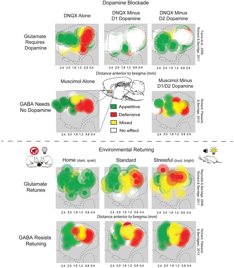
The nucleus accumbens (NAc or NAcc; also known as the accumbens nucleus, or formerly as the ''nucleus accumbens septi'', Latin for "nucleus adjacent to the septum") is a region in the basal forebrain rostral to the preoptic area of the hypothalamus. The nucleus accumbens and the olfactory tubercle collectively form the ventral striatum. The ventral striatum and dorsal striatum collectively form the striatum, which is the main component of the basal ganglia. The dopaminergic neurons of the mesolimbic pathway project onto the GABAergic medium spiny neurons of the nucleus accumbens and olfactory tubercle. Each cerebral hemisphere has its own nucleus accumbens, which can be divided into two structures: the nucleus accumbens core and the nucleus accumbens shell. These substructures have different morphology and functions. Different NAcc subregions (core vs shell) and neuron subpopulations within each region (D1-type vs D2-type medium spiny neurons) are responsible for different ... [...More Info...] [...Related Items...] OR: [Wikipedia] [Google] [Baidu] |
Septal Nuclei
The septal area (medial olfactory area), consisting of the lateral septum and medial septum, is an area in the lower, posterior part of the medial surface of the frontal lobe, and refers to the nearby septum pellucidum. The septal nuclei are located in this area. The septal nuclei are composed of medium-size neurons which are classified into dorsal, ventral, medial, and caudal groups. The septal nuclei receive reciprocal connections from the olfactory bulb, hippocampus, amygdala, hypothalamus, midbrain, habenula, cingulate gyrus, and thalamus. The septal nuclei are essential in generating the theta rhythm of the hippocampus. The septal area (medial olfactory area) has no relation to the sense of smell, but it is considered a pleasure zone in animals. The septal nuclei play a role in reward and reinforcement along with the nucleus accumbens. In the 1950s, Olds & Milner showed that rats with electrodes implanted in this area will self-stimulate repeatedly (i.e., press a bar to rece ... [...More Info...] [...Related Items...] OR: [Wikipedia] [Google] [Baidu] |
Olfactory
The sense of smell, or olfaction, is the special sense through which smells (or odors) are perceived. The sense of smell has many functions, including detecting desirable foods, hazards, and pheromones, and plays a role in taste. In humans, it occurs when an odor binds to a receptor within the nasal cavity, transmitting a signal through the olfactory system. Glomeruli aggregate signals from these receptors and transmit them to the olfactory bulb, where the sensory input will start to interact with parts of the brain responsible for smell identification, memory, and emotion. There are many different causes for alteration, lack, or disturbance to a normal sense of smell, and can include damage to the nose or smell receptors, or central problems affecting the brain. Some causes include upper respiratory infections, traumatic brain injury, and neurodegenerative disease. History of study Early scientific study of the sense of smell includes the extensive doctoral dissertation of ... [...More Info...] [...Related Items...] OR: [Wikipedia] [Google] [Baidu] |
Axons
An axon (from Greek ἄξων ''áxōn'', axis), or nerve fiber (or nerve fibre: see American and British English spelling differences#-re, -er, spelling differences), is a long, slender projection of a nerve cell, or neuron, in vertebrates, that typically conducts electrical impulses known as action potentials away from the Soma (biology), nerve cell body. The function of the axon is to transmit information to different neurons, muscles, and glands. In certain sensory neurons (pseudounipolar neurons), such as those for touch and warmth, the axons are called afferent nerve fibers and the electrical impulse travels along these from the peripheral nervous system, periphery to the cell body and from the cell body to the spinal cord along another branch of the same axon. Axon dysfunction can be the cause of many inherited and acquired neurological disorders that affect both the Peripheral nervous system, peripheral and Central nervous system, central neurons. Nerve fibers are Axon#Cl ... [...More Info...] [...Related Items...] OR: [Wikipedia] [Google] [Baidu] |
Pons
The pons (from Latin , "bridge") is part of the brainstem that in humans and other bipeds lies inferior to the midbrain, superior to the medulla oblongata and anterior to the cerebellum. The pons is also called the pons Varolii ("bridge of Varolius"), after the Italian anatomist and surgeon Costanzo Varolio (1543–75). This region of the brainstem includes neural pathways and tracts that conduct signals from the brain down to the cerebellum and medulla, and tracts that carry the sensory signals up into the thalamus.Saladin Kenneth S.(2007) Anatomy & physiology the unity of form and function. Dubuque, IA: McGraw-Hill Structure The pons is in the brainstem situated between the midbrain and the medulla oblongata, and in front of the cerebellum. A separating groove between the pons and the medulla is the inferior pontine sulcus. The superior pontine sulcus separates the pons from the midbrain. The pons can be broadly divided into two parts: the basilar part of the pons (ventral ... [...More Info...] [...Related Items...] OR: [Wikipedia] [Google] [Baidu] |
Ventral Tegmental Area
The ventral tegmental area (VTA) (tegmentum is Latin for ''covering''), also known as the ventral tegmental area of Tsai, or simply ventral tegmentum, is a group of neurons located close to the midline on the floor of the midbrain. The VTA is the origin of the dopaminergic cell bodies of the mesocorticolimbic dopamine system and other dopamine pathways; it is widely implicated in the drug and natural reward circuitry of the brain. The VTA plays an important role in a number of processes, including reward cognition ( motivational salience, associative learning, and positively-valenced emotions) and orgasm, among others, as well as several psychiatric disorders. Neurons in the VTA project to numerous areas of the brain, ranging from the prefrontal cortex to the caudal brainstem and several regions in between. Structure Neurobiologists have often had great difficulty distinguishing the VTA in humans and other primate brains from the substantia nigra (SN) and surrounding nucl ... [...More Info...] [...Related Items...] OR: [Wikipedia] [Google] [Baidu] |
Primate
Primates are a diverse order of mammals. They are divided into the strepsirrhines, which include the lemurs, galagos, and lorisids, and the haplorhines, which include the tarsiers and the simians (monkeys and apes, the latter including humans). Primates arose 85–55 million years ago first from small terrestrial mammals, which adapted to living in the trees of tropical forests: many primate characteristics represent adaptations to life in this challenging environment, including large brains, visual acuity, color vision, a shoulder girdle allowing a large degree of movement in the shoulder joint, and dextrous hands. Primates range in size from Madame Berthe's mouse lemur, which weighs , to the eastern gorilla, weighing over . There are 376–524 species of living primates, depending on which classification is used. New primate species continue to be discovered: over 25 species were described in the 2000s, 36 in the 2010s, and three in the 2020s. Primates have large bra ... [...More Info...] [...Related Items...] OR: [Wikipedia] [Google] [Baidu] |
Telencephalic
The cerebrum, telencephalon or endbrain is the largest part of the brain containing the cerebral cortex (of the two cerebral hemispheres), as well as several subcortical structures, including the hippocampus, basal ganglia, and olfactory bulb. In the human brain, the cerebrum is the uppermost region of the central nervous system. The cerebrum prenatal development, develops prenatally from the forebrain (prosencephalon). In mammals, the Dorsum (biology), dorsal telencephalon, or Pallium (neuroanatomy), pallium, develops into the cerebral cortex, and the ventral telencephalon, or Pallium (neuroanatomy), subpallium, becomes the basal ganglia. The cerebrum is also divided into approximately symmetric Lateralization of brain function, left and right cerebral hemispheres. With the assistance of the cerebellum, the cerebrum controls all voluntary actions in the human body. Structure The cerebrum is the largest part of the brain. Depending upon the position of the animal it lies eithe ... [...More Info...] [...Related Items...] OR: [Wikipedia] [Google] [Baidu] |


.jpg)
