|
Uterus-like Mass
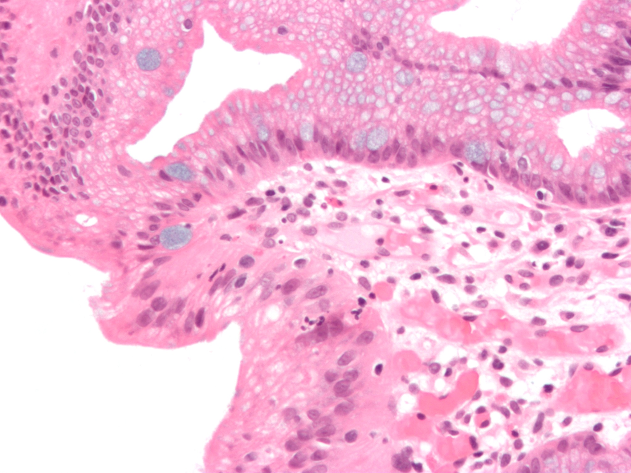
The uterus-like mass (ULM) is a tumorlike anatomical entity originally described in the ovary in 1981 and thereafter reported in several locations of the pelvis and abdominal cavity including broad ligament, greater omentum, cervix, small intestine, mesentery and conus medullaris. Basically, it is represented by a miniature uterus comprising a smooth muscle wall lined by endometrium thus outlining a uterus anatomical structure. Some of the reported cases have been associated to urinary tract and internal genitalia malformations whereas others appeared as a solitary finding. The term endomyometriosis has also been applied to this lesion. Different pathogenetic views have been suggested for this anomaly: a) a metaplastic change in endometriosis foci bringing about smooth muscle hyperplasia; b) a congenital anomaly due to fusion defects of the Müllerian ducts; and c) a sub-coelomic transformation of the mesenchyme. ULM has also been reported associated to endometrial carcinoma and br ... [...More Info...] [...Related Items...] OR: [Wikipedia] [Google] [Baidu] |
Ovary
The ovary is an organ in the female reproductive system that produces an ovum. When released, this travels down the fallopian tube into the uterus, where it may become fertilized by a sperm. There is an ovary () found on each side of the body. The ovaries also secrete hormones that play a role in the menstrual cycle and fertility. The ovary progresses through many stages beginning in the prenatal period through menopause. It is also an endocrine gland because of the various hormones that it secretes. Structure The ovaries are considered the female gonads. Each ovary is whitish in color and located alongside the lateral wall of the uterus in a region called the ovarian fossa. The ovarian fossa is the region that is bounded by the external iliac artery and in front of the ureter and the internal iliac artery. This area is about 4 cm x 3 cm x 2 cm in size.Daftary, Shirish; Chakravarti, Sudip (2011). Manual of Obstetrics, 3rd Edition. Elsevier. pp. 1-16. . The ovari ... [...More Info...] [...Related Items...] OR: [Wikipedia] [Google] [Baidu] |
Hyperplasia
Hyperplasia (from ancient Greek ὑπέρ ''huper'' 'over' + πλάσις ''plasis'' 'formation'), or hypergenesis, is an enlargement of an organ or tissue caused by an increase in the amount of organic tissue that results from cell proliferation. It may lead to the gross enlargement of an organ, and the term is sometimes confused with benign neoplasia or benign tumor. Hyperplasia is a common preneoplastic response to stimulus. Microscopically, cells resemble normal cells but are increased in numbers. Sometimes cells may also be increased in size (hypertrophy). Hyperplasia is different from hypertrophy in that the adaptive cell change in hypertrophy is an increase in the ''size'' of cells, whereas hyperplasia involves an increase in the ''number'' of cells. Causes Hyperplasia may be due to any number of causes, including proliferation of basal layer of epidermis to compensate skin loss, chronic inflammatory response, hormonal dysfunctions, or compensation for damage o ... [...More Info...] [...Related Items...] OR: [Wikipedia] [Google] [Baidu] |
Metaplasia
Metaplasia ( gr, "change in form") is the transformation of one differentiated cell type to another differentiated cell type. The change from one type of cell to another may be part of a normal maturation process, or caused by some sort of abnormal stimulus. In simplistic terms, it is as if the original cells are not robust enough to withstand their environment, so they transform into another cell type better suited to their environment. If the stimulus causing metaplasia is removed or ceases, tissues return to their normal pattern of differentiation. Metaplasia is not synonymous with dysplasia, and is not considered to be an actual cancer. It is also contrasted with heteroplasia, which is the spontaneous abnormal growth of cytologic and histologic elements. Today, metaplastic changes are usually considered to be an early phase of carcinogenesis, specifically for those with a history of cancers or who are known to be susceptible to carcinogenic changes. Metaplastic change is t ... [...More Info...] [...Related Items...] OR: [Wikipedia] [Google] [Baidu] |
Hysterectomy
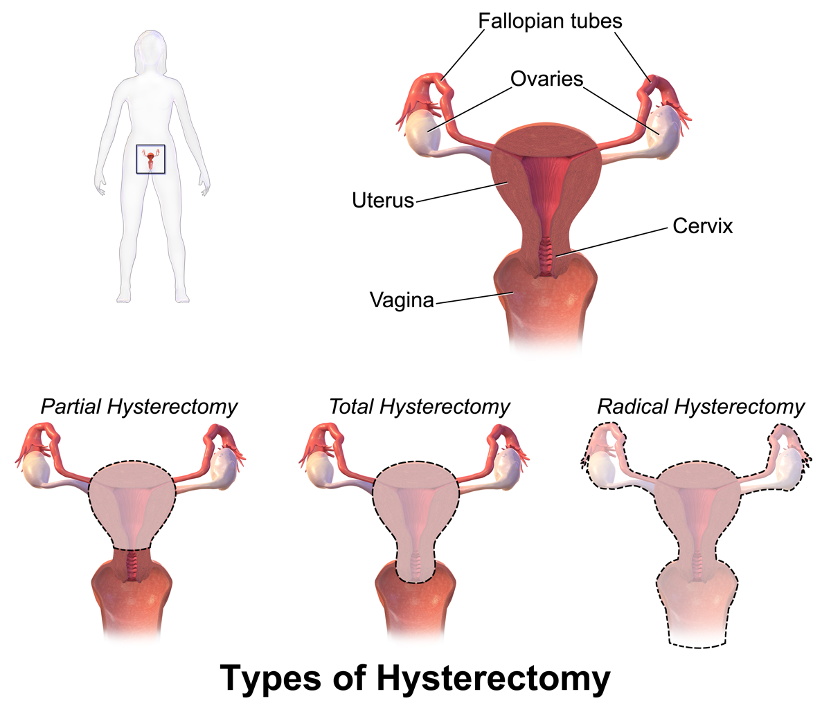
Hysterectomy is the surgical removal of the uterus. It may also involve removal of the cervix, ovaries (oophorectomy), Fallopian tubes (salpingectomy), and other surrounding structures. Usually performed by a gynecologist, a hysterectomy may be total (removing the body, fundus, and cervix of the uterus; often called "complete") or partial (removal of the uterine body while leaving the cervix intact; also called "supracervical"). Removal of the uterus renders the patient unable to bear children (as does removal of ovaries and fallopian tubes) and has surgical risks as well as long-term effects, so the surgery is normally recommended only when other treatment options are not available or have failed. It is the second most commonly performed gynecological surgical procedure, after cesarean section, in the United States. Nearly 68 percent were performed for conditions such as endometriosis, irregular bleeding, and uterine fibroids. It is expected that the frequency of hysterectom ... [...More Info...] [...Related Items...] OR: [Wikipedia] [Google] [Baidu] |
CA-125
Mucin-16 (MUC-16) also known as Ovarian cancer-related tumor marker CA125 is a protein that in humans is encoded by the ''MUC16'' gene. MUC-16 is a member of the mucin family glycoproteins. MUC-16 has found application as a tumor marker or biomarker that may be elevated in the blood of some patients with specific types of cancers, most notably ovarian cancer, or other conditions that are benign. Structure Mucin 16 is a membrane associated mucin that possesses a single transmembrane domain. A unique property of MUC16 is its large size. MUC16 is more than twice as long as MUC1 and MUC4 and contains about 22,000 amino acids, making it the largest membrane-associated mucin. MUC16 is composed of three different domains: * An N-terminal domain * A tandem repeat domain * A C-terminal domain The N-terminal and tandem repeat domains are both entirely extracellular and highly O-glycosylated. All mucins contain a tandem repeat domain that has repeating amino acid sequences high in ... [...More Info...] [...Related Items...] OR: [Wikipedia] [Google] [Baidu] |
Deletion (genetics)
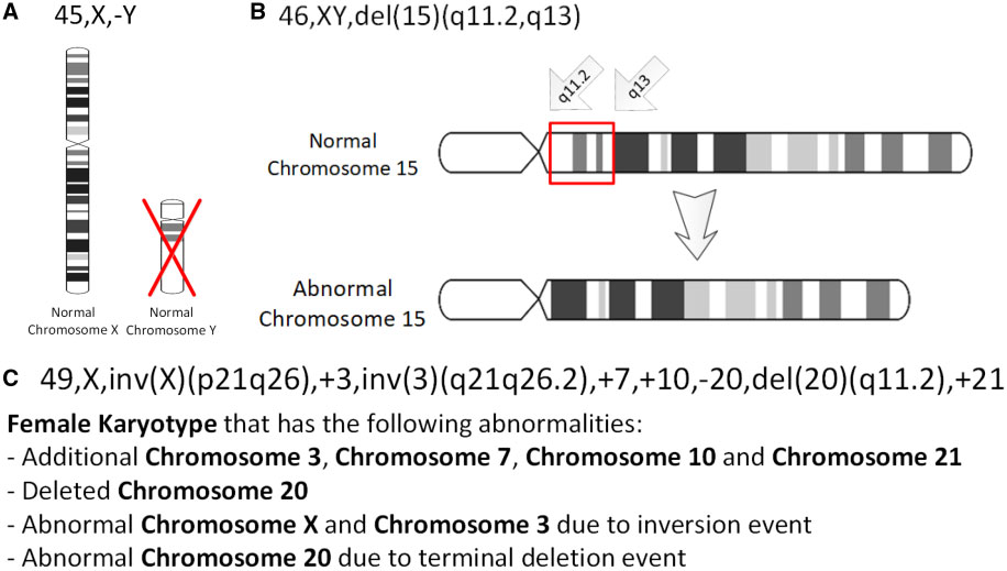
In genetics, a deletion (also called gene deletion, deficiency, or deletion mutation) (sign: Δ) is a mutation (a genetic aberration) in which a part of a chromosome or a sequence of DNA is left out during DNA replication. Any number of nucleotides can be deleted, from a single base to an entire piece of chromosome. Some chromosomes have fragile spots where breaks occur which result in the deletion of a part of chromosome. The breaks can be induced by heat, viruses, radiations, chemicals. When a chromosome breaks, a part of it is deleted or lost, the missing piece of chromosome is referred to as deletion or a deficiency. For synapsis to occur between a chromosome with a large intercalary deficiency and a normal complete homolog, the unpaired region of the normal homolog must loop out of the linear structure into a deletion or compensation loop. The smallest single base deletion mutations occur by a single base flipping in the template DNA, followed by template DNA strand sli ... [...More Info...] [...Related Items...] OR: [Wikipedia] [Google] [Baidu] |
Breast Cancer
Breast cancer is cancer that develops from breast tissue. Signs of breast cancer may include a lump in the breast, a change in breast shape, dimpling of the skin, milk rejection, fluid coming from the nipple, a newly inverted nipple, or a red or scaly patch of skin. In those with distant spread of the disease, there may be bone pain, swollen lymph nodes, shortness of breath, or yellow skin. Risk factors for developing breast cancer include obesity, a lack of physical exercise, alcoholism, hormone replacement therapy during menopause, ionizing radiation, an early age at first menstruation, having children late in life or not at all, older age, having a prior history of breast cancer, and a family history of breast cancer. About 5–10% of cases are the result of a genetic predisposition inherited from a person's parents, including BRCA1 and BRCA2 among others. Breast cancer most commonly develops in cells from the lining of milk ducts and the lobules that supply these ... [...More Info...] [...Related Items...] OR: [Wikipedia] [Google] [Baidu] |
Endometrial Cancer
Endometrial cancer is a cancer that arises from the endometrium (the lining of the uterus or womb). It is the result of the abnormal growth of cells that have the ability to invade or spread to other parts of the body. The first sign is most often vaginal bleeding not associated with a menstrual period. Other symptoms include pain with urination, pain during sexual intercourse, or pelvic pain. Endometrial cancer occurs most commonly after menopause. Approximately 40% of cases are related to obesity. Endometrial cancer is also associated with excessive estrogen exposure, high blood pressure and diabetes. Whereas taking estrogen alone increases the risk of endometrial cancer, taking both estrogen and a progestogen in combination, as in most birth control pills, decreases the risk. Between two and five percent of cases are related to genes inherited from the parents. Endometrial cancer is sometimes loosely referred to as "uterine cancer", although it is distinct from other fo ... [...More Info...] [...Related Items...] OR: [Wikipedia] [Google] [Baidu] |
Mesenchyme
Mesenchyme () is a type of loosely organized animal embryonic connective tissue of undifferentiated cells that give rise to most tissues, such as skin, blood or bone. The interactions between mesenchyme and epithelium help to form nearly every organ in the developing embryo. Vertebrates Structure Mesenchyme is characterized morphologically by a prominent ground substance matrix containing a loose aggregate of reticular fibers and unspecialized mesenchymal stem cells. Mesenchymal cells can migrate easily (in contrast to epithelial cells, which lack mobility), are organized into closely adherent sheets, and are polarized in an apical-basal orientation. Development The mesenchyme originates from the mesoderm. From the mesoderm, the mesenchyme appears as an embryologically primitive "soup". This "soup" exists as a combination of the mesenchymal cells plus serous fluid plus the many different tissue proteins. Serous fluid is typically stocked with the many serous elements, such a ... [...More Info...] [...Related Items...] OR: [Wikipedia] [Google] [Baidu] |
Coelom
The coelom (or celom) is the main body cavity in most animals and is positioned inside the body to surround and contain the digestive tract and other organs. In some animals, it is lined with mesothelium. In other animals, such as molluscs, it remains undifferentiated. In the past, and for practical purposes, coelom characteristics have been used to classify bilaterian animal phyla into informal groups. Etymology The term ''coelom'' derives from the Ancient Greek word (), meaning 'cavity'. Structure Development The coelom is the mesodermally lined cavity between the gut and the outer body wall. During the development of the embryo, coelom formation begins in the gastrulation stage. The developing digestive tube of an embryo forms as a blind pouch called the archenteron. In Protostomes, the coelom forms by a process known as schizocoely. The archenteron initially forms, and the mesoderm splits into two layers: the first attaches to the body wall or ectoderm, forming ... [...More Info...] [...Related Items...] OR: [Wikipedia] [Google] [Baidu] |
Paramesonephric Duct
Paramesonephric ducts (or Müllerian ducts) are paired ducts of the embryo that run down the lateral sides of the genital ridge and terminate at the sinus tubercle in the primitive urogenital sinus. In the female, they will develop to form the fallopian tubes, uterus, cervix, and the upper one-third of the vagina. Development The female reproductive system is composed of two embryological segments: the urogenital sinus and the paramesonephric ducts. The two are conjoined at the sinus tubercle. Paramesonephric ducts are present on the embryo of both sexes. Only in females do they develop into reproductive organs. They degenerate in males of certain species, but the adjoining mesonephric ducts develop into male reproductive organs. The sex based differences in the contributions of the paramesonephric ducts to reproductive organs is based on the presence, and degree of presence, of Anti-Müllerian hormone. During the formation of the reproductive system, the paramesonephric ducts ar ... [...More Info...] [...Related Items...] OR: [Wikipedia] [Google] [Baidu] |
Congenital Disorder
A birth defect, also known as a congenital disorder, is an abnormal condition that is present at birth regardless of its cause. Birth defects may result in disabilities that may be physical, intellectual, or developmental. The disabilities can range from mild to severe. Birth defects are divided into two main types: structural disorders in which problems are seen with the shape of a body part and functional disorders in which problems exist with how a body part works. Functional disorders include metabolic and degenerative disorders. Some birth defects include both structural and functional disorders. Birth defects may result from genetic or chromosomal disorders, exposure to certain medications or chemicals, or certain infections during pregnancy. Risk factors include folate deficiency, drinking alcohol or smoking during pregnancy, poorly controlled diabetes, and a mother over the age of 35 years old. Many are believed to involve multiple factors. Birth defects may be vi ... [...More Info...] [...Related Items...] OR: [Wikipedia] [Google] [Baidu] |


