|
Tuber Cinereum
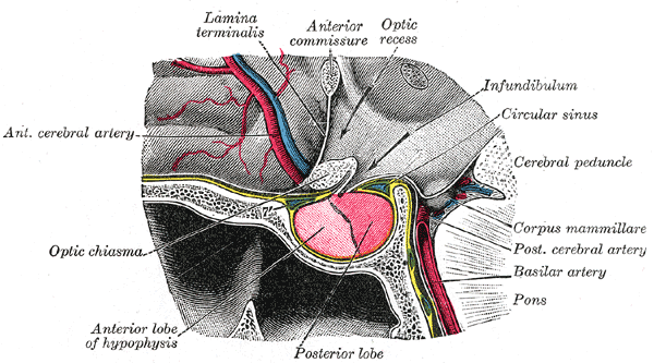
The tuber cinereum is a hollow eminence of the middle–ventral hypothalamus, specifically the arcuate nucleus, situated between the mammillary bodies and the optic chiasm. In addition to the ventral hypothalamus, the tuber cinereum includes the median eminence and pituitary gland. Together with the hollow itself, it is sometimes referred to as the pituitary stalk. Structure The tuber cinereum is an inferior distention of the floor of the third ventricle; the conical hollow formed by the distention (a continuation of the ventricle itself), is known as the infundibulum (''funnel''). Thus, the tuber cinereum is anteriorly continuous with the lamina terminalis, while laterally it is continuous with the anterior perforated substances of the hypothalamus. The inferior end adjoins the posterior lobe of the pituitary gland. Capillaries of the tuber cinereum are specialized and confluent to enable rapid communication via brain- or blood-borne factors between compartments of the t ... [...More Info...] [...Related Items...] OR: [Wikipedia] [Google] [Baidu] |
Tuberculum Cinereum
The trigeminal tubercle, or tuberculum cinereum is a raised area between the rootlets of the accessory nerve and Posterolateral sulcus of medulla oblongata, posterolateral sulcus. It overlies the spinal tract of the trigeminal nerve. It is an elevation in the lower part of medulla oblongata, medulla, lateral to the cuneate fasciculus, produced by a mass of grey matter called the spinal trigeminal nucleus. References [...More Info...] [...Related Items...] OR: [Wikipedia] [Google] [Baidu] |
Tuber Cinereum Hamartoma
Tuber cinereum hamartoma is a benign tumor in which a disorganized collection of neurons and glia accumulate at the tuber cinereum of the hypothalamus on the floor of the third ventricle. It is a congenital malformation, included on the spectrum of gray matter heterotopias. Formation occurs during embryogenesis, typically between days 33 and 41 of gestation. Size of the tumor varies from one to three centimeters in diameter, with the mean being closer to the low end of this range. It is estimated to occur at a frequency of one in one million individuals. Signs and symptoms The classic presentation is gelastic or laughing epilepsy, a disorder characterized by spells of involuntary laughter with interval irritability and depressed mood. The tumor can be associated with other seizure types as well as precocious puberty and behavioral disorders. Gelastic epilepsy has been more classically associated with sessile lesions and precocious puberty reported with pedunculated morphology. ... [...More Info...] [...Related Items...] OR: [Wikipedia] [Google] [Baidu] |
Hormone
A hormone (from the Greek participle , "setting in motion") is a class of signaling molecules in multicellular organisms that are sent to distant organs by complex biological processes to regulate physiology and behavior. Hormones are required for the correct development of animals, plants and fungi. Due to the broad definition of a hormone (as a signaling molecule that exerts its effects far from its site of production), numerous kinds of molecules can be classified as hormones. Among the substances that can be considered hormones, are eicosanoids (e.g. prostaglandins and thromboxanes), steroids (e.g. oestrogen and brassinosteroid), amino acid derivatives (e.g. epinephrine and auxin), protein or peptides (e.g. insulin and CLE peptides), and gases (e.g. ethylene and nitric oxide). Hormones are used to communicate between organs and tissues. In vertebrates, hormones are responsible for regulating a variety of physiological processes and behavioral activities such as diges ... [...More Info...] [...Related Items...] OR: [Wikipedia] [Google] [Baidu] |
Perivascular Space
A perivascular space, also known as a Virchow–Robin space, is a fluid-filled space surrounding certain blood vessels in several organs, including the brain, potentially having an immunological function, but more broadly a dispersive role for neural and blood-derived messengers. The brain pia mater is reflected from the surface of the brain onto the surface of blood vessels in the subarachnoid space. In the brain, ''perivascular cuffs'' are regions of leukocyte aggregation in the perivascular spaces, usually found in patients with viral encephalitis. Perivascular spaces vary in dimension according to the type of blood vessel. In the brain where most capillaries have an imperceptible perivascular space, select structures of the brain, such as the circumventricular organs, are notable for having large perivascular spaces surrounding highly permeable capillaries, as observed by microscopy. The median eminence, a brain structure at the base of the hypothalamus, contains capillar ... [...More Info...] [...Related Items...] OR: [Wikipedia] [Google] [Baidu] |
Vascular Permeability
Vascular permeability, often in the form of capillary permeability or microvascular permeability, characterizes the capacity of a blood vessel wall to allow for the flow of small molecules (drugs, nutrients, water, ions) or even whole cells (lymphocytes on their way to the site of inflammation) in and out of the vessel. Blood vessel walls are lined by a single layer of endothelial cells. The gaps between endothelial cells (cell junctions) are strictly regulated depending on the type and physiological state of the tissue. There are several techniques to measure vascular permeability to certain molecules. For instance, the cannulation of a single microvessel with a micropipette, the microvessel is perfused with a certain pressure, occluded downstream and then the velocity of some cells will be related to the permeability.Michel, C. C., Mason, J. C., Curry, F. E. & Tooke, J. E. Development of Landis Technique for Measuring Filtration Coefficient of Individual Capillaries in Frog M ... [...More Info...] [...Related Items...] OR: [Wikipedia] [Google] [Baidu] |
Hypophyseal Portal System
The hypophyseal portal system is a system of blood vessels in the microcirculation at the base of the brain, connecting the hypothalamus with the anterior pituitary. Its main function is to quickly transport and exchange hormones between the hypothalamus arcuate nucleus and anterior pituitary gland. The capillaries in the portal system are fenestrated (have many small channels with high vascular permeability) which allows a rapid exchange between the hypothalamus and the pituitary. The main hormones transported by the system include gonadotropin-releasing hormone, corticotropin-releasing hormone, growth hormone–releasing hormone, and thyrotropin-releasing hormone. Structure The blood supply and direction of flow in the hypophyseal portal system has been studied over many years on laboratory animals and human cadaver specimens with injection and vascular corrosion casting methods. Short portal vessels between the neural and anterior pituitary lobes provide an avenue for rapid ho ... [...More Info...] [...Related Items...] OR: [Wikipedia] [Google] [Baidu] |
Confluence
In geography, a confluence (also: ''conflux'') occurs where two or more flowing bodies of water join to form a single channel. A confluence can occur in several configurations: at the point where a tributary joins a larger river (main stem); or where two streams meet to become the source of a river of a new name (such as the confluence of the Monongahela and Allegheny rivers at Pittsburgh, forming the Ohio); or where two separated channels of a river (forming a river island) rejoin at the downstream end. Scientific study of confluences Confluences are studied in a variety of sciences. Hydrology studies the characteristic flow patterns of confluences and how they give rise to patterns of erosion, bars, and scour pools. The water flows and their consequences are often studied with mathematical models. Confluences are relevant to the distribution of living organisms (i.e., ecology) as well; "the general pattern ownstream of confluencesof increasing stream flow and decreasing s ... [...More Info...] [...Related Items...] OR: [Wikipedia] [Google] [Baidu] |
Capillary
A capillary is a small blood vessel from 5 to 10 micrometres (μm) in diameter. Capillaries are composed of only the tunica intima, consisting of a thin wall of simple squamous endothelial cells. They are the smallest blood vessels in the body: they convey blood between the arterioles and venules. These microvessels are the site of exchange of many substances with the interstitial fluid surrounding them. Substances which cross capillaries include water, oxygen, carbon dioxide, urea, glucose, uric acid, lactic acid and creatinine. Lymph capillaries connect with larger lymph vessels to drain lymphatic fluid collected in the microcirculation. During early embryonic development, new capillaries are formed through vasculogenesis, the process of blood vessel formation that occurs through a '' de novo'' production of endothelial cells that then form vascular tubes. The term '' angiogenesis'' denotes the formation of new capillaries from pre-existing blood vessels and already present ... [...More Info...] [...Related Items...] OR: [Wikipedia] [Google] [Baidu] |
Anterior Perforated Substance
The anterior perforated substance is a part of the brain. It is bilateral. It is irregular and quadrilateral. It lies in front of the optic tract and behind the olfactory trigone. Structure The anterior perforated substance is bilateral. It lies in front of the optic tract. It lies behind the olfactory trigone, separated by the fissure prima. Medially and in front, it is continuous with the subcallosal gyrus. Laterally, it is bounded by the lateral stria of the olfactory tract, and is continued into the uncus. Its gray substance is confluent above with that of the corpus striatum, and is perforated anteriorly by numerous small blood vessels that supply such areas as the internal capsule. The anterior cerebral artery arises just below the anterior perforated substance. The middle cerebral artery passes through its lateral two thirds. Blood supply The anterior perforated substance is supplied by lenticulostriate arteries, which branch from the middle cerebral artery. It i ... [...More Info...] [...Related Items...] OR: [Wikipedia] [Google] [Baidu] |
Lamina Terminalis
The median portion of the wall of the forebrain consists of a thin lamina, the lamina terminalis, which stretches from the interventricular foramen (Foramen of Monro) to the recess at the base of the optic stalk (optic nerve) and contains the vascular organ of the lamina terminalis, which regulates the osmotic concentration of the blood. The lamina terminalis is immediately anterior to the tuber cinereum; together they form the pituitary stalk. The lamina terminalis can be opened via endoscopic neurosurgery in an attempt to create a path that cerebrospinal fluid can flow through when a person has hydrocephalus and when it is not possible to perform an Endoscopic third ventriculostomy, but the effectiveness of this technique is not certain. This is the rostral end (tip) of the neural tube (embryological central nervous system) in the early weeks of development. Failure of the lamina terminalis to close properly at this stage of development will result in anencephaly or meroencep ... [...More Info...] [...Related Items...] OR: [Wikipedia] [Google] [Baidu] |
Third Ventricle
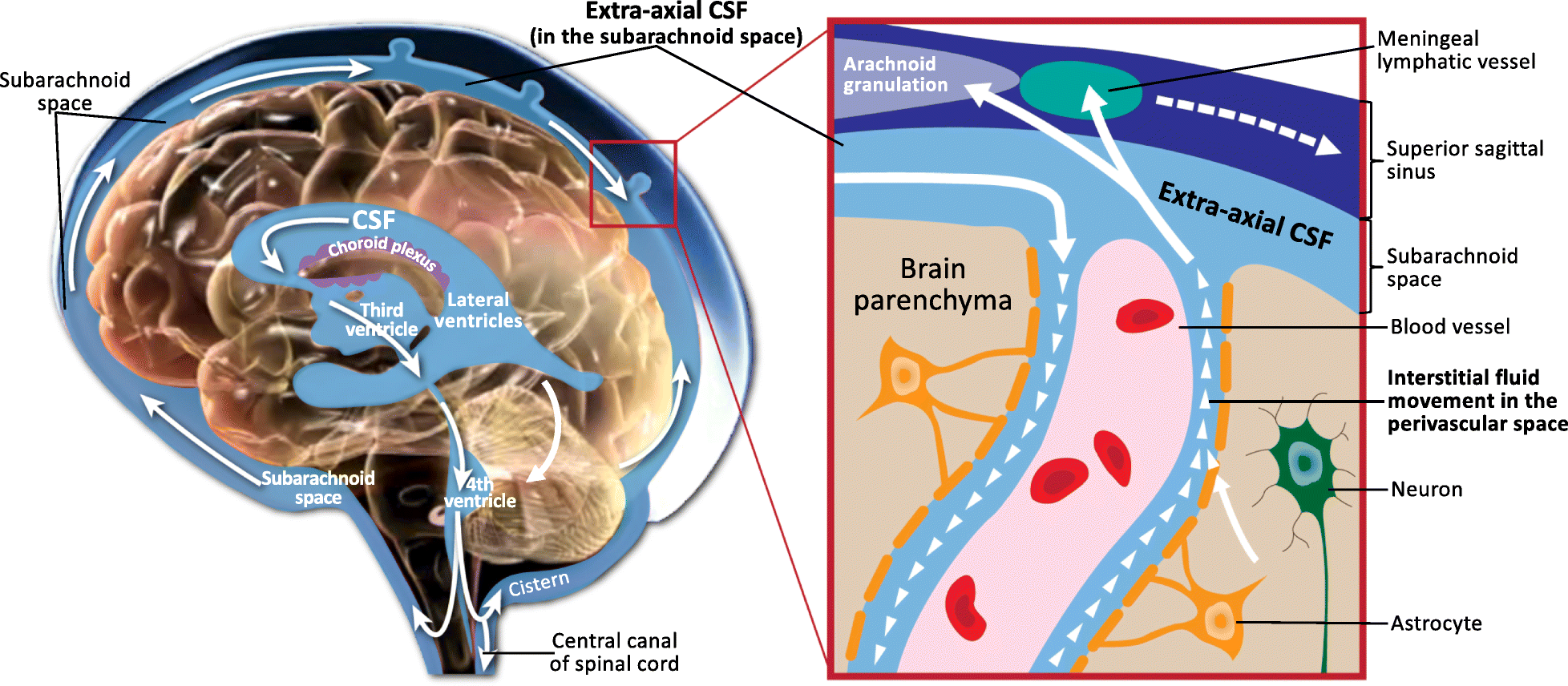
The third ventricle is one of the four connected ventricles of the ventricular system within the mammalian brain. It is a slit-like cavity formed in the diencephalon between the two thalami, in the midline between the right and left lateral ventricles, and is filled with cerebrospinal fluid (CSF). Running through the third ventricle is the interthalamic adhesion, which contains thalamic neurons and fibers that may connect the two thalami. Structure The third ventricle is a narrow, laterally flattened, vaguely rectangular region, filled with cerebrospinal fluid, and lined by ependyma. It is connected at the superior anterior corner to the lateral ventricles, by the interventricular foramina, and becomes the cerebral aqueduct (''aqueduct of Sylvius'') at the posterior caudal corner. Since the interventricular foramina are on the lateral edge, the corner of the third ventricle itself forms a bulb, known as the ''anterior recess'' (it is also known as the ''bulb of the ventricl ... [...More Info...] [...Related Items...] OR: [Wikipedia] [Google] [Baidu] |


_effect.png)
