|
Tomography, Emission-computed, Single-photon
Single-photon emission computed tomography (SPECT, or less commonly, SPET) is a nuclear medicine tomographic imaging technique using gamma rays. It is very similar to conventional nuclear medicine planar imaging using a gamma camera (that is, scintigraphy), but is able to provide true 3D information. This information is typically presented as cross-sectional slices through the patient, but can be freely reformatted or manipulated as required. The technique needs delivery of a gamma-emitting radioisotope (a radionuclide) into the patient, normally through injection into the bloodstream. On occasion, the radioisotope is a simple soluble dissolved ion, such as an isotope of gallium(III). Most of the time, though, a marker radioisotope is attached to a specific ligand to create a radioligand, whose properties bind it to certain types of tissues. This marriage allows the combination of ligand and radiopharmaceutical to be carried and bound to a place of interest in the bod ... [...More Info...] [...Related Items...] OR: [Wikipedia] [Google] [Baidu] |
Technetium Exametazime
Technetium (99mTc) exametazime is a radiopharmaceutical sold under the trade name Ceretec, and is used by nuclear medicine physicians for the detection of altered regional cerebral perfusion in stroke and other cerebrovascular diseases. It can also be used for the labelling of leukocytes to localise intra-abdominal infections and inflammatory bowel disease. Exametazime (the part without technetium) is sometimes referred to as ''hexamethylpropylene amine oxime'' or ''HMPAO'', although correct chemical names are: *(NE)-N- 3R)-3-3-(2R,3E)-3-hydroxyiminobutan-2-ylmino">3-(2R,3E)-3-hydroxyiminobutan-2-yl.html" ;"title="3R)-3-3-(2R,3E)-3-hydroxyiminobutan-2-yl">3R)-3-3-(2R,3E)-3-hydroxyiminobutan-2-ylmino2,2-dimethylpropyl]amino]butan-2-ylidene]hydroxylamine *or 3,3'-((2,2,-dimethyl-1,3-propanediyl)diimino)bis-2-butanone dioxime. Chemistry The drug consists of exametazime as a chelating agent for the radioisotope technetium-99m. Both enantiomeric forms of exametazime are used—the dr ... [...More Info...] [...Related Items...] OR: [Wikipedia] [Google] [Baidu] |
Tomographic Reconstruction
Tomographic reconstruction is a type of multidimensional inverse problem where the challenge is to yield an estimate of a specific system from a finite number of projections. The mathematical basis for tomographic imaging was laid down by Johann Radon. A notable example of applications is the reconstruction of computed tomography (CT) where cross-sectional images of patients are obtained in non-invasive manner. Recent developments have seen the Radon transform and its inverse used for tasks related to realistic object insertion required for testing and evaluating computed tomography use in airport security. This article applies in general to reconstruction methods for all kinds of tomography, but some of the terms and physical descriptions refer directly to the reconstruction of X-ray computed tomography. Introducing formula The projection of an object, resulting from the tomographic measurement process at a given angle \theta, is made up of a set of line integrals (see F ... [...More Info...] [...Related Items...] OR: [Wikipedia] [Google] [Baidu] |
Myocardium
Cardiac muscle (also called heart muscle, myocardium, cardiomyocytes and cardiac myocytes) is one of three types of vertebrate muscle tissues, with the other two being skeletal muscle and smooth muscle. It is an involuntary, striated muscle that constitutes the main tissue of the wall of the heart. The cardiac muscle (myocardium) forms a thick middle layer between the outer layer of the heart wall (the pericardium) and the inner layer (the endocardium), with blood supplied via the coronary circulation. It is composed of individual cardiac muscle cells joined by intercalated discs, and encased by collagen fibers and other substances that form the extracellular matrix. Cardiac muscle contracts in a similar manner to skeletal muscle, although with some important differences. Electrical stimulation in the form of a cardiac action potential triggers the release of calcium from the cell's internal calcium store, the sarcoplasmic reticulum. The rise in calcium causes the cell's m ... [...More Info...] [...Related Items...] OR: [Wikipedia] [Google] [Baidu] |
Ischemic Heart Disease
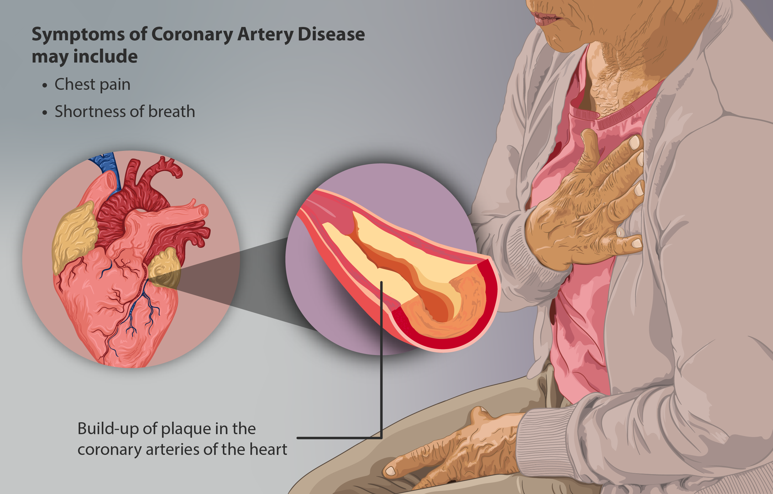
Coronary artery disease (CAD), also called coronary heart disease (CHD), ischemic heart disease (IHD), myocardial ischemia, or simply heart disease, involves the reduction of blood flow to the heart muscle due to build-up of atherosclerotic plaque in the arteries of the heart. It is the most common of the cardiovascular diseases. Types include stable angina, unstable angina, myocardial infarction, and sudden cardiac death. A common symptom is chest pain or discomfort which may travel into the shoulder, arm, back, neck, or jaw. Occasionally it may feel like heartburn. Usually symptoms occur with exercise or emotional stress, last less than a few minutes, and improve with rest. Shortness of breath may also occur and sometimes no symptoms are present. In many cases, the first sign is a heart attack. Other complications include heart failure or an abnormal heartbeat. Risk factors include high blood pressure, smoking, diabetes, lack of exercise, obesity, high blood cholesterol, po ... [...More Info...] [...Related Items...] OR: [Wikipedia] [Google] [Baidu] |
Bone Scintigraphy
A bone scan or bone scintigraphy is a nuclear medicine imaging technique of the bone. It can help diagnose a number of bone conditions, including cancer of the bone or metastasis, location of bone inflammation and fractures (that may not be visible in traditional X-ray images), and bone infection (osteomyelitis). Nuclear medicine provides functional imaging and allows visualisation of bone metabolism or bone remodeling, which most other imaging techniques (such as X-ray computed tomography, CT) cannot. Bone scintigraphy competes with positron emission tomography (PET) for imaging of abnormal metabolism in bones, but is considerably less expensive. Bone scintigraphy has higher sensitivity but lower specificity than CT or MRI for diagnosis of scaphoid fractures following negative plain radiography. History Some of the earliest investigations into skeletal metabolism were carried out by George de Hevesy in the 1930s, using phosphorus-32 and by Charles Pecher in the 1940s. In t ... [...More Info...] [...Related Items...] OR: [Wikipedia] [Google] [Baidu] |
Leukocyte
White blood cells, also called leukocytes or leucocytes, are the cells of the immune system that are involved in protecting the body against both infectious disease and foreign invaders. All white blood cells are produced and derived from multipotent cells in the bone marrow known as hematopoietic stem cells. Leukocytes are found throughout the body, including the blood and lymphatic system. All white blood cells have nuclei, which distinguishes them from the other blood cells, the anucleated red blood cells (RBCs) and platelets. The different white blood cells are usually classified by cell lineage (myeloid cells or lymphoid cells). White blood cells are part of the body's immune system. They help the body fight infection and other diseases. Types of white blood cells are granulocytes (neutrophils, eosinophils, and basophils), and agranulocytes (monocytes, and lymphocytes (T cells and B cells)). Myeloid cells (myelocytes) include neutrophils, eosinophils, mast cells, bas ... [...More Info...] [...Related Items...] OR: [Wikipedia] [Google] [Baidu] |
Ejection Fraction
An ejection fraction (EF) is the volumetric fraction (or portion of the total) of fluid (usually blood) ejected from a chamber (usually the heart) with each contraction (or heartbeat). It can refer to the cardiac atrium, ventricle, gall bladder, or leg veins, although if unspecified it usually refers to the left ventricle of the heart. EF is widely used as a measure of the pumping efficiency of the heart and is used to classify heart failure types. It is also used as an indicator of the severity of heart failure, although it has recognized limitations. The EF of the left heart, known as the left ventricular ejection fraction (LVEF), is calculated by dividing the volume of blood pumped from the left ventricle per beat (stroke volume) by the volume of blood collected in the left ventricle at the end of diastolic filling (end-diastolic volume). LVEF is an indicator of the effectiveness of pumping into the systemic circulation. The EF of the right heart, or right ventricular ejection ... [...More Info...] [...Related Items...] OR: [Wikipedia] [Google] [Baidu] |
Electrocardiography
Electrocardiography is the process of producing an electrocardiogram (ECG or EKG), a recording of the heart's electrical activity. It is an electrogram of the heart which is a graph of voltage versus time of the electrical activity of the heart using electrodes placed on the skin. These electrodes detect the small electrical changes that are a consequence of cardiac muscle depolarization followed by repolarization during each cardiac cycle (heartbeat). Changes in the normal ECG pattern occur in numerous cardiac abnormalities, including cardiac rhythm disturbances (such as atrial fibrillation and ventricular tachycardia), inadequate coronary artery blood flow (such as myocardial ischemia and myocardial infarction), and electrolyte disturbances (such as hypokalemia and hyperkalemia). Traditionally, "ECG" usually means a 12-lead ECG taken while lying down as discussed below. However, other devices can record the electrical activity of the heart such as a Holter monitor but also s ... [...More Info...] [...Related Items...] OR: [Wikipedia] [Google] [Baidu] |
Radionuclide Angiography
Radionuclide angiography is an area of nuclear medicine which specialises in imaging to show the functionality of the right and left ventricles of the heart, thus allowing informed diagnostic intervention in heart failure. It involves use of a radiopharmaceutical, injected into a patient, and a gamma camera for acquisition. A MUGA scan (multigated acquisition) involves an acquisition triggered (gated) at different points of the cardiac cycle. MUGA scanning is also called equilibrium radionuclide angiocardiography, radionuclide ventriculography (RNVG), or gated blood pool imaging, as well as SYMA scanning (synchronized multigated acquisition scanning). This mode of imaging uniquely provides a cine type of image of the beating heart, and allows the interpreter to determine the efficiency of the individual heart valves and chambers. MUGA/Cine scanning represents a robust adjunct to the now more common echocardiogram. Mathematics regarding acquisition of cardiac output (''Q'') is ... [...More Info...] [...Related Items...] OR: [Wikipedia] [Google] [Baidu] |
Gated SPECT
Gated SPECT is a nuclear medicine imaging technique, typically for the heart in myocardial perfusion imagery. An electrocardiogram (ECG) guides the image acquisition, and the resulting set of single-photon emission computed tomography (SPECT) images shows the heart as it contracts over the interval from one R wave to the next. Gated myocardial perfusion imaging has been shown to have high prognostic value and sensitivity for critical stenosis. The acquisition computer defines the number of time bins or frames to divide the R to R interval of the patient's electrocardiogram. A "window" may be set which discards data from R to R intervals which deviate from some amount from the patient's average R to R wave duration. This discards preventricular contractions and arrhythmias from the acquisition and improves the quality of the resulting study. The gamma camera will take a series of pictures around the patient, dividing each 'step' of the camera head's motion into the predetermined ... [...More Info...] [...Related Items...] OR: [Wikipedia] [Google] [Baidu] |
SPECT CT
Single-photon emission computed tomography (SPECT, or less commonly, SPET) is a nuclear medicine tomographic imaging technique using gamma rays. It is very similar to conventional nuclear medicine planar imaging using a gamma camera (that is, scintigraphy), but is able to provide true 3D information. This information is typically presented as cross-sectional slices through the patient, but can be freely reformatted or manipulated as required. The technique needs delivery of a gamma-emitting radioisotope (a radionuclide) into the patient, normally through injection into the bloodstream. On occasion, the radioisotope is a simple soluble dissolved ion, such as an isotope of gallium(III). Most of the time, though, a marker radioisotope is attached to a specific ligand to create a radioligand, whose properties bind it to certain types of tissues. This marriage allows the combination of ligand and radiopharmaceutical to be carried and bound to a place of interest in the body, where ... [...More Info...] [...Related Items...] OR: [Wikipedia] [Google] [Baidu] |
Radiopharmacology
Radiopharmacology is radiochemistry applied to medicine and thus the pharmacology of radiopharmaceuticals (medicinal radiocompounds, that is, pharmaceutical drugs that are radioactive). Radiopharmaceuticals are used in the field of nuclear medicine as radioactive tracers in medical imaging and in therapy for many diseases (for example, brachytherapy). Many radiopharmaceuticals use technetium-99m (Tc-99m) which has many useful properties as a gamma-emitting tracer nuclide. In the book ''Technetium'' a total of 31 different radiopharmaceuticals based on Tc-99m are listed for imaging and functional studies of the brain, myocardium, thyroid, lungs, liver, gallbladder, kidneys, skeleton, blood and tumors. The term ''radioisotope'', which in its general sense refers to any radioactive isotope (radionuclide), has historically been used to refer to all radiopharmaceuticals, and this usage remains common. Technically, however, many radiopharmaceuticals incorporate a radioactive tracer a ... [...More Info...] [...Related Items...] OR: [Wikipedia] [Google] [Baidu] |


