|
Sulforhodamine 101
Sulforhodamine 101 (SR101) is a red fluorescent dye. In neurophysiological experiments which comprise calcium imaging methods, it can be used for a counterstaining of astrocytes to be able to analyze data from neurons separately. However, in addition to labeling astrocytes, SR101 labels myelinating oligodendrocytes. SR101 has been reported to affect excitability of neurons and should therefore be used with caution. A sulfonyl chloride derivative of sulforhodamine 101, known as Texas Red Texas Red or sulforhodamine 101 acid chloride is a red fluorescent dye, used in histology for staining cell specimens, for sorting cells with fluorescent-activated cell sorting machines, in fluorescence microscopy applications, and in immunohis ..., is used for conjugation to a number of functional groups, especially primary amines. References {{Reflist Benzenesulfonates Rhodamine dyes Nitrogen heterocycles Oxygen heterocycles ... [...More Info...] [...Related Items...] OR: [Wikipedia] [Google] [Baidu] |
Fluorescent
Fluorescence is the emission of light by a substance that has absorbed light or other electromagnetic radiation. It is a form of luminescence. In most cases, the emitted light has a longer wavelength, and therefore a lower photon energy, than the absorbed radiation. A perceptible example of fluorescence occurs when the absorbed radiation is in the ultraviolet region of the electromagnetic spectrum (invisible to the human eye), while the emitted light is in the visible region; this gives the fluorescent substance a distinct color that can only be seen when the substance has been exposed to UV light. Fluorescent materials cease to glow nearly immediately when the radiation source stops, unlike phosphorescent materials, which continue to emit light for some time after. Fluorescence has many practical applications, including mineralogy, gemology, medicine, chemical sensors (fluorescence spectroscopy), fluorescent labelling, dyes, biological detectors, cosmic-ray detection, vacuu ... [...More Info...] [...Related Items...] OR: [Wikipedia] [Google] [Baidu] |
Neurophysiological
Neurophysiology is a branch of physiology and neuroscience that studies nervous system function rather than nervous system architecture. This area aids in the diagnosis and monitoring of neurological diseases. Historically, it has been dominated by electrophysiology—the electrical recording of neural activity ranging from the molar (the electroencephalogram, EEG) to the cellular (intracellular recording of the properties of single neurons), such as patch clamp, voltage clamp, extracellular single-unit recording and recording of local field potentials. However, since the neurone is an electrochemical machine, it is difficult to isolate electrical events from the metabolic and molecular processes that cause them. Thus, neurophysiologists currently utilise tools from chemistry (calcium imaging), physics (functional magnetic resonance imaging, fMRI), and molecular biology (site directed mutations) to examine brain activity. The word originates from the Greek word meaning "nerve" and ... [...More Info...] [...Related Items...] OR: [Wikipedia] [Google] [Baidu] |
Calcium Imaging
Calcium imaging is a microscopy technique to optically measure the calcium (Ca2+) status of an isolated cell, tissue or medium. Calcium imaging takes advantage of calcium indicators, fluorescent molecules that respond to the binding of Ca2+ ions by fluorescence properties. Two main classes of calcium indicators exist: chemical indicators and genetically encoded calcium indicators (GECI). This technique has allowed studies of calcium signalling in a wide variety of cell types. In neurons, electrical activity is always accompanied by an influx of Ca2+ ions. Thus, calcium imaging can be used to monitor the electrical activity in hundreds of neurons in cell culture or in living animals, which has made it possible to dissect the function of neuronal circuits. Chemical indicators Chemical indicators are small molecules that can chelate calcium ions. All these molecules are based on an EGTA homologue called BAPTA, with high selectivity for calcium (Ca2+) ions versus magnesium (Mg2+) io ... [...More Info...] [...Related Items...] OR: [Wikipedia] [Google] [Baidu] |
Counterstain
A counterstain is a stain with colour contrasting to the principal stain, making the stained structure easily visible using a microscope. Examples include the malachite green counterstain to the fuchsine stain in the Gimenez staining technique and the eosin counterstain to haematoxylin in the H&E stain. In Gram staining, crystal violet stains only Gram-positive bacteria, and safranin counterstain is applied which stains all cells, allowing the identification of Gram-negative bacteria as well. An alternative method uses dilute carbofluozide. Counterstains are sometimes used to separate animals from organic detritus In biology, detritus () is dead particulate organic material, as distinguished from dissolved organic material. Detritus typically includes the bodies or fragments of bodies of dead organisms, and fecal material. Detritus typically hosts commun ... in microbiology studies. References Staining {{pathology-stub ... [...More Info...] [...Related Items...] OR: [Wikipedia] [Google] [Baidu] |
Astrocyte
Astrocytes (from Ancient Greek , , "star" + , , "cavity", "cell"), also known collectively as astroglia, are characteristic star-shaped glial cells in the brain and spinal cord. They perform many functions, including biochemical control of endothelial cells that form the blood–brain barrier, provision of nutrients to the nervous tissue, maintenance of extracellular ion balance, regulation of cerebral blood flow, and a role in the repair and scarring process of the brain and spinal cord following infection and traumatic injuries. The proportion of astrocytes in the brain is not well defined; depending on the counting technique used, studies have found that the astrocyte proportion varies by region and ranges from 20% to 40% of all glia. Another study reports that astrocytes are the most numerous cell type in the brain. Astrocytes are the major source of cholesterol in the central nervous system. Apolipoprotein E transports cholesterol from astrocytes to neurons and other glial ... [...More Info...] [...Related Items...] OR: [Wikipedia] [Google] [Baidu] |
Neuron
A neuron, neurone, or nerve cell is an electrically excitable cell that communicates with other cells via specialized connections called synapses. The neuron is the main component of nervous tissue in all animals except sponges and placozoa. Non-animals like plants and fungi do not have nerve cells. Neurons are typically classified into three types based on their function. Sensory neurons respond to stimuli such as touch, sound, or light that affect the cells of the sensory organs, and they send signals to the spinal cord or brain. Motor neurons receive signals from the brain and spinal cord to control everything from muscle contractions to glandular output. Interneurons connect neurons to other neurons within the same region of the brain or spinal cord. When multiple neurons are connected together, they form what is called a neural circuit. A typical neuron consists of a cell body (soma), dendrites, and a single axon. The soma is a compact structure, and the axon and dend ... [...More Info...] [...Related Items...] OR: [Wikipedia] [Google] [Baidu] |
Oligodendrocyte
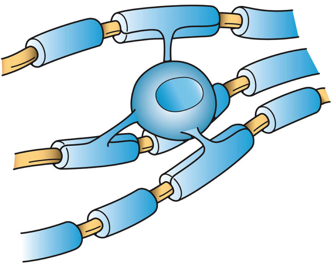
Oligodendrocytes (), or oligodendroglia, are a type of neuroglia whose main functions are to provide support and insulation to axons in the central nervous system of jawed vertebrates, equivalent to the function performed by Schwann cells in the peripheral nervous system. Oligodendrocytes do this by creating the myelin sheath. A single oligodendrocyte can extend its processes to 50 axons, wrapping approximately 1 μm of myelin sheath around each axon; Schwann cells, on the other hand, can wrap around only one axon. Each oligodendrocyte forms one segment of myelin for several adjacent axons. Oligodendrocytes are found only in the central nervous system, which comprises the brain and spinal cord. These cells were originally thought to have been produced in the ventral neural tube; however, research now shows oligodendrocytes originate from the ventral ventricular zone of the embryonic spinal cord and possibly have some concentrations in the forebrain. They are the last cell ... [...More Info...] [...Related Items...] OR: [Wikipedia] [Google] [Baidu] |
Sulfonyl Chloride
In inorganic chemistry, sulfonyl halide groups occur when a sulfonyl () functional group is singly bonded to a halogen atom. They have the general formula , where X is a halogen. The stability of sulfonyl halides decreases in the order fluorides > chlorides > bromides > iodides, all four types being well known. The sulfonyl chlorides and fluorides are of dominant importance in this series. Structure Sulfonyl halides have tetrahedral sulfur centres attached to two oxygen atoms, an organic radical, and a halide. In a representative example, methanesulfonyl chloride, the S=O, S−C, and S−Cl bond distances are respectively 142.4, 176.3, and 204.6 pm. Sulfonyl chlorides Sulfonic acid chlorides, or sulfonyl chlorides, are a sulfonyl halide with the general formula . Production Arylsulfonyl chlorides are made industrially in a two-step, one-pot reaction from an arene (in this case, benzene) and chlorosulfuric acid: :C6H6 + HOSO2Cl -> C6H5SO3H + HCl :C6H5SO3H + HOSO2Cl -> C6 ... [...More Info...] [...Related Items...] OR: [Wikipedia] [Google] [Baidu] |
Texas Red
Texas Red or sulforhodamine 101 acid chloride is a red fluorescent dye, used in histology for staining cell specimens, for sorting cells with fluorescent-activated cell sorting machines, in fluorescence microscopy applications, and in immunohistochemistry. Texas Red fluoresces at about 615 nm, and the peak of its absorption spectrum is at 589 nm. The powder is dark purple. Solutions can be excited by a dye laser tuned to 595-605 nm, or less efficiently a krypton laser at 567 nm. The absorption extinction coefficient at 596 nm is about 85,000 M−1cm−1. The compound is usually a mixture of two monosulfonyl chlorides, i.e., as pictured, or with the SO3 and SO2Cl groups exchanged. It can be used as a marker of proteins, with which it easily forms conjugates via the sulfonyl chloride (SO2Cl) group. In water, the sulfonyl chloride group of unreacted Texas Red molecules hydrolyses to sulfonate and the molecule becomes the very water-soluble sulforhodamine ... [...More Info...] [...Related Items...] OR: [Wikipedia] [Google] [Baidu] |
Rhodamine Dyes
Rhodamine is a family of related dyes, a subset of the triarylmethane dyes. They are derivatives of xanthene. Important members of the rhodamine family are Rhodamine 6G, Rhodamine 123, and Rhodamine B. They are mainly used to dye paper and inks, but they lack the lightfastness for fabric dyeing. Use Aside from their major applications, they are often used as a tracer dye, e.g. to determine the rate and direction of flow and transport of water. Rhodamine dyes fluoresce and can thus be detected easily and inexpensively with instruments called fluorometers. Rhodamine dyes are used extensively in biotechnology applications such as fluorescence microscopy, flow cytometry, fluorescence correlation spectroscopy and ELISA. Rhodamine 123 is used in biochemistry to inhibit mitochondrion function. Rhodamine 123 appears to bind to the mitochondrial membranes and inhibit transport processes, especially the electron transport chain, thus slowing down cellular respiration. It is a substrate ... [...More Info...] [...Related Items...] OR: [Wikipedia] [Google] [Baidu] |


