|
Counterstain
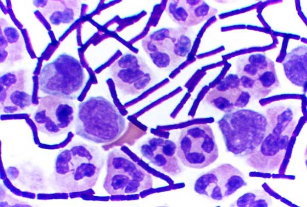
A counterstain is a stain with colour contrasting to the principal stain, making the stained structure easily visible using a microscope. Examples include the malachite green counterstain to the fuchsine stain in the Gimenez staining technique and the eosin counterstain to haematoxylin in the H&E stain. In Gram staining, crystal violet stains only Gram-positive bacteria, and safranin counterstain is applied which stains all cells, allowing the identification of Gram-negative bacteria as well. An alternative method uses dilute carbofluozide. Counterstains are sometimes used to separate animals from organic detritus In biology, detritus ( or ) is organic matter made up of the decomposition, decomposing remains of organisms and plants, and also of feces. Detritus usually hosts communities of microorganisms that colonize and decomposition, decompose (Reminera ... in microbiology studies. References Staining {{pathology-stub ... [...More Info...] [...Related Items...] OR: [Wikipedia] [Google] [Baidu] |
Staining (biology)
Staining is a technique used to enhance contrast in samples, generally at the microscopic level. Stains and dyes are frequently used in histology (microscopic study of biological tissues), in cytology (microscopic study of cells), and in the medical fields of histopathology, hematology, and cytopathology that focus on the study and diagnoses of diseases at the microscopic level. Stains may be used to define biological tissues (highlighting, for example, muscle fibers or connective tissue), cell populations (classifying different blood cells), or organelles within individual cells. In biochemistry, it involves adding a class-specific (DNA, proteins, lipids, carbohydrates) dye to a substrate to qualify or quantify the presence of a specific compound. Staining and fluorescent tagging can serve similar purposes. Biological staining is also used to mark cells in flow cytometry, and to flag proteins or nucleic acids in gel electrophoresis. Light microscopes are used for vi ... [...More Info...] [...Related Items...] OR: [Wikipedia] [Google] [Baidu] |
Gram Staining
Gram stain (Gram staining or Gram's method), is a method of staining used to classify bacterial species into two large groups: gram-positive bacteria and gram-negative bacteria. It may also be used to diagnose a fungal infection. The name comes from the Danish bacteriologist Hans Christian Gram, who developed the technique in 1884. Gram staining differentiates bacteria by the chemical and physical properties of their cell walls. Gram-positive cells have a thick layer of peptidoglycan in the cell wall that retains the primary stain, crystal violet. Gram-negative cells have a thinner peptidoglycan layer that allows the crystal violet to wash out on addition of ethanol. They are stained pink or red by the counterstain, commonly safranin or fuchsine. Lugol's iodine solution is always added after addition of crystal violet to form a stable complex with crystal violet that strengthens the bonds of the stain with the cell wall. Gram staining is almost always the first step in the ... [...More Info...] [...Related Items...] OR: [Wikipedia] [Google] [Baidu] |
Haematoxylin
Haematoxylin American and British English spelling differences#ae and oe, or hematoxylin (), also called natural black 1 or Colour Index International, C.I. 75290, is a chemical compound, compound extracted from wood#Heartwood and sapwood, heartwood of the logwood tree (''Haematoxylum campechianum'') with a chemical formula of . This Natural dye, naturally derived dye has been used as a staining, histologic stain, as an ink and as a dye in the textile and leather industry. As a dye, haematoxylin has been called palo de Campeche, logwood extract, bluewood and blackwood. In histology, haematoxylin staining is commonly followed by counterstain, counterstaining with eosin. When paired, this staining procedure is known as H&E staining and is one of the most commonly used combinations in histology. In addition to its use in the H&E stain, haematoxylin is also a component of the Papanicolaou stain (or Pap stain) which is widely used in the study of cytology specimens. Although the stain ... [...More Info...] [...Related Items...] OR: [Wikipedia] [Google] [Baidu] |
Malachite Green
Malachite green is an organic compound that is used as a dyestuff and controversially as an antimicrobial in aquaculture. Malachite green is traditionally used as a dye for materials such as silk, leather, and paper. Despite its name the dye is not prepared from the mineral malachite; the name just comes from the similarity of color. Structures and properties Malachite green is classified in the dyestuff industry as a triarylmethane dye and also using in pigment industry. Formally, malachite green refers to the chloride salt , although the term malachite green is used loosely and often just refers to the colored cation. The oxalate salt is also marketed. The anions have no effect on the color. The intense green color of the cation results from a strong absorption band at 621 nm ( extinction coefficient of ). Malachite green is prepared by the condensation of benzaldehyde and dimethylaniline to give leuco malachite green (LMG): : Second, this colorless leuco compound, ... [...More Info...] [...Related Items...] OR: [Wikipedia] [Google] [Baidu] |
Safranin
Safranin (Safranin O or basic red 2) is a biological stain used in histology and cytology. Safranin is used as a counterstain in some staining protocols, colouring cell nuclei red. This is the classic counterstain in both Gram stains and endospore staining. It can also be used for the detection of cartilage, mucin and mast cell granules. Safranin typically has the chemical structure shown at right (sometimes described as dimethyl safranin). There is also trimethyl safranin, which has an added methyl group in the ''ortho-'' position (see Arene substitution pattern) of the lower ring. Both compounds behave essentially identically in biological staining applications, and most manufacturers of safranin do not distinguish between the two. Commercial safranin preparations often contain a blend of both types. Safranin is also used as redox indicator in analytical chemistry. Safranines Safranines are the azonium compounds of symmetrical 2,8-dimethyl-3,7-diaminophenazine. They ... [...More Info...] [...Related Items...] OR: [Wikipedia] [Google] [Baidu] |
Gimenez Stain
The Gimenez staining technique uses biological stains to detect and identify bacterial infections in tissue samples. Although largely superseded by techniques like Giemsa staining, the Gimenez technique may be valuable for detecting certain slow-growing or fastidious bacteria. Basic fuchsin stain in aqueous solution with phenol and ethanol colours many bacteria (both gram positive and Gram negative) red, magenta, or pink. A malachite green counterstain gives a blue-green background cast to the surrounding tissue. See also *Histology * List of common staining protocols *Microscopy Microscopy is the technical field of using microscopes to view subjects too small to be seen with the naked eye (objects that are not within the resolution range of the normal eye). There are three well-known branches of microscopy: optical mic ... References * P. Bruneval ''et al.''.Detection of fastidious bacteria in cardiac valves in cases of blood culture negative endocarditis. ''Journal o ... [...More Info...] [...Related Items...] OR: [Wikipedia] [Google] [Baidu] |
Eosin
Eosin is the name of several fluorescent acidic compounds which bind to and from salts with basic, or eosinophilic, compounds like proteins containing basic amino acid residues such as histidine, arginine and lysine, and stains them dark red or pink as a result of the actions of bromine on eosin. In addition to staining proteins in the cytoplasm, it can be used to stain collagen and muscle fibers for examination under the microscope. Structures that stain readily with eosin are termed eosinophilic. In the field of histology, Eosin Y is the form of eosin used most often as a histologic stain. History and etymology Eosin was named by its inventor Heinrich Caro after the nickname (Eos) of a childhood friend, Anna Peters. It was commercialized (mainly for the textile industry) in 1874, in the same year when it was invented. Variants There are actually two very closely related compounds commonly referred to as ''eosin''. Most often used is in histology is eosin Y, which is a t ... [...More Info...] [...Related Items...] OR: [Wikipedia] [Google] [Baidu] |
H&E Stain
Hematoxylin and eosin stain ( or haematoxylin and eosin stain or hematoxylin–eosin stain; often abbreviated as H&E stain or HE stain) is one of the principal tissue stains used in histology. It is the most widely used stain in medical diagnosis and is often the ''gold standard.'' For example, when a pathologist looks at a biopsy of a suspected cancer, the histological section is likely to be stained with H&E. H&E is the combination of two histological stains: hematoxylin and eosin. The hematoxylin stains cell nuclei a purplish blue, and eosin stains the extracellular matrix and cytoplasm pink, with other structures taking on different shades, hues, and combinations of these colors. Hence a pathologist can easily differentiate between the nuclear and cytoplasmic parts of a cell, and additionally, the overall patterns of coloration from the stain show the general layout and distribution of cells and provides a general overview of a tissue sample's structure. Thus, patte ... [...More Info...] [...Related Items...] OR: [Wikipedia] [Google] [Baidu] |
Crystal Violet
Crystal violet or gentian violet, also known as methyl violet 10B or hexamethyl pararosaniline chloride, is a triphenylmethane, triarylmethane dye used as a histological stain and in Gram staining, Gram's method of classifying bacteria. Crystal violet has antibacterial, Antifungal medication, antifungal, and anthelmintic (Anthelmintic, vermicide) properties and was formerly important as a topical antiseptic. The medical use of the dye has been largely superseded by more modern drugs, although it is still listed by the World Health Organization. The name ''gentian violet'' was originally used for a mixture of methyl pararosaniline dyes (methyl violet), but is now often considered a synonym for ''crystal violet''. The name refers to its colour, being like that of the petals of certain Gentiana, gentian flowers; it is not made from gentians or list of plants known as violet, violets. Production A number of possible routes can be used to prepare crystal violet. The original procedur ... [...More Info...] [...Related Items...] OR: [Wikipedia] [Google] [Baidu] |
Gram-positive
In bacteriology, gram-positive bacteria are bacteria that give a positive result in the Gram stain test, which is traditionally used to quickly classify bacteria into two broad categories according to their type of cell wall. The Gram stain is used by microbiologists to place bacteria into two main categories, gram-positive (+) and gram-negative bacteria, gram-negative (−). Gram-positive bacteria have a thick layer of peptidoglycan within the cell wall, and gram-negative bacteria have a thin layer of peptidoglycan. Gram-positive bacteria retain the crystal violet stain used in the test, resulting in a purple color when observed through an optical microscope. The thick layer of peptidoglycan in the bacterial cell wall retains the Stain (biology), stain after it has been fixed in place by iodine. During the decolorization step, the decolorizer removes crystal violet from all other cells. Conversely, gram-negative bacteria cannot retain the violet stain after the decolorization ... [...More Info...] [...Related Items...] OR: [Wikipedia] [Google] [Baidu] |
Gram Stain Anthrax
The gram (originally gramme; SI unit symbol g) is a unit of mass in the International System of Units (SI) equal to one thousandth of a kilogram. Originally defined in 1795 as "the absolute weight of a volume of pure water equal to the cube of the hundredth part of a metre melting ice", the defining temperature (0 °C) was later changed to the temperature of maximum density of water (approximately 4 °C). Subsequent redefinitions agree with this original definition to within 30 parts per million (0.003%), with the maximum density of water remaining very close to 1 g/cm3, as shown by modern measurements. By the late 19th century, there was an effort to make the base unit the kilogram and the gram a derived unit. In 1960, the new International System of Units defined a ''gram'' as one thousandth of a kilogram (i.e., one gram is ). The kilogram, as of 2019, is defined by the International Bureau of Weights and Measures from the metre, the second, and from the ... [...More Info...] [...Related Items...] OR: [Wikipedia] [Google] [Baidu] |
Microscope
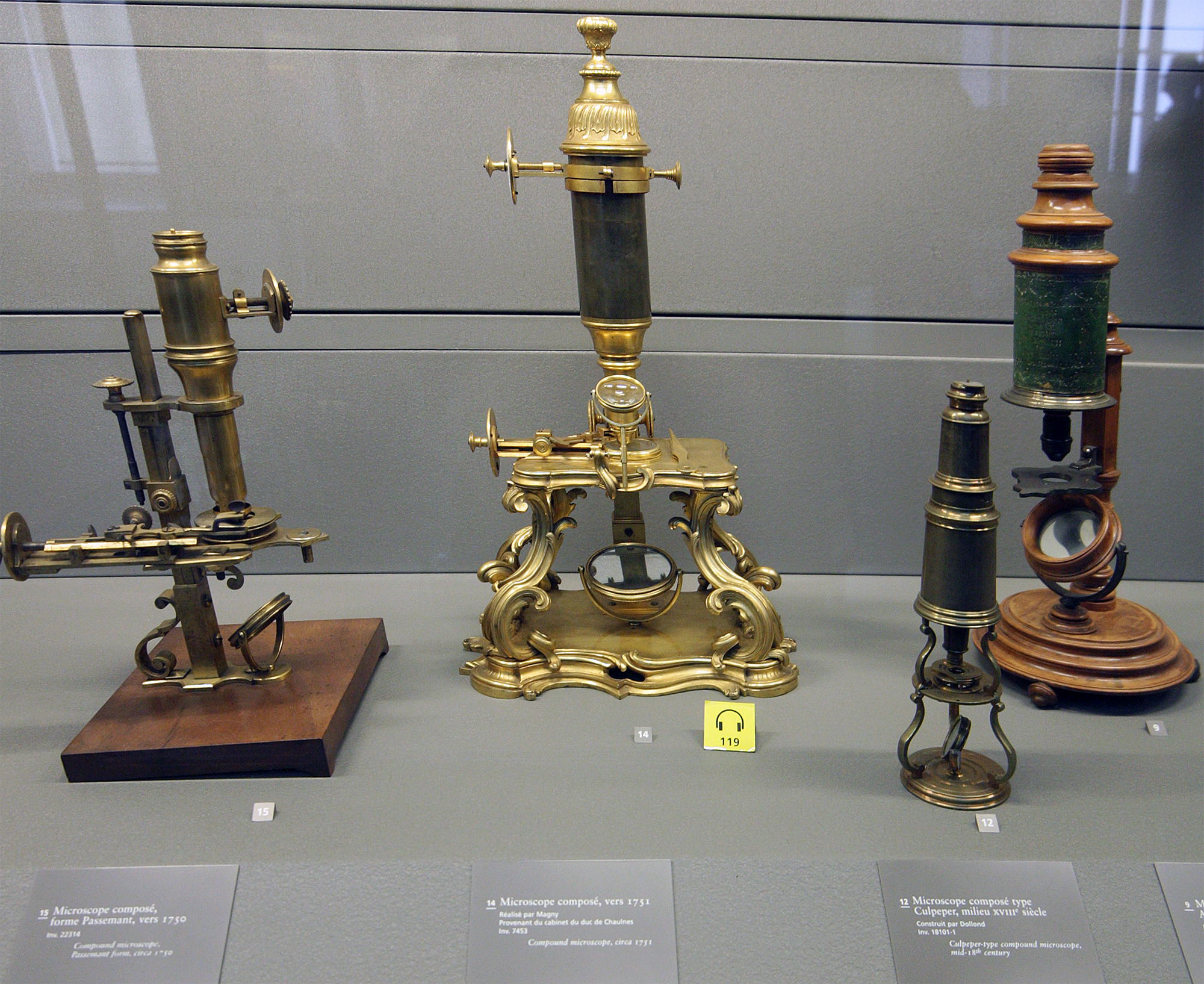
A microscope () is a laboratory equipment, laboratory instrument used to examine objects that are too small to be seen by the naked eye. Microscopy is the science of investigating small objects and structures using a microscope. Microscopic means being invisible to the eye unless aided by a microscope. There are many types of microscopes, and they may be grouped in different ways. One way is to describe the method an instrument uses to interact with a sample and produce images, either by sending a beam of light or electrons through a sample in its optical path, by detecting fluorescence, photon emissions from a sample, or by scanning across and a short distance from the surface of a sample using a probe. The most common microscope (and the first to be invented) is the optical microscope, which uses lenses to refract visible light that passed through a microtome, thinly sectioned sample to produce an observable image. Other major types of microscopes are the fluorescence micro ... [...More Info...] [...Related Items...] OR: [Wikipedia] [Google] [Baidu] |


