|
Microscope
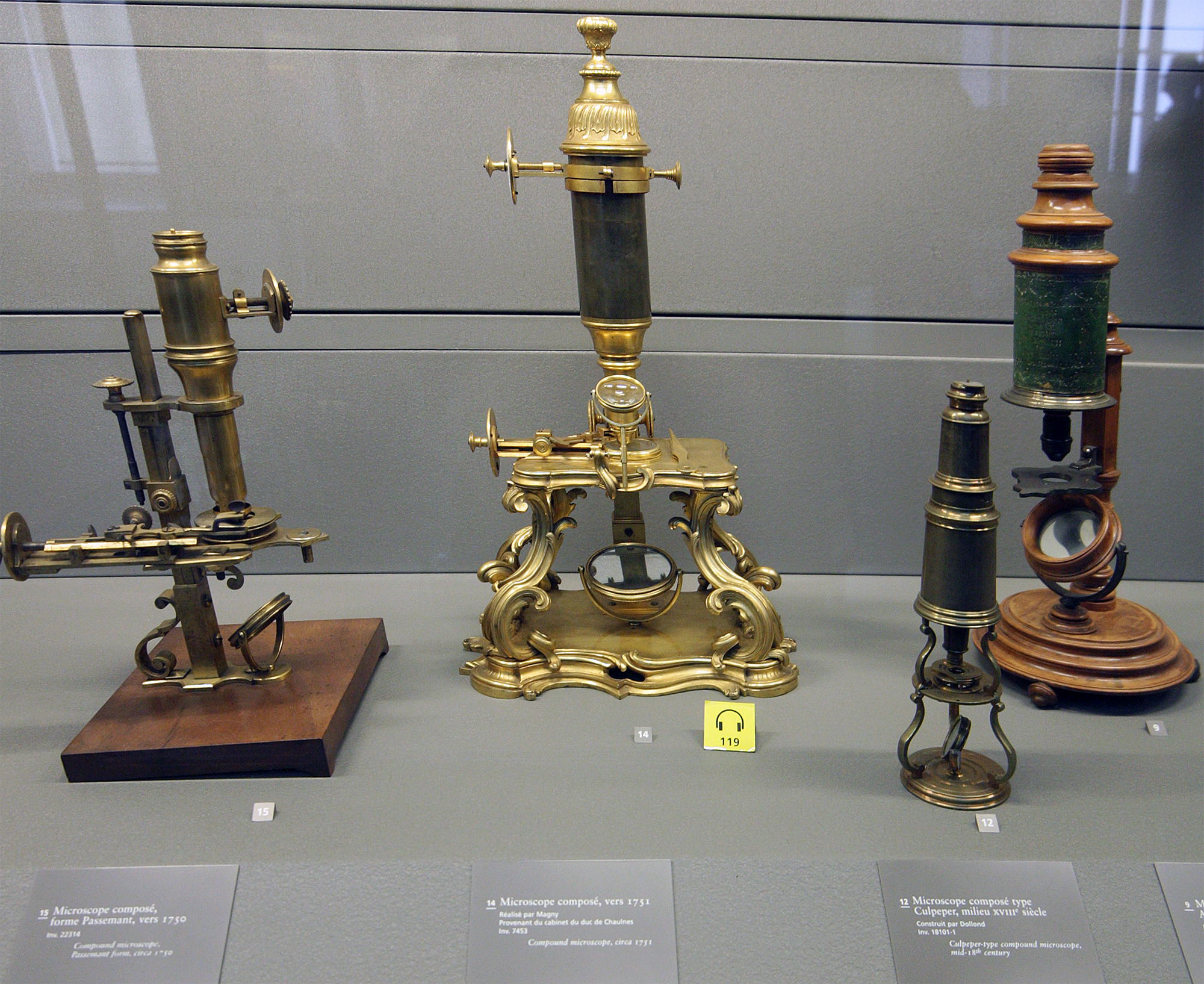
A microscope () is a laboratory instrument used to examine objects that are too small to be seen by the naked eye. Microscopy is the science of investigating small objects and structures using a microscope. Microscopic means being invisible to the eye unless aided by a microscope. There are many types of microscopes, and they may be grouped in different ways. One way is to describe the method an instrument uses to interact with a sample and produce images, either by sending a beam of light or electrons through a sample in its optical path, by detecting photon emissions from a sample, or by scanning across and a short distance from the surface of a sample using a probe. The most common microscope (and the first to be invented) is the optical microscope, which uses lenses to refract visible light that passed through a thinly sectioned sample to produce an observable image. Other major types of microscopes are the fluorescence microscope, electron microscope (both the t ... [...More Info...] [...Related Items...] OR: [Wikipedia] [Google] [Baidu] |
Optical Microscope
The optical microscope, also referred to as a light microscope, is a type of microscope that commonly uses visible light and a system of lenses to generate magnified images of small objects. Optical microscopes are the oldest design of microscope and were possibly invented in their present compound form in the 17th century. Basic optical microscopes can be very simple, although many complex designs aim to improve resolution and sample contrast. The object is placed on a stage and may be directly viewed through one or two eyepieces on the microscope. In high-power microscopes, both eyepieces typically show the same image, but with a stereo microscope, slightly different images are used to create a 3-D effect. A camera is typically used to capture the image (micrograph). The sample can be lit in a variety of ways. Transparent objects can be lit from below and solid objects can be lit with light coming through ( bright field) or around ( dark field) the objective lens. Polar ... [...More Info...] [...Related Items...] OR: [Wikipedia] [Google] [Baidu] |
Microscopy
Microscopy is the technical field of using microscopes to view objects and areas of objects that cannot be seen with the naked eye (objects that are not within the resolution range of the normal eye). There are three well-known branches of microscopy: optical, electron, and scanning probe microscopy, along with the emerging field of X-ray microscopy. Optical microscopy and electron microscopy involve the diffraction, reflection, or refraction of electromagnetic radiation/electron beams interacting with the specimen, and the collection of the scattered radiation or another signal in order to create an image. This process may be carried out by wide-field irradiation of the sample (for example standard light microscopy and transmission electron microscopy) or by scanning a fine beam over the sample (for example confocal laser scanning microscopy and scanning electron microscopy). Scanning probe microscopy involves the interaction of a scanning probe with the surface of the ... [...More Info...] [...Related Items...] OR: [Wikipedia] [Google] [Baidu] |
Electron Microscope
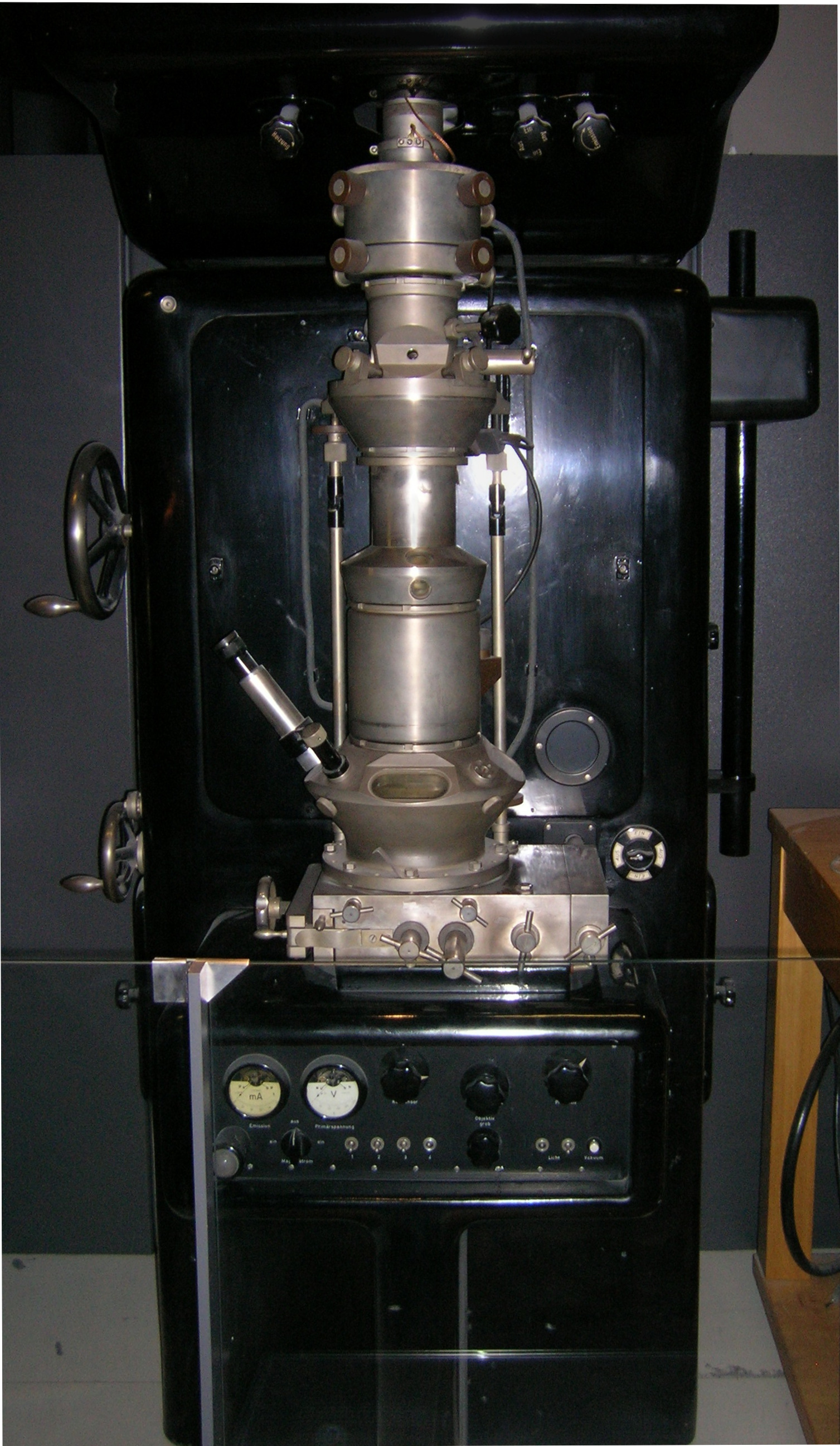
An electron microscope is a microscope that uses a beam of accelerated electrons as a source of illumination. As the wavelength of an electron can be up to 100,000 times shorter than that of visible light photons, electron microscopes have a higher resolving power than light microscopes and can reveal the structure of smaller objects. A scanning transmission electron microscope has achieved better than 50 pm resolution in annular dark-field imaging mode and magnifications of up to about 10,000,000× whereas most light microscopes are limited by diffraction to about 200 nm resolution and useful magnifications below 2000×. Electron microscopes use shaped magnetic fields to form electron optical lens systems that are analogous to the glass lenses of an optical light microscope. Electron microscopes are used to investigate the ultrastructure of a wide range of biological and inorganic specimens including microorganisms, cells, large molecules, biopsy samples, ... [...More Info...] [...Related Items...] OR: [Wikipedia] [Google] [Baidu] |
Transmission Electron Microscope
Transmission electron microscopy (TEM) is a microscopy technique in which a beam of electrons is transmitted through a specimen to form an image. The specimen is most often an ultrathin section less than 100 nm thick or a suspension on a grid. An image is formed from the interaction of the electrons with the sample as the beam is transmitted through the specimen. The image is then magnified and focused onto an imaging device, such as a fluorescent screen, a layer of photographic film, or a sensor such as a scintillator attached to a charge-coupled device. Transmission electron microscopes are capable of imaging at a significantly higher resolution than light microscopes, owing to the smaller de Broglie wavelength of electrons. This enables the instrument to capture fine detail—even as small as a single column of atoms, which is thousands of times smaller than a resolvable object seen in a light microscope. Transmission electron microscopy is a major analytical method i ... [...More Info...] [...Related Items...] OR: [Wikipedia] [Google] [Baidu] |
Fluorescence Microscope
A fluorescence microscope is an optical microscope that uses fluorescence instead of, or in addition to, scattering, reflection, and attenuation or absorption, to study the properties of organic or inorganic substances. "Fluorescence microscope" refers to any microscope that uses fluorescence to generate an image, whether it is a simple set up like an epifluorescence microscope or a more complicated design such as a confocal microscope, which uses optical sectioning to get better resolution of the fluorescence image. Principle The specimen is illuminated with light of a specific wavelength (or wavelengths) which is absorbed by the fluorophores, causing them to emit light of longer wavelengths (i.e., of a different color than the absorbed light). The illumination light is separated from the much weaker emitted fluorescence through the use of a spectral emission filter. Typical components of a fluorescence microscope are a light source ( xenon arc lamp or mercury-vapor lam ... [...More Info...] [...Related Items...] OR: [Wikipedia] [Google] [Baidu] |
Microscopic
The microscopic scale () is the scale of objects and events smaller than those that can easily be seen by the naked eye, requiring a lens or microscope to see them clearly. In physics, the microscopic scale is sometimes regarded as the scale between the macroscopic scale and the quantum scale. Microscopic units and measurements are used to classify and describe very small objects. One common microscopic length scale unit is the micrometre (also called a ''micron'') (symbol: μm), which is one millionth of a metre. History Whilst compound microscopes were first developed in the 1590s, the significance of the microscopic scale was only truly established in the 1600s when Marcello Malphigi and Antonie van Leeuwenhoek microscopically observed frog lungs and microorganisms. As microbiology was established, the significance of making scientific observations at a microscopic level increased. Published in 1665, Robert Hooke’s book Micrographia details his microscopic observa ... [...More Info...] [...Related Items...] OR: [Wikipedia] [Google] [Baidu] |
Eyepiece
An eyepiece, or ocular lens, is a type of lens that is attached to a variety of optical devices such as telescopes and microscopes. It is named because it is usually the lens that is closest to the eye when someone looks through the device. The objective lens or mirror collects light and brings it to focus creating an image. The eyepiece is placed near the focal point of the objective to magnify this image. The amount of magnification depends on the focal length of the eyepiece. An eyepiece consists of several "lens elements" in a housing, with a "barrel" on one end. The barrel is shaped to fit in a special opening of the instrument to which it is attached. The image can be focused by moving the eyepiece nearer and further from the objective. Most instruments have a focusing mechanism to allow movement of the shaft in which the eyepiece is mounted, without needing to manipulate the eyepiece directly. The eyepieces of binoculars are usually permanently mounted in the binocular ... [...More Info...] [...Related Items...] OR: [Wikipedia] [Google] [Baidu] |
Zacharias Janssen
Zacharias Janssen; also Zacharias Jansen or Sacharias Jansen; 1585 – pre-1632) was a Dutch spectacle-maker who lived most of his life in Middelburg. He is associated with the invention of the first optical telescope and/or the first truly compound microscope, but these claims (made 20 years after his death) may be fabrications put forward by his son. Biography Zacharias Janssen was born in The Hague. Local records seem to indicate he was born in 1585 although a date of birth as early as 1580 or as late as 1588 are also given. His parents were Hans MartensSource: a book by Huib J. Zuidervaart which is to be published in the spring of 2008. The in Middelburg has already been shown a first version of this book. (who may have had the occupation of a peddler) and Maeyken Meertens, both probably from Antwerp, Belgium. He grew up with his sister Sara in Middelburg, at the time the second most important city of the Netherlands. He was known as a "street seller" who was constantly ... [...More Info...] [...Related Items...] OR: [Wikipedia] [Google] [Baidu] |
Objective (optics)
In optical engineering, the objective is the optical element that gathers light from the object being observed and focuses the light rays to produce a real image. Objectives can be a single lens or mirror, or combinations of several optical elements. They are used in microscopes, binoculars, telescopes, cameras, slide projectors, CD players and many other optical instruments. Objectives are also called object lenses, object glasses, or objective glasses. Microscope objectives The objective lens of a microscope is the one at the bottom near the sample. At its simplest, it is a very high-powered magnifying glass, with very short focal length. This is brought very close to the specimen being examined so that the light from the specimen comes to a focus inside the microscope tube. The objective itself is usually a cylinder containing one or more lenses that are typically made of glass; its function is to collect light from the sample. Magnification One of the most important prop ... [...More Info...] [...Related Items...] OR: [Wikipedia] [Google] [Baidu] |
Electron
The electron (, or in nuclear reactions) is a subatomic particle with a negative one elementary electric charge. Electrons belong to the first generation of the lepton particle family, and are generally thought to be elementary particles because they have no known components or substructure. The electron's mass is approximately 1/1836 that of the proton. Quantum mechanical properties of the electron include an intrinsic angular momentum ( spin) of a half-integer value, expressed in units of the reduced Planck constant, . Being fermions, no two electrons can occupy the same quantum state, per the Pauli exclusion principle. Like all elementary particles, electrons exhibit properties of both particles and waves: They can collide with other particles and can be diffracted like light. The wave properties of electrons are easier to observe with experiments than those of other particles like neutrons and protons because electrons have a lower mass and hence a longer ... [...More Info...] [...Related Items...] OR: [Wikipedia] [Google] [Baidu] |


