|
Subscapularis Muscle
The subscapularis is a large triangular muscle which fills the subscapular fossa and inserts into the lesser tubercle of the humerus and the front of the capsule of the shoulder-joint. Structure It arises from its medial two-thirds and Some fibers arise from tendinous laminae, which intersect the muscle and are attached to ridges on the bone; others from an aponeurosis, which separates the muscle from the teres major and the long head of the triceps brachii. The fibers pass laterally and coalesce into a tendon that is inserted into the lesser tubercle of the humerus and the anterior part of the shoulder-joint capsule. Tendinous fibers extend to the greater tubercle with insertions into the bicipital groove. Relations The tendon of the muscle is separated from the neck of the scapula by a large bursa, which communicates with the cavity of the shoulder-joint through an aperture in the capsule. The subscapularis is separated from the serratus anterior books.google.com/books ... [...More Info...] [...Related Items...] OR: [Wikipedia] [Google] [Baidu] |
Subscapular Fossa
The scapula (plural scapulae or scapulas), also known as the shoulder blade, is the bone that connects the humerus (upper arm bone) with the clavicle (collar bone). Like their connected bones, the scapulae are paired, with each scapula on either side of the body being roughly a mirror image of the other. The name derives from the Classical Latin word for trowel or small shovel, which it was thought to resemble. In compound terms, the prefix omo- is used for the shoulder blade in medical terminology. This prefix is derived from ὦμος (ōmos), the Ancient Greek word for shoulder, and is cognate with the Latin , which in Latin signifies either the shoulder or the upper arm bone. The scapula forms the back of the shoulder girdle. In humans, it is a flat bone, roughly triangular in shape, placed on a posterolateral aspect of the thoracic cage. Structure The scapula is a thick, flat bone lying on the thoracic wall that provides an attachment for three groups of muscles: intrin ... [...More Info...] [...Related Items...] OR: [Wikipedia] [Google] [Baidu] |
Teres Major
The teres major muscle is a muscle of the upper limb. It attaches to the scapula and the humerus and is one of the seven scapulohumeral muscles. It is a thick but somewhat flattened muscle. The teres major muscle (from Latin ''teres'', meaning "rounded") is positioned above the latissimus dorsi muscle and assists in the Anatomical terms of motion#Flexion and extension, extension and Anatomical terms of motion#Rotation, medial rotation of the humerus. This muscle is commonly confused as a rotator cuff muscle, but it is not because it does not attach to the capsule of the shoulder joint, unlike the teres minor muscle for example. Structure The teres major muscle originates on the dorsal surface of the Scapula, inferior angle and the lower part of the Scapula, lateral border of the scapula. The fibers of teres major insert into the medial lip of the Bicipital groove, intertubercular sulcus of the humerus. Relations The tendon, at its insertion, lies behind that of the latissimus ... [...More Info...] [...Related Items...] OR: [Wikipedia] [Google] [Baidu] |
Muscles Of The Upper Limb
Skeletal muscles (commonly referred to as muscles) are organs of the vertebrate muscular system and typically are attached by tendons to bones of a skeleton. The muscle cells of skeletal muscles are much longer than in the other types of muscle tissue, and are often known as muscle fibers. The muscle tissue of a skeletal muscle is striated – having a striped appearance due to the arrangement of the sarcomeres. Skeletal muscles are voluntary muscles under the control of the somatic nervous system. The other types of muscle are cardiac muscle which is also striated and smooth muscle which is non-striated; both of these types of muscle tissue are classified as involuntary, or, under the control of the autonomic nervous system. A skeletal muscle contains multiple fascicles – bundles of muscle fibers. Each individual fiber, and each muscle is surrounded by a type of connective tissue layer of fascia. Muscle fibers are formed from the fusion of developmental myoblasts in a p ... [...More Info...] [...Related Items...] OR: [Wikipedia] [Google] [Baidu] |
Biceps
The biceps or biceps brachii ( la, musculus biceps brachii, "two-headed muscle of the arm") is a large muscle that lies on the front of the upper arm between the shoulder and the elbow. Both heads of the muscle arise on the scapula and join to form a single muscle belly which is attached to the upper forearm. While the biceps crosses both the shoulder and elbow joints, its main function is at the elbow where it flexes the forearm and supinates the forearm. Both these movements are used when opening a bottle with a corkscrew: first biceps screws in the cork (supination), then it pulls the cork out (flexion). Structure The biceps is one of three muscles in the anterior compartment of the upper arm, along with the brachialis muscle and the coracobrachialis muscle, with which the biceps shares a nerve supply. The biceps muscle has two heads, the short head and the long head, distinguished according to their origin at the coracoid process and supraglenoid tubercle of the sc ... [...More Info...] [...Related Items...] OR: [Wikipedia] [Google] [Baidu] |
Christian Gerber
Christian Gerber (born 24 May 1952, in Switzerland) is an orthopedic surgeon, physician, scientist and medical teacher in orthopedic surgery. He is the current Chairman of the Department of Orthopedics at the University of Zürich, Switzerland. After studying medicine at the University of Berne, he was trained in orthopedic surgery in several Swiss hospitals, including the Davos Hospital and the University Hospital of Berne. He specialized in shoulder surgery at the University of Texas, San Antonio in 1984 and subsequently trained on tumor foot and ankle surgery as well as pediatric orthopedics in Paris in 1985. From 1985 to 1991 he served as a consultant at the Department of Orthopedics at the University of Berne, where in 1991 he was promoted to associate professor in orthopedic surgery. In 1992, he was appointed chief of the Department of Orthopedics in Fribourg and since 1995 he has worked in thBalgristUniversity Hospital in Zürich, where he holds the position of medical direc ... [...More Info...] [...Related Items...] OR: [Wikipedia] [Google] [Baidu] |
Subluxation
A subluxation is an incomplete or partial dislocation of a joint or organ. According to the World Health Organization (WHO), a subluxation is a "significant structural displacement", and is therefore always visible on static imaging studies, such as X-rays. In contrast, the chiropractic belief of "vertebral subluxation" may or may not involve a significant displacement or even pain or clear dysfunction.WHO guidelines on basic training and safety in chiropractic p. 4, including footnote. Joints [...More Info...] [...Related Items...] OR: [Wikipedia] [Google] [Baidu] |
Shoulder-joint
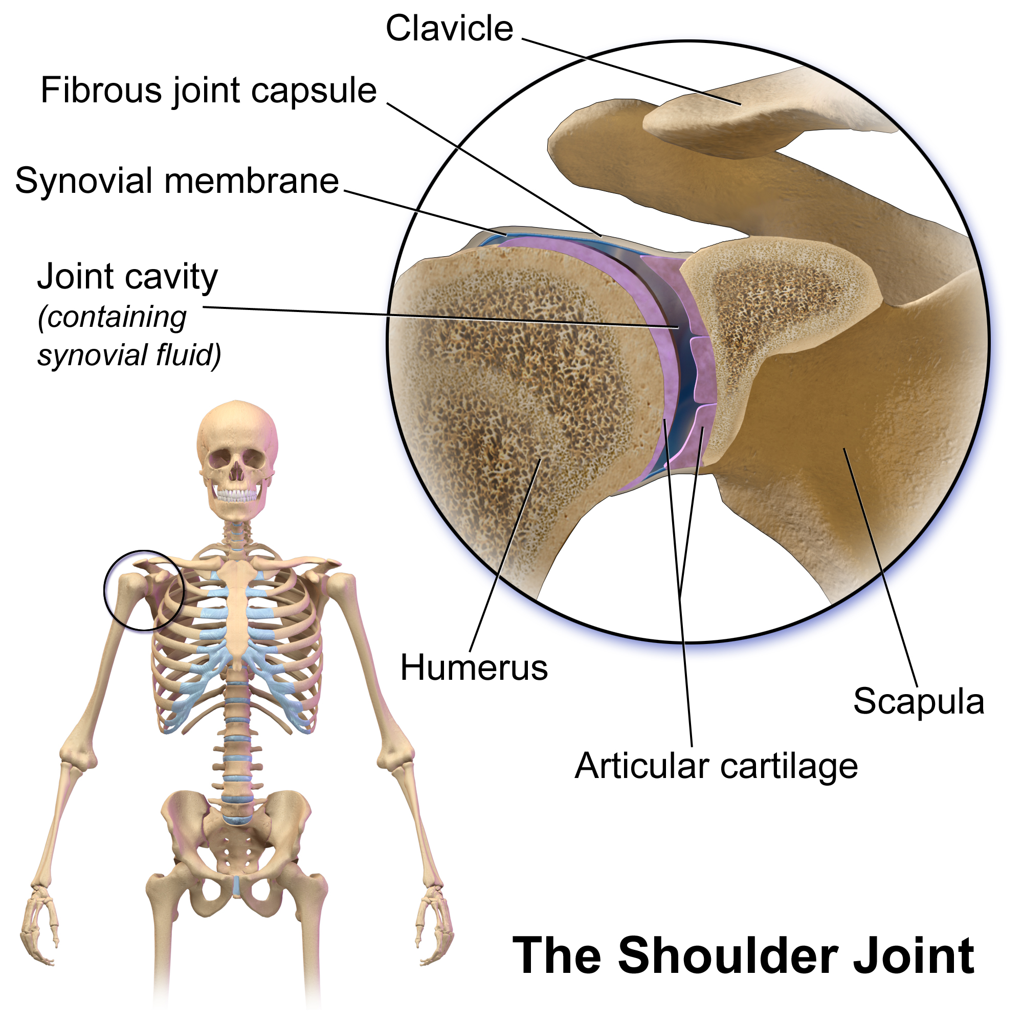
The shoulder joint (or glenohumeral joint from Greek ''glene'', eyeball, + -''oid'', 'form of', + Latin ''humerus'', shoulder) is structurally classified as a synovial ball-and-socket joint and functionally as a diarthrosis and multiaxial joint. It involves an articulation between the glenoid fossa of the scapula (shoulder blade) and the head of the humerus (upper arm bone). Due to the very loose joint capsule that gives a limited interface of the humerus and scapula, it is the most mobile joint of the human body. Structure The shoulder joint is a ball-and-socket joint between the scapula and the humerus. The socket of the glenoid fossa of the scapula is itself quite shallow, but it is made deeper by the addition of the glenoid labrum. The glenoid labrum is a ring of cartilaginous fibre attached to the circumference of the cavity. This ring is continuous with the tendon of the biceps brachii above. Spaces Significant joint spaces are: * The normal glenohumeral space is 4–5 ... [...More Info...] [...Related Items...] OR: [Wikipedia] [Google] [Baidu] |
Head Of The Humerus
The humerus (; ) is a long bone in the arm that runs from the shoulder to the elbow. It connects the scapula and the two bones of the lower arm, the radius and ulna, and consists of three sections. The humeral upper extremity consists of a rounded head, a narrow neck, and two short processes (tubercles, sometimes called tuberosities). The body is cylindrical in its upper portion, and more prismatic below. The lower extremity consists of 2 epicondyles, 2 processes (trochlea & capitulum), and 3 fossae (radial fossa, coronoid fossa, and olecranon fossa). As well as its true anatomical neck, the constriction below the greater and lesser tubercles of the humerus is referred to as its surgical neck due to its tendency to fracture, thus often becoming the focus of surgeons. Etymology The word "humerus" is derived from la, humerus, umerus meaning upper arm, shoulder, and is linguistically related to Gothic ''ams'' shoulder and Greek ''ōmos''. Structure Upper extremity The upper or pro ... [...More Info...] [...Related Items...] OR: [Wikipedia] [Google] [Baidu] |
Brachial Plexus
The brachial plexus is a network () of nerves formed by the anterior rami of the lower four cervical nerves and first thoracic nerve ( C5, C6, C7, C8, and T1). This plexus extends from the spinal cord, through the cervicoaxillary canal in the neck, over the first rib, and into the armpit, it supplies afferent and efferent nerve fibers the to chest, shoulder, arm, forearm, and hand. Structure The brachial plexus is divided into five ''roots'', three ''trunks'', six ''divisions'' (three anterior and three posterior), three ''cords'', and five ''branches''. There are five "terminal" branches and numerous other "pre-terminal" or "collateral" branches, such as the subscapular nerve, the thoracodorsal nerve, and the long thoracic nerve, that leave the plexus at various points along its length. A common structure used to identify part of the brachial plexus in cadaver dissections is the M or W shape made by the musculocutaneous nerve, lateral cord, median nerve, medial cord, and ... [...More Info...] [...Related Items...] OR: [Wikipedia] [Google] [Baidu] |
Posterior Cord
The posterior cord is a part of the brachial plexus The brachial plexus is a network () of nerves formed by the anterior rami of the lower four cervical nerves and first thoracic nerve ( C5, C6, C7, C8, and T1). This plexus extends from the spinal cord, through the cervicoaxillary canal in th .... It consists of contributions from all of the roots of the brachial plexus. The posterior cord gives rise to the following nerves: Additional images File:PLEXUS BRACHIALIS.jpg, Brachial plexus File:Slide12OOO.JPG, Posterior cord File:Slide1SSS.JPG, Posterior cord File:Slide1cord.JPG, Brachial plexus.Deep dissection. File:Slide1ecc.JPG, Brachial plexus.Deep dissection.Anterolateral view References MBBS resources http://mbbsbasic.googlepages.com/ External links * - "Axilla, dissection, anterior view" Nerves of the upper limb {{neuroscience-stub ... [...More Info...] [...Related Items...] OR: [Wikipedia] [Google] [Baidu] |
Upper Subscapular Nerve
The upper (superior) subscapular nerve is the first branch of the posterior cord of the brachial plexus. The upper subscapular nerve contains axons from the ventral rami of the C5 and C6 cervical spinal nerves. It innervates the superior portion of the subscapularis muscle. The inferior portion of the subscapularis is innervated by the lower subscapular nerve. Structure The axons which form the upper subscapular nerve travel from the ventral rami of C5 and C6. They join at the upper trunk and move through its posterior division to form the posterior cord, along with the other two posterior divisions of the middle and lower trunks. The axons then branch from the posterior cord and form the upper subscapular nerve. Function The upper subscapular nerve innervates the superior portion of the subscapularis muscle The subscapularis is a large triangular muscle which fills the subscapular fossa and inserts into the lesser tubercle of the humerus and the front of the capsul ... [...More Info...] [...Related Items...] OR: [Wikipedia] [Google] [Baidu] |
Serratus Anterior
The serratus anterior is a muscle that originates on the surface of the 1st to 8th ribs at the side of the chest and inserts along the entire anterior length of the medial border of the scapula. The serratus anterior acts to pull the scapula forward around the thorax. The muscle is named from Latin: ''serrare'' = to saw, referring to the shape, ''anterior'' = on the front side of the body. Structure Serratus anterior normally originates by nine or ten muscle slips – branches from either the first to ninth ribs or the first to eighth ribs. Because two slips usually arise from the second rib, the number of slips is greater than the number of ribs from which they originate. The muscle is inserted along the medial border of the scapula between the superior and inferior angles along with being inserted along the thoracic vertebrae. The muscle is divided into three named parts depending on their points of insertions: #the serratus anterior superior is inserted near the superior a ... [...More Info...] [...Related Items...] OR: [Wikipedia] [Google] [Baidu] |


