|
Biceps
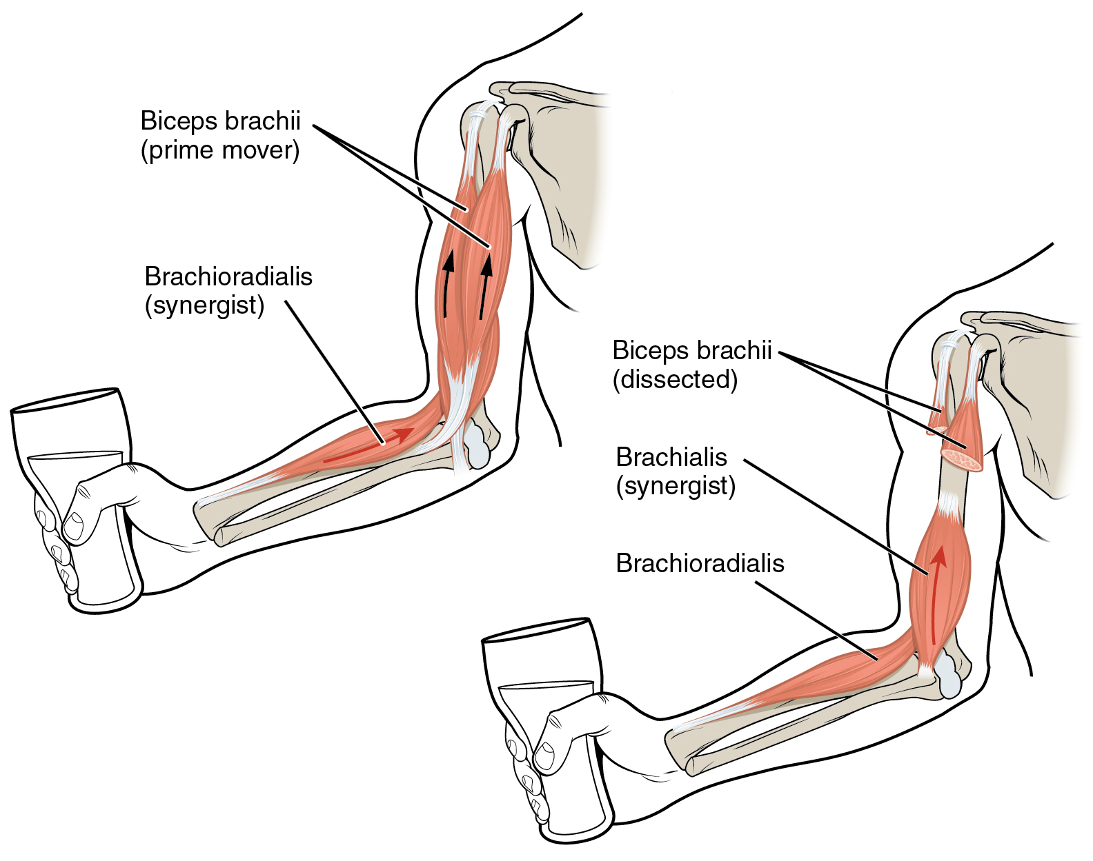
The biceps or biceps brachii (, "two-headed muscle of the arm") is a large muscle that lies on the front of the upper arm between the shoulder and the elbow. Both heads of the muscle arise on the scapula and join to form a single muscle belly which is attached to the upper forearm. While the long head of the biceps crosses both the shoulder and elbow joints, its main function is at the elbow where it flexes and supinates the forearm. Both these movements are used when opening a bottle with a corkscrew: first biceps screws in the cork (supination), then it pulls the cork out (flexion). Structure The biceps is one of three muscles in the anterior compartment of the upper arm, along with the brachialis muscle and the coracobrachialis muscle, with which the biceps shares a nerve supply. The biceps muscle has two heads, the short head and the long head, distinguished according to their origin at the coracoid process and supraglenoid tubercle of the scapula, respectivel ... [...More Info...] [...Related Items...] OR: [Wikipedia] [Google] [Baidu] |
Musculocutaneous Nerve
The musculocutaneous nerve is a Mixed nerve, mixed branch of the lateral cord of the brachial plexus derived from cervical spinal nerves C5-C7. It arises opposite the lower border of the pectoralis minor. It provides motor innervation to the muscles of the anterior compartment of the arm: the Coracobrachialis muscle, coracobrachialis, Biceps brachi, biceps brachii, and Brachialis muscle, brachialis. It provides sensory innervation to the lateral forearm (via its Lateral cutaneous nerve of forearm, terminal branch). It courses through the anterior part of the arm, terminating 2 cm above elbow; after passing the lateral edge of the tendon of Biceps brachi, biceps brachii it is becomes known as the Lateral cutaneous nerve of forearm, lateral cutaneous nerve of the forearm. Structure Course Musculocutaneous nerve arises from the lateral cord of the brachial plexus with root value of C5 to C7 of the spinal cord. It follows the course of the third part of the axillary artery ... [...More Info...] [...Related Items...] OR: [Wikipedia] [Google] [Baidu] |
Shoulder
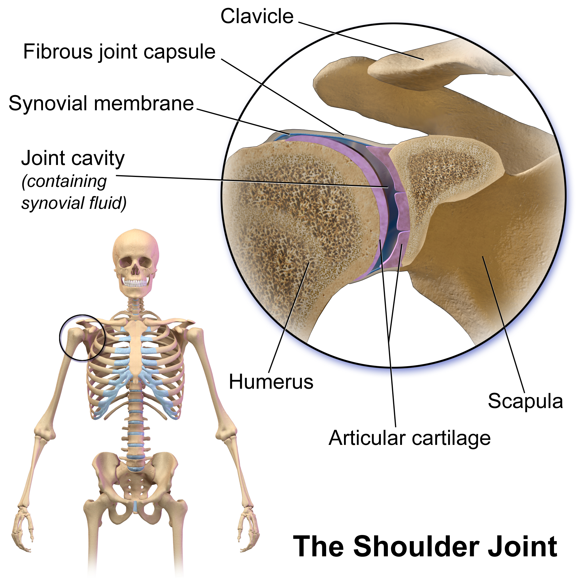
The human shoulder is made up of three bones: the clavicle (collarbone), the scapula (shoulder blade), and the humerus (upper arm bone) as well as associated muscles, ligaments and tendons. The articulations between the bones of the shoulder make up the shoulder joints. The shoulder joint, also known as the glenohumeral joint, is the major joint of the shoulder, but can more broadly include the acromioclavicular joint. In human anatomy, the shoulder joint comprises the part of the body where the humerus attaches to the scapula, and the head sits in the glenoid cavity. The shoulder is the group of structures in the region of the joint. The shoulder joint is the main joint of the shoulder. It is a ball and socket joint that allows the arm to rotate in a circular fashion or to hinge out and up away from the body. The joint capsule is a soft tissue envelope that encircles the glenohumeral joint and attaches to the scapula, humerus, and head of the biceps. It is lined by a ... [...More Info...] [...Related Items...] OR: [Wikipedia] [Google] [Baidu] |
Muscle Head
Anatomical terminology is used to uniquely describe aspects of skeletal muscle, cardiac muscle, and smooth muscle such as their actions, structure, size, and location. Types There are three types of muscle tissue in the body: skeletal, smooth, and cardiac. Skeletal muscle Skeletal muscle, or "voluntary muscle", is a striated muscle tissue that primarily joins to bone with tendons. Skeletal muscle enables movement of bones, and maintains posture. The widest part of a muscle that pulls on the tendons is known as the belly. Muscle slip A muscle slip is a slip of muscle that can either be an anatomical variant, or a branching of a muscle as in rib connections of the serratus anterior muscle. Smooth muscle Smooth muscle is involuntary and found in parts of the body where it conveys action without conscious intent. The majority of this type of muscle tissue is found in the digestive and urinary systems where it acts by propelling forward food, chyme, and feces in the former and ur ... [...More Info...] [...Related Items...] OR: [Wikipedia] [Google] [Baidu] |
Supraglenoid Tubercle
The supraglenoid tubercle is a region of the scapula from which the long head of the biceps brachii muscle originates. It is a small, rough projection superior to the glenoid cavity near the base of the coracoid process. The term supraglenoid is from the Latin ''supra,'' meaning above, and ''glenoid,'' meaning socket or cavity. Clinical relevance Biceps tendonitis Biceps tendonitis originates on the long head of the biceps brachii at the supraglenoid tubercle in 30% of cases. The main symptom is generally anterior biceps instability, but the disease can also be characterized by chronic anterior shoulder pain which radiates towards the lateral part of the elbow. In cases of biceps tendinitis, steroids can be injected fluoroscopically at the supraglenoid tubercle to reduce pain associated with the pathology. Avulsion The supraglenoid tubercle ossifies separately from the rest of the scapula, so may not be as strong as the rest of the bone. It can be pulled off ( avulsed), of ... [...More Info...] [...Related Items...] OR: [Wikipedia] [Google] [Baidu] |
Brachialis Muscle
The brachialis (brachialis anticus) is a muscle in the upper arm that flexes the elbow. It lies beneath the biceps brachii, and makes up part of the floor of the region known as the cubital fossa (elbow pit). It originates from the anterior aspect of the distal humerus; it inserts onto the tuberosity of the ulna. It is innervated by the musculocutaneous nerve, and commonly also receives additional innervation from the radial nerve."Brachialis Muscle." Kenhub. Kenhub, Aug. 2001 The brachialis is the prime mover of elbow flexion generating about 50% more power than the biceps.Saladin, Kenneth S, Stephen J. Sullivan, and Christina A. Gan. Anatomy & Physiology: The Unity of Form and Function. 2015. Print. Structure Origin The brachialis originates from the anterior surface of the distal half of the humerus, near the insertion of the deltoid muscle, which it embraces by two angular processes. Its origin extends below to within 2.5 cm of the margin of the articular surface o ... [...More Info...] [...Related Items...] OR: [Wikipedia] [Google] [Baidu] |
Triceps Brachii Muscle
The triceps, or triceps brachii (Latin for "three-headed muscle of the arm"), is a large muscle on the back of the upper limb of many vertebrates. It consists of three parts: the medial, lateral, and long head. All three heads cross the elbow joint. However, the long head also crosses the shoulder joint. The triceps muscle contracts when the elbow is straightened and expands when the elbow is bent. The long head gets a further contraction when the arm is behind the torso due to how it crosses the shoulder joint. It is the muscle principally responsible for extension of the elbow joint (straightening of the arm). Structure * The long head arises from the infraglenoid tubercle of the scapula. It extends distally anterior to the teres minor and posterior to the teres major. * The medial head arises proximally in the humerus, just inferior to the groove of the radial nerve; from the dorsal (back) surface of the humerus; from the medial intermuscular septum; and its dista ... [...More Info...] [...Related Items...] OR: [Wikipedia] [Google] [Baidu] |
Bicipital Aponeurosis
The bicipital aponeurosis (also known as lacertus fibrosus) is a broad aponeurosis of the biceps brachii, which is located in the cubital fossa of the elbow. It separates superficial from deep structures in much of the fossa. Structure The bicipital aponeurosis originates from the distal insertion of the biceps brachii, and inserts into the deep fascia of the forearm. The biceps tendon inserts on the radial tuberosity, and the bicipital aponeurosis lies medially to it. It reinforces the cubital fossa, helping to protect the brachial artery and the median nerve running underneath. Variations Some individuals (about 3% of the population) have a ''superficial ulnar artery'' that runs superficially to the bicipital aponeurosis instead of underneath it. These individuals are at risk for accidental injury to the ulnar artery during venipuncture. Clinical significance The bicipital aponeurosis is superficial to the brachial artery and the median nerve, but deep to the median cu ... [...More Info...] [...Related Items...] OR: [Wikipedia] [Google] [Baidu] |
Coracobrachialis
The coracobrachialis muscle muscle in the upper medial part of the arm. It is located within the anterior compartment of the arm. It originates from the coracoid process of the scapula; it inserts onto the middle of the medial aspect of the body of the humerus. It is innervated by the musculocutaneous nerve. It acts to adduct and flex the arm. Structure Origin Coracobrachialis muscle arises from the (deep surface of the) apex of the coracoid process of the scapula (a common origin with the short head of the biceps brachii). It additionally also arises from the proximal portion of tendon of origin of the biceps brachii muscle. Insertion It is inserted (by means of a flat tendon) into an impression at the middle of the medial border of the body of the humerus (shaft of the humerus) between the attachments of the medial head of the triceps brachii and the brachialis. Innervation Coracobrachialis muscle is perforated by and innervated by the musculocutaneous nerve, which ari ... [...More Info...] [...Related Items...] OR: [Wikipedia] [Google] [Baidu] |
Humerus
The humerus (; : humeri) is a long bone in the arm that runs from the shoulder to the elbow. It connects the scapula and the two bones of the lower arm, the radius (bone), radius and ulna, and consists of three sections. The humeral upper extremity of humerus, upper extremity consists of a rounded head, a narrow neck, and two short processes (tubercles, sometimes called tuberosities). The body of humerus, body is cylindrical in its upper portion, and more prism (geometry), prismatic below. The lower extremity of humerus, lower extremity consists of 2 epicondyles, 2 processes (trochlea of the humerus, trochlea and capitulum of the humerus, capitulum), and 3 fossae (radial fossa, coronoid fossa, and olecranon fossa). As well as its true anatomical neck, the constriction below the greater and lesser tubercles of the humerus is referred to as its Surgical neck of the humerus, surgical neck due to its tendency to fracture, thus often becoming the focus of surgeons. Etymology The word ... [...More Info...] [...Related Items...] OR: [Wikipedia] [Google] [Baidu] |
Intertubercular Groove
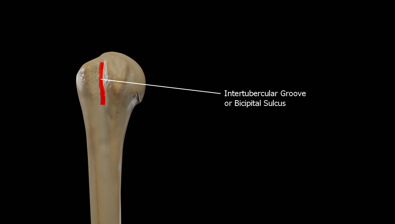
The bicipital groove (intertubercular groove, sulcus intertubercularis) is a deep groove on the humerus that separates the greater tubercle from the lesser tubercle. It allows for the long tendon of the biceps brachii muscle to pass. Structure The bicipital groove separates the greater tubercle from the lesser tubercle. It is usually around 8 cm long and 1 cm wide in adults. The groove lodges the long tendon of the biceps brachii muscle, positioned between the tendon of the pectoralis major muscle on the lateral lip and the tendon of the teres major muscle on the medial lip. It also transmits a branch of the anterior humeral circumflex artery to the shoulder joint. The insertion of the latissimus dorsi muscle is found along the floor of the bicipital groove. The teres major muscle inserts on the medial lip of the groove. It runs obliquely downward, and ends near the junction of the upper with the middle third of the bone. It is the lateral wall of the axilla. Function The ... [...More Info...] [...Related Items...] OR: [Wikipedia] [Google] [Baidu] |


