|
Semilunar Hiatus
The semilunar hiatus or hiatus semilunaris, is a crescent-shaped groove in the lateral wall of the nasal cavity just inferior to the ethmoid bulla. It is the location of the openings of the maxillary sinuses. It is bounded inferiorly and anteriorly by the sharp concave margin of the uncinate process of the ethmoid bone, superiorly by the ethmoid bulla, and posteriorly by the middle nasal concha. Sinus drainage Following the curve anteriorly leads into the infundibulum of the frontonasal duct, which drains the frontal sinus. The anterior ethmoidal cells of the ethmoidal sinus open into the front part of the infundibulum as well. In slightly over 50% of subjects, this is directly continuous with the frontonasal duct from the frontal air sinus. When the anterior end of the uncinate process fuses with the front part of the bulla, however, this continuity is interrupted and the frontonasal duct then drains directly into the anterior end of the middle meatus. The ostium for the ... [...More Info...] [...Related Items...] OR: [Wikipedia] [Google] [Baidu] |
Nasal Cavity
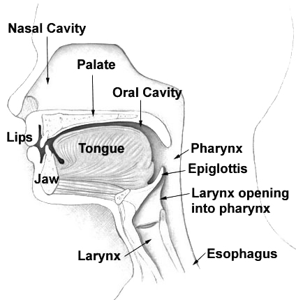
The nasal cavity is a large, air-filled space above and behind the nose in the middle of the face. The nasal septum divides the cavity into two cavities, also known as fossae. Each cavity is the continuation of one of the two nostrils. The nasal cavity is the uppermost part of the respiratory system and provides the nasal passage for inhaled air from the nostrils to the nasopharynx and rest of the respiratory tract. The paranasal sinuses surround and drain into the nasal cavity. Structure The term "nasal cavity" can refer to each of the two cavities of the nose, or to the two sides combined. The lateral wall of each nasal cavity mainly consists of the maxilla. However, there is a deficiency that is compensated for by the perpendicular plate of the palatine bone, the medial pterygoid plate, the labyrinth of ethmoid and the inferior concha. The paranasal sinuses are connected to the nasal cavity through small orifices called ostia. Most of these ostia communicate with ... [...More Info...] [...Related Items...] OR: [Wikipedia] [Google] [Baidu] |
Ethmoid Bulla
The ethmoid bulla (or ethmoidal bulla) is an elevation on the lateral wall of the middle meatus of the nose. It is produced by middle ethmoidal cells. It develops during the first trimester of gestation, and varies significantly based on the size of air cells. Structure The ethmoid bulla is on the lateral wall of the middle meatus of the nose. It is produced by middle ethmoidal cells, which are contained within this bulla, and open on or near to it (often just below it). Just below the bulla is a curved fissure, the hiatus semilunaris. The maxillary sinus also opens below the bulla. It is the largest among the middle ethmoidal cells. Development The ethmoid bulla begins to develop between 8 weeks and 12 weeks of gestation Gestation is the period of development during the carrying of an embryo, and later fetus, inside viviparous animals (the embryo develops within the parent). It is typical for mammals, but also occurs for some non-mammals. Mammals during preg .... ... [...More Info...] [...Related Items...] OR: [Wikipedia] [Google] [Baidu] |
Uncinate Process Of The Ethmoid Bone
In the ethmoid bone, a sickle shaped projection, the uncinate process, projects posteroinferiorly from the ethmoid labyrinth. Between the posterior edge of this process and the anterior surface of the ethmoid bulla, there is a two-dimensional space, resembling a crescent shape. This space continues laterally as a three-dimensional slit-like space - the ethmoidal infundibulum. This is bounded by the uncinate process, medially, the orbital lamina of ethmoid bone The orbital lamina of ethmoid bone, (or lamina papyracea or orbital lamina) is a smooth, oblong bone plate which forms the lateral surface of the labyrinth of the ethmoid bone in the skull. The plate covers in the middle and posterior ethmoidal ... (lamina papyracea), laterally, and the ethmoidal bulla, posterosuperiorly. This concept is easier to understand if one imagine the infundibulum as a prism so that its medial face is the hiatus semilunaris. The "lateral face" of this infundibulum contains the ostium of the max ... [...More Info...] [...Related Items...] OR: [Wikipedia] [Google] [Baidu] |
Nasal Concha
In anatomy, a nasal concha (), plural conchae (), also called a nasal turbinate or turbinal, is a long, narrow, curled shelf of bone that protrudes into the breathing passage of the nose in humans and various animals. The conchae are shaped like an elongated seashell, which gave them their name (Latin ''concha'' from Greek ''κόγχη''). A concha is any of the scrolled spongy bones of the nasal passages in vertebrates.''Anatomy of the Human Body'' Gray, Henry (1918) The Nasal Cavity. In humans, the conchae divide the nasal airway into four groove-like air passages, and are responsible for forcing inhaled air to flow in a steady, regular pattern around the largest possible of [...More Info...] [...Related Items...] OR: [Wikipedia] [Google] [Baidu] |
Frontonasal Duct
The frontonasal duct is a communication between the frontal air sinuses and their corresponding nasal cavity. The duct is lined by mucous membrane A mucous membrane or mucosa is a membrane that lines various cavities in the body of an organism and covers the surface of internal organs. It consists of one or more layers of epithelial cells overlying a layer of loose connective tissue. It i .... The duct empties into the nasal cavity middle nasal meatus through the infundibulum of the semilunar hiatus. Bones of the head and neck {{Respiratory-stub ... [...More Info...] [...Related Items...] OR: [Wikipedia] [Google] [Baidu] |
Frontal Sinus
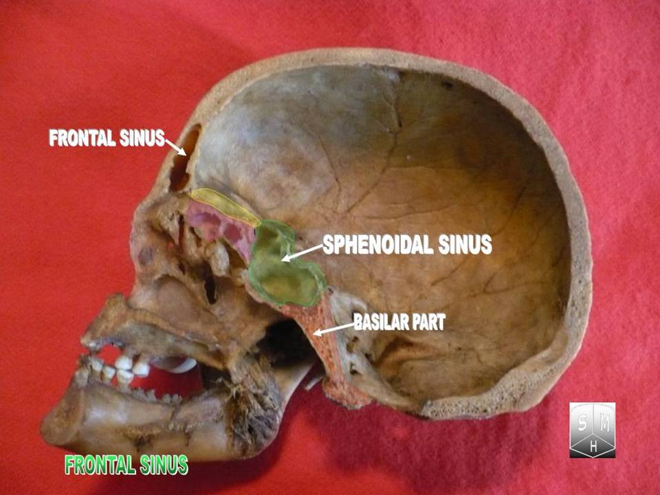
The frontal sinuses are one of the four pairs of paranasal sinuses that are situated behind the brow ridges. Sinuses are mucosa-lined airspaces within the bones of the face and skull. Each opens into the anterior part of the corresponding middle nasal meatus of the nose through the frontonasal duct which traverses the anterior part of the labyrinth of the ethmoid. These structures then open into the semilunar hiatus in the middle meatus. Structure Each frontal sinus is situated between the external and internal plates of the frontal bone.Frontal sinuses are rarely symmetrical. Their average measurements are as follows: height 28 mm, breadth 24 mm, depth 20 mm, creating a space of 6-7 ml. Blood supply The mucous membrane of the frontal sinuses receives arterial supply via the supraorbital artery, and anterior ethmoidal artery. Innervation The mucous membrane in this sinus is innervated by the supraorbital nerve, which contains the postganglionic par ... [...More Info...] [...Related Items...] OR: [Wikipedia] [Google] [Baidu] |
Anterior Ethmoidal Cells
The ethmoid sinuses or ethmoid air cells of the ethmoid bone are one of the four paired paranasal sinuses. The cells are variable in both size and number in the lateral mass of each of the ethmoid bones and cannot be palpated during an extraoral examination. They are divided into anterior and posterior groups. The ethmoid air cells are numerous thin-walled cavities situated in the ethmoidal labyrinth and completed by the frontal, maxilla, lacrimal, sphenoidal, and palatine bones. They lie between the upper parts of the nasal cavities and the orbits, and are separated from these cavities by thin bony lamellae. Groups of sinuses The groups of the ethmoidal air cells drain into the nasal meatuses.Otorhinolaryngology, Head and Neck Surgery, Anniko, Springer, 2010, page 188 * The posterior group the ''posterior ethmoidal sinus'' drains into the superior meatus above the middle nasal concha; sometimes one or more opens into the sphenoidal sinus. * The anterior group the ''anterior e ... [...More Info...] [...Related Items...] OR: [Wikipedia] [Google] [Baidu] |
Frontal Air Sinus
The frontal sinuses are one of the four pairs of paranasal sinuses that are situated behind the brow ridges. Sinuses are mucosa-lined airspaces within the bones of the face and skull. Each opens into the anterior part of the corresponding middle nasal meatus of the nose through the frontonasal duct which traverses the anterior part of the labyrinth of the ethmoid. These structures then open into the semilunar hiatus in the middle meatus. Structure Each frontal sinus is situated between the external and internal plates of the frontal bone.Frontal sinuses are rarely symmetrical. Their average measurements are as follows: height 28 mm, breadth 24 mm, depth 20 mm, creating a space of 6-7 ml. Blood supply The mucous membrane of the frontal sinuses receives arterial supply via the supraorbital artery, and anterior ethmoidal artery. Innervation The mucous membrane in this sinus is innervated by the supraorbital nerve, which contains the postganglionic parasympathet ... [...More Info...] [...Related Items...] OR: [Wikipedia] [Google] [Baidu] |
Middle Meatus
In anatomy, the term nasal meatus can refer to any of the three meatuses (passages) through the skulls nasal cavity: the superior meatus (''meatus nasi superior''), middle meatus (''meatus nasi medius''), and inferior meatus (''meatus nasi inferior''). The nasal meatuses are located beneath each of the corresponding nasal conchae. In the case where a fourth, supreme nasal concha is present, there is a fourth supreme nasal meatus. Structure The superior meatus is the smallest of the three. It is a narrow cavity located obliquely below the superior concha. This meatus is short, lies above and extends from the middle part of the middle concha below. From behind, the sphenopalatine foramen opens into the cavity of the superior meatus and the meatus communicates with the posterior ethmoidal cells. Above and at the back of the superior concha is the sphenoethmoidal recess which the sphenoidal sinus opens into. The superior meatus occupies the middle third of the nasal cavity’s ... [...More Info...] [...Related Items...] OR: [Wikipedia] [Google] [Baidu] |
Maxillary Sinus
The pyramid-shaped maxillary sinus (or antrum of Nathaniel Highmore (surgeon), Highmore) is the largest of the paranasal sinuses, and drains into the middle meatus of the nose through the osteomeatal complex.Human Anatomy, Jacobs, Elsevier, 2008, page 209-210 Structure It is the largest air sinus in the body. Found in the body of the maxilla, this sinus has three recesses: an alveolar recess pointed inferiorly, bounded by the alveolar process of the maxilla; a zygomatic recess pointed laterally, bounded by the zygomatic bone; and an infraorbital recess pointed superiorly, bounded by the inferior Orbital surface of the body of the maxilla, orbital surface of the maxilla. The medial wall is composed primarily of cartilage. The ostia for drainage are located high on the medial wall and open into the semilunar hiatus of the lateral nasal cavity; because of the position of the ostia, gravity cannot drain the maxillary sinus contents when the head is erect (see pathology). The ostium of ... [...More Info...] [...Related Items...] OR: [Wikipedia] [Google] [Baidu] |


