|
Anterior Ethmoidal Cells
The ethmoid sinuses or ethmoid air cells of the ethmoid bone are one of the four paired paranasal sinuses. The cells are variable in both size and number in the lateral mass of each of the ethmoid bones and cannot be palpated during an extraoral examination. They are divided into anterior and posterior groups. The ethmoid air cells are numerous thin-walled cavities situated in the ethmoidal labyrinth and completed by the frontal, maxilla, lacrimal, sphenoidal, and palatine bones. They lie between the upper parts of the nasal cavities and the orbits, and are separated from these cavities by thin bony lamellae. Groups of sinuses The groups of the ethmoidal air cells drain into the nasal meatuses.Otorhinolaryngology, Head and Neck Surgery, Anniko, Springer, 2010, page 188 * The posterior group the ''posterior ethmoidal sinus'' drains into the superior meatus above the middle nasal concha; sometimes one or more opens into the sphenoidal sinus. * The anterior group the ''anterior e ... [...More Info...] [...Related Items...] OR: [Wikipedia] [Google] [Baidu] |
Paranasal Sinus
Paranasal sinuses are a group of four paired air-filled spaces that surround the nasal cavity. The maxillary sinuses are located under the eyes; the frontal sinuses are above the eyes; the ethmoidal sinuses are between the eyes and the sphenoidal sinuses are behind the eyes. The sinuses are named for the facial bones in which they are located. Structure Humans possess four pairs of paranasal sinuses, divided into subgroups that are named according to the bones within which the sinuses lie. They are all innervated by branches of the trigeminal nerve (CN V). * The maxillary sinuses, the largest of the paranasal sinuses, are under the eyes, in the maxillary bones (open in the back of the semilunar hiatus of the nose). They are innervated by the maxillary nerve (CN V2). * The frontal sinuses, superior to the eyes, in the frontal bone, which forms the hard part of the forehead. They are innervated by the ophthalmic nerve (CN V1). * The ethmoidal sinuses, which are formed fro ... [...More Info...] [...Related Items...] OR: [Wikipedia] [Google] [Baidu] |
Middle Meatus
In anatomy, the term nasal meatus can refer to any of the three meatuses (passages) through the skulls nasal cavity: the superior meatus (''meatus nasi superior''), middle meatus (''meatus nasi medius''), and inferior meatus (''meatus nasi inferior''). The nasal meatuses are located beneath each of the corresponding nasal conchae. In the case where a fourth, supreme nasal concha is present, there is a fourth supreme nasal meatus. Structure The superior meatus is the smallest of the three. It is a narrow cavity located obliquely below the superior concha. This meatus is short, lies above and extends from the middle part of the middle concha below. From behind, the sphenopalatine foramen opens into the cavity of the superior meatus and the meatus communicates with the posterior ethmoidal cells. Above and at the back of the superior concha is the sphenoethmoidal recess which the sphenoidal sinus opens into. The superior meatus occupies the middle third of the nasal cavity’s ... [...More Info...] [...Related Items...] OR: [Wikipedia] [Google] [Baidu] |
Proptosis
Exophthalmos (also called exophthalmus, exophthalmia, proptosis, or exorbitism) is a bulging of the eye anteriorly out of the orbit. Exophthalmos can be either bilateral (as is often seen in Graves' disease) or unilateral (as is often seen in an orbital tumor). Complete or partial dislocation from the orbit is also possible from trauma or swelling of surrounding tissue resulting from trauma. In the case of Graves' disease, the displacement of the eye results from abnormal connective tissue deposition in the orbit and extraocular muscles, which can be visualized by CT or MRI. If left untreated, exophthalmos can cause the eyelids to fail to close during sleep, leading to corneal dryness and damage. Another possible complication is a form of redness or irritation called superior limbic keratoconjunctivitis, in which the area above the cornea becomes inflamed as a result of increased friction when blinking. The process that is causing the displacement of the eye may also compress ... [...More Info...] [...Related Items...] OR: [Wikipedia] [Google] [Baidu] |
Lamina Papyracea
The orbital lamina of ethmoid bone, (or lamina papyracea or orbital lamina) is a smooth, oblong bone plate which forms the lateral surface of the labyrinth of the ethmoid bone in the skull. The plate covers in the middle and posterior ethmoidal cells and forms a large part of the medial wall of the orbit. It articulates above with the orbital plate of the frontal bone, below with the maxilla and the orbital process of palatine bone, in front with the lacrimal, and behind with the sphenoid. Its name lamina papyracea is an appropriate description, as this part of the ethmoid bone is paper-thin and fractures easily. A fracture here could cause entrapment of the medial rectus muscle. Additional Images File:Slide4fen.JPG, Orbital lamina of ethmoid bone References External links * - "Orbits and Eye: Bones" * Bones of the head and neck {{Portal bar, Anatomy ... [...More Info...] [...Related Items...] OR: [Wikipedia] [Google] [Baidu] |
Haller Cell
The ethmoid sinuses or ethmoid air cells of the ethmoid bone are one of the four paired paranasal sinuses. The cells are variable in both size and number in the lateral mass of each of the ethmoid bones and cannot be palpated during an extraoral examination. They are divided into anterior and posterior groups. The ethmoid air cells are numerous thin-walled cavities situated in the ethmoidal labyrinth and completed by the frontal, maxilla, lacrimal, sphenoidal, and palatine bones. They lie between the upper parts of the nasal cavities and the orbits, and are separated from these cavities by thin bony lamellae. Groups of sinuses The groups of the ethmoidal air cells drain into the nasal meatuses.Otorhinolaryngology, Head and Neck Surgery, Anniko, Springer, 2010, page 188 * The posterior group the ''posterior ethmoidal sinus'' drains into the superior meatus above the middle nasal concha; sometimes one or more opens into the sphenoidal sinus. * The anterior group the ''anterior ... [...More Info...] [...Related Items...] OR: [Wikipedia] [Google] [Baidu] |
Facial Nerve
The facial nerve, also known as the seventh cranial nerve, cranial nerve VII, or simply CN VII, is a cranial nerve that emerges from the pons of the brainstem, controls the muscles of facial expression, and functions in the conveyance of taste sensations from the anterior two-thirds of the tongue. The nerve typically travels from the pons through the facial canal in the temporal bone and exits the skull at the stylomastoid foramen. It arises from the brainstem from an area posterior to the cranial nerve VI (abducens nerve) and anterior to cranial nerve VIII (vestibulocochlear nerve). The facial nerve also supplies preganglionic parasympathetic fibers to several head and neck ganglia. The facial and intermediate nerves can be collectively referred to as the nervus intermediofacialis. The path of the facial nerve can be divided into six segments: # intracranial (cisternal) segment # meatal (canalicular) segment (within the internal auditory canal) # labyrinthine segmen ... [...More Info...] [...Related Items...] OR: [Wikipedia] [Google] [Baidu] |
Secretion
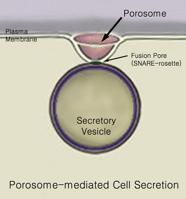
440px Secretion is the movement of material from one point to another, such as a secreted chemical substance from a cell or gland. In contrast, excretion is the removal of certain substances or waste products from a cell or organism. The classical mechanism of cell secretion is via secretory portals at the plasma membrane called porosomes. Porosomes are permanent cup-shaped lipoprotein structures embedded in the cell membrane, where secretory vesicles transiently dock and fuse to release intra-vesicular contents from the cell. Secretion in bacterial species means the transport or translocation of effector molecules for example: proteins, enzymes or toxins (such as cholera toxin in pathogenic bacteria e.g. '' Vibrio cholerae'') from across the interior (cytoplasm or cytosol) of a bacterial cell to its exterior. Secretion is a very important mechanism in bacterial functioning and operation in their natural surrounding environment for adaptation and survival. In eukaryotic ... [...More Info...] [...Related Items...] OR: [Wikipedia] [Google] [Baidu] |
Mucus
Mucus ( ) is a slippery aqueous secretion produced by, and covering, mucous membranes. It is typically produced from cells found in mucous glands, although it may also originate from mixed glands, which contain both serous and mucous cells. It is a viscous colloid containing inorganic salts, antimicrobial enzymes (such as lysozymes), immunoglobulins (especially IgA), and glycoproteins such as lactoferrin and mucins, which are produced by goblet cells in the mucous membranes and submucosal glands. Mucus serves to protect epithelial cells in the linings of the respiratory, digestive, and urogenital systems, and structures in the visual and auditory systems from pathogenic fungi, bacteria and viruses. Most of the mucus in the body is produced in the gastrointestinal tract. Amphibians, fish, snails, slugs, and some other invertebrates also produce external mucus from their epidermis as protection against pathogens, and to help in movement and is also produced in ... [...More Info...] [...Related Items...] OR: [Wikipedia] [Google] [Baidu] |
Parasympathetic
The parasympathetic nervous system (PSNS) is one of the three divisions of the autonomic nervous system, the others being the sympathetic nervous system and the enteric nervous system. The enteric nervous system is sometimes considered part of the autonomic nervous system, and sometimes considered an independent system. The autonomic nervous system is responsible for regulating the body's unconscious actions. The parasympathetic system is responsible for stimulation of "rest-and-digest" or "feed and breed" activities that occur when the body is at rest, especially after eating, including sexual arousal, salivation, lacrimation (tears), urination, digestion, and defecation. Its action is described as being complementary to that of the sympathetic nervous system, which is responsible for stimulating activities associated with the fight-or-flight response. Nerve fibres of the parasympathetic nervous system arise from the central nervous system. Specific nerves include severa ... [...More Info...] [...Related Items...] OR: [Wikipedia] [Google] [Baidu] |
Postganglionic
In the autonomic nervous system, fibers from the ganglion to the effector organ are called postganglionic fibers. Neurotransmitters The neurotransmitters of postganglionic fibers differ: * In the parasympathetic division, neurons are ''cholinergic''. That is to say acetylcholine is the primary neurotransmitter responsible for the communication between neurons on the parasympathetic pathway. * In the sympathetic division, neurons are mostly ''adrenergic'' (that is, epinephrine and norepinephrine function as the primary neurotransmitters). Notable exceptions to this rule include the sympathetic innervation of sweat glands and arrectores pilorum muscles where the neurotransmitter at both pre and post ganglionic synapses is acetylcholine. Another notable structure is the medulla of the adrenal gland, where chromaffin cells function as modified post-ganglionic nerves. Instead of releasing epinephrine and norepinephrine into a synaptic cleft, these cells of the adrenal medulla ... [...More Info...] [...Related Items...] OR: [Wikipedia] [Google] [Baidu] |
Pterygopalatine Ganglion
The pterygopalatine ganglion (aka Meckel's ganglion, nasal ganglion, or sphenopalatine ganglion) is a parasympathetic ganglion found in the pterygopalatine fossa. It is largely innervated by the greater petrosal nerve (a branch of the facial nerve); and its postsinaptic axons project to the lacrimal glands and nasal mucosa. The flow of blood to the nasal mucosa, in particular the venous plexus of the conchae, is regulated by the pterygopalatine ganglion and heats or cools the air in the nose. It is one of four parasympathetic ganglia of the head and neck, the others being the submandibular ganglion, otic ganglion, and ciliary ganglion. Structure The pterygopalatine ganglion (of Meckel), the largest of the parasympathetic ganglia associated with the branches of the maxillary nerve, is deeply placed in the pterygopalatine fossa, close to the sphenopalatine foramen. It is triangular or heart-shaped, of a reddish-gray color, and is situated just below the maxillary nerve as ... [...More Info...] [...Related Items...] OR: [Wikipedia] [Google] [Baidu] |
Ethmoidal Nerves
The ethmoidal nerves, which arise from the nasociliary nerve, supply the ethmoidal cells; the posterior branch leaves the orbital cavity through the posterior ethmoidal foramen Lateral to either olfactory groove are the internal openings of the anterior and posterior ethmoidal foramina (or canals). The posterior ethmoidal foramen opens at the back part of this margin under cover of the projecting lamina of the sphenoi ... and gives some filaments to the sphenoidal sinus. There are two ethmoidal nerves on each side of the face: * posterior ethmoidal nerve * anterior ethmoidal nerve References External links ufl.edu Trigeminal nerve {{neuroanatomy-stub ... [...More Info...] [...Related Items...] OR: [Wikipedia] [Google] [Baidu] |


