|
Radial Collateral Ligament (elbow)
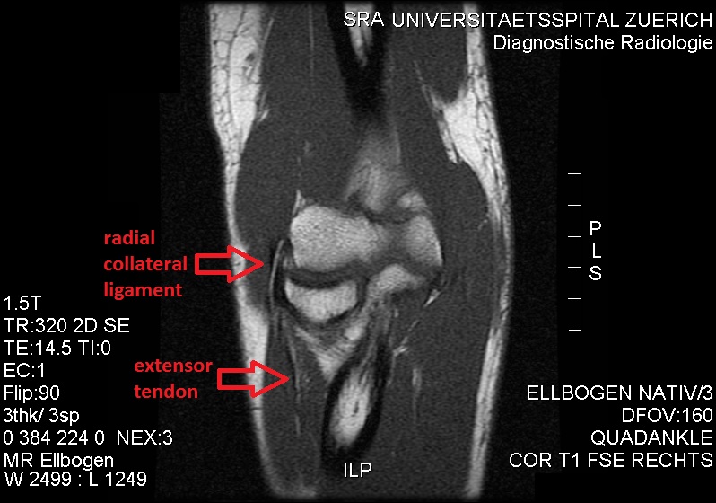
The radial collateral ligament (RCL), lateral collateral ligament (LCL), or external lateral ligamentAs opposed to the "internal lateral ligament", better known as the medial or ulnar collateral ligament is a ligament in the elbow on the side of the radius. Structure The composition of the triangular ligamentous structure on the lateral side of the elbow varies widely between individuals, see alsFigure 4/ref> and can be considered either a single ligament, in which case multiple distal attachments are generally mentioned and the annular ligament is described separately, or as several separate ligaments, in which case parts of those ligaments are often described as indistinguishable from each other. In the latter case, the ligaments are collectively referred to as the lateral collateral ligament complex (LCLC), consisting of four ligaments: * the radial collateral ligament roper(RCL), from the lateral epicondyle to the annular ligament deep to the common extensor tendon * the la ... [...More Info...] [...Related Items...] OR: [Wikipedia] [Google] [Baidu] |
Elbow
The elbow is the region between the arm and the forearm that surrounds the elbow joint. The elbow includes prominent landmarks such as the olecranon, the cubital fossa (also called the chelidon, or the elbow pit), and the lateral and the medial epicondyles of the humerus. The elbow joint is a hinge joint between the arm and the forearm; more specifically between the humerus in the upper arm and the radius and ulna in the forearm which allows the forearm and hand to be moved towards and away from the body. The term ''elbow'' is specifically used for humans and other primates, and in other vertebrates forelimb plus joint is used. The name for the elbow in Latin is ''cubitus'', and so the word cubital is used in some elbow-related terms, as in ''cubital nodes'' for example. Structure Joint The elbow joint has three different portions surrounded by a common joint capsule. These are joints between the three bones of the elbow, the humerus of the upper arm, and the radius and ... [...More Info...] [...Related Items...] OR: [Wikipedia] [Google] [Baidu] |
Lateral Epicondyle Of The Humerus
The lateral epicondyle of the humerus is a large, tuberculated eminence, curved a little forward, and giving attachment to the radial collateral ligament of the elbow joint, and to a tendon common to the origin of the supinator and some of the extensor muscles. Specifically, these extensor muscles include the anconeus muscle, the supinator, extensor carpi radialis brevis, extensor digitorum, extensor digiti minimi, and extensor carpi ulnaris. In birds, where the arm is somewhat rotated compared to other tetrapods, it is termed dorsal epicondyle of the humerus. In comparative anatomy, the term ''ectepicondyle'' is sometimes used. A common injury associated with the lateral epicondyle of the humerus is lateral epicondylitis also known as tennis elbow. Repetitive overuse of the forearm, as seen in tennis or other sports, can result in inflammation of "the tendons that join the forearm muscles on the outside of the elbow. The forearm muscles and tendons become damaged from overus ... [...More Info...] [...Related Items...] OR: [Wikipedia] [Google] [Baidu] |
Annular Ligament Of Radius
The annular ligament (orbicular ligament) is a strong band of fibers that encircles the head of the radius, and retains it in contact with the radial notch of the ulna.''Gray's Anatomy'' (1918), see infobox Per '' Terminologia Anatomica 1998'', the spelling is "anular", but the spelling "annular" is frequently encountered. Indeed, the most recent version of ''Terminologia Anatomica'' (2019) uses "annular" as the preferred English spelling. Anatomy The annular ligament is attached by both its ends to the anterior and posterior margins of the radial notch of the ulna, together with which it forms the articular surface that surrounds the head and neck of the radius. The ligament is strong and well defined, yet its flexibility permits the slightly oval head of the radius to rotate freely during pronation and supination. The head of the radius is wider than the bone's neck, and, because the annular ligament embraces both, the radial head is "trapped" inside the ligament which thus act ... [...More Info...] [...Related Items...] OR: [Wikipedia] [Google] [Baidu] |
Ulnar Collateral Ligament Of Elbow Joint
The ulnar collateral ligament (UCL) or internal lateral ligament is a thick triangular ligament at the medial aspect of the elbow uniting the distal aspect of the humerus to the proximal aspect of the ulna. Structure It consists of two portions, an anterior and posterior united by a thinner intermediate portion. Note that this ligament is also referred to as the medial collateral ligament and should not be confused with the lateral ulnar collateral ligament (LUCL). The ''anterior portion'', directed obliquely forward, is attached, above, by its apex, to the front part of the medial epicondyle of the humerus; and, below, by its broad base to the medial margin of the coronoid process of the ulna. The ''posterior portion'', also of triangular form, is attached, above, by its apex, to the lower and back part of the medial epicondyle; below, to the medial margin of the olecranon. Between these two bands a few intermediate fibers descend from the medial epicondyle to blend with a ... [...More Info...] [...Related Items...] OR: [Wikipedia] [Google] [Baidu] |
Ligament
A ligament is the fibrous connective tissue that connects bones to other bones. It is also known as ''articular ligament'', ''articular larua'', ''fibrous ligament'', or ''true ligament''. Other ligaments in the body include the: * Peritoneal ligament: a fold of peritoneum or other membranes. * Fetal remnant ligament: the remnants of a fetal tubular structure. * Periodontal ligament: a group of fibers that attach the cementum of teeth to the surrounding alveolar bone. Ligaments are similar to tendons and fasciae as they are all made of connective tissue. The differences among them are in the connections that they make: ligaments connect one bone to another bone, tendons connect muscle to bone, and fasciae connect muscles to other muscles. These are all found in the skeletal system of the human body. Ligaments cannot usually be regenerated naturally; however, there are periodontal ligament stem cells located near the periodontal ligament which are involved in the adult regener ... [...More Info...] [...Related Items...] OR: [Wikipedia] [Google] [Baidu] |
Radius (bone)
The radius or radial bone is one of the two large bones of the forearm, the other being the ulna. It extends from the lateral side of the elbow to the thumb side of the wrist and runs parallel to the ulna. The ulna is usually slightly longer than the radius, but the radius is thicker. Therefore the radius is considered to be the larger of the two. It is a long bone, prism-shaped and slightly curved longitudinally. The radius is part of two joints: the elbow and the wrist. At the elbow, it joins with the capitulum of the humerus, and in a separate region, with the ulna at the radial notch. At the wrist, the radius forms a joint with the ulna bone. The corresponding bone in the lower leg is the fibula. Structure The long narrow medullary cavity is enclosed in a strong wall of compact bone. It is thickest along the interosseous border and thinnest at the extremities, same over the cup-shaped articular surface (fovea) of the head. The trabeculae of the spongy tissue are some ... [...More Info...] [...Related Items...] OR: [Wikipedia] [Google] [Baidu] |
Common Extensor Tendon
The common extensor tendon is a tendon that attaches to the lateral epicondyle of the humerus. Structure The common extensor tendon serves as the upper attachment (in part) for the superficial muscles that are located on the posterior aspect of the forearm: * Extensor carpi radialis brevis * Extensor digitorum * Extensor digiti minimi * Extensor carpi ulnaris The tendon of extensor carpi radialis brevis is usually the most major tendon to which the other tendons merge. Function The common extensor tendon is the major attachment point for extensor muscles of the forearm. This enables finger extension and aids in forearm supination. Clinical significance Lateral elbow pain can be caused by various pathologies of the common extensor tendon. Overuse injuries can lead to inflammation. Tennis elbow is a common issue with the common extensor tendon. See also * Common flexor tendon The common flexor tendon is a tendon that attaches to the medial epicondyle of the humerus (lowe ... [...More Info...] [...Related Items...] OR: [Wikipedia] [Google] [Baidu] |
Supinator Crest
The ulna (''pl''. ulnae or ulnas) is a long bone found in the forearm that stretches from the elbow to the smallest finger, and when in anatomical position, is found on the medial side of the forearm. That is, the ulna is on the same side of the forearm as the little finger. It runs parallel to the radius, the other long bone in the forearm. The ulna is usually slightly longer than the radius, but the radius is thicker. Therefore, the radius is considered to be the larger of the two. Structure The ulna is a long bone found in the forearm that stretches from the elbow to the smallest finger, and when in anatomical position, is found on the medial side of the forearm. It is broader close to the elbow, and narrows as it approaches the wrist. Close to the elbow, the ulna has a bony process, the olecranon process, a hook-like structure that fits into the olecranon fossa of the humerus. This prevents hyperextension and forms a hinge joint with the trochlea of the humerus. There is ... [...More Info...] [...Related Items...] OR: [Wikipedia] [Google] [Baidu] |
Radial Notch
The radial notch of the ulna (lesser sigmoid cavity) is a narrow, oblong, articular depression on the lateral side of the coronoid process; it receives the circumferential articular surface of the head of the radius. It is concave from before backward, and its prominent extremities serve for the attachment of the annular ligament. Additional images File:Gray333.png, Annular ligament of radius, from above. References External links * *elbow/elbowbones/bones3at the Dartmouth Medical School The Geisel School of Medicine at Dartmouth is the graduate medical school of Dartmouth College in Hanover, New Hampshire. The fourth oldest medical school in the United States, it was founded in 1797 by New England physician Nathan Smith. It is o ...'s Department of Anatomy Upper limb anatomy Ulna {{musculoskeletal-stub ... [...More Info...] [...Related Items...] OR: [Wikipedia] [Google] [Baidu] |
Head Of Radius
The head of the radius has a cylindrical form, and on its upper surface is a shallow cup or fovea for articulation with the capitulum of the humerus. The circumference of the head is smooth; it is broad medially where it articulates with the radial notch of the ulna, narrow in the rest of its extent, which is embraced by the annular ligament.''Gray's Anatomy'' (1918), see infobox Articular surfaces The head of the radius is shaped to articulate with a complex of articular surfaces during both flexion-extension at the elbow and supination-pronation in the forearm: Humeroradial joint The head's proximal surface is concave and cup-shaped to correspond to the spherical surface of the capitulum of the humerus. The radius can thus glide on the capitulum during elbow flexion-extension while simultaneously rotate about its own main axis during supination-pronation. Between the capitulum and the trochlea of the humerus is the capitulotrochlear groove. A semi-lunar surface around the ... [...More Info...] [...Related Items...] OR: [Wikipedia] [Google] [Baidu] |
Tennis Elbow
Tennis elbow, also known as lateral epicondylitis or enthesopathy of the extensor carpi radialis origin, is a condition in which the outer part of the elbow becomes painful and tender. The pain may also extend into the back of the forearm. Onset of symptoms is generally gradual although they can seem sudden and be misinterpreted as an injury. Golfer's elbow is a similar condition that affects the inside of the elbow. Enthesopathies are idiopathic, meaning science has not yet determined the cause. Enthesopathies are most common in middle age (ages 35 to 60). It is often stated that the condition is caused by excessive use of the muscles of the back of the forearm, but this is not supported by experimental evidence and is a common misinterpretation or unhelpful thought about symptoms. It may be associated with work or sports, classically racquet sports, but most people with the condition are not exposed to these activities. The diagnosis is based on the symptoms and examination ... [...More Info...] [...Related Items...] OR: [Wikipedia] [Google] [Baidu] |


