|
Ulnar Collateral Ligament Of Elbow Joint
The ulnar collateral ligament (UCL) or internal lateral ligament is a thick triangular ligament at the medial aspect of the elbow uniting the distal aspect of the humerus to the proximal aspect of the ulna. Structure It consists of two portions, an anterior and posterior united by a thinner intermediate portion. Note that this ligament is also referred to as the medial collateral ligament and should not be confused with the lateral ulnar collateral ligament (LUCL). The ''anterior portion'', directed obliquely forward, is attached, above, by its apex, to the front part of the medial epicondyle of the humerus; and, below, by its broad base to the medial margin of the coronoid process of the ulna. The ''posterior portion'', also of triangular form, is attached, above, by its apex, to the lower and back part of the medial epicondyle; below, to the medial margin of the olecranon. Between these two bands a few intermediate fibers descend from the medial epicondyle to blend with a ... [...More Info...] [...Related Items...] OR: [Wikipedia] [Google] [Baidu] |
Medial Epicondyle Of The Humerus
The medial epicondyle of the humerus is an epicondyle of the humerus bone of the upper arm in humans. It is larger and more prominent than the lateral epicondyle and is directed slightly more posteriorly in the anatomical position. In birds, where the arm is somewhat rotated compared to other tetrapods, it is called the ventral epicondyle of the humerus. In comparative anatomy, the more neutral term entepicondyle is used. The medial epicondyle gives attachment to the ulnar collateral ligament of elbow joint, to the pronator teres, and to a common tendon of origin (the common flexor tendon) of some of the flexor muscles of the forearm: the flexor carpi radialis, the flexor carpi ulnaris, the flexor digitorum superficialis, and the palmaris longus. The medial epicondyle is located on the distal end of the humerus. Additionally, the medial epicondyle is inferior to the medial supracondylar ridge. It is also proximal to the olecranon fossa. The medial epicondyle protects the uln ... [...More Info...] [...Related Items...] OR: [Wikipedia] [Google] [Baidu] |
Intermediate Fibers
Intermediate fibers, also known as fast oxidative-glycolytic fibers, are fast twitch muscle fibers which have been converted via endurance training. These fibers are slightly larger in diameter, have more mitochondria A mitochondrion (; ) is an organelle found in the Cell (biology), cells of most Eukaryotes, such as animals, plants and Fungus, fungi. Mitochondria have a double lipid bilayer, membrane structure and use aerobic respiration to generate adenosi ... as well as a greater blood supply and more endurance than typical fast twitch fibers. Most of the body's muscles are composed of these intermediate fibers. References * Visualizing Human Biology, Kathleen Anne Ireland, David J. Tenenbaum, raken jilani {{muscle-stub Muscular system ... [...More Info...] [...Related Items...] OR: [Wikipedia] [Google] [Baidu] |
Cubitus Valgus
Cubitus valgus is a medical deformity in which the forearm is angled away from the body to a greater degree than normal when fully extended. A small degree of cubitus valgus (known as the carrying angle) is acceptable and occurs in the general population. When present at birth, it can be an indication of Turner syndromeChapter on Amenorrhea in: or Noonan syndrome. It can also be acquired through fracture or other trauma. The physiological cubitus valgus varies from 3° to 29°. Women usually have a more pronounced Cubitus valgus than men. The deformity can also occur as a complication of fracture of the lateral condyle of the humerus, which may lead to tardy/delayed ulnar nerve palsy.The opposite condition is cubitus varus (). See also * Valgus deformity * Varus deformity Varus may refer to: * ... [...More Info...] [...Related Items...] OR: [Wikipedia] [Google] [Baidu] |
Baseball Pitching
In baseball, the pitcher is the player who throws ("pitches") the baseball from the pitcher's mound toward the catcher to begin each play, with the goal of retiring a batter, who attempts to either make contact with the pitched ball or draw a walk. In the numbering system used to record defensive plays, the pitcher is assigned the number 1. The pitcher is often considered the most important player on the defensive side of the game, and as such is situated at the right end of the defensive spectrum. There are many different types of pitchers, such as the starting pitcher, relief pitcher, middle reliever, lefty specialist, setup man, and the closer. Traditionally, the pitcher also bats. Starting in 1973 with the American League(and later the National League) and spreading to further leagues throughout the 1980s and 1990s, the hitting duties of the pitcher have generally been given over to the position of designated hitter, a cause of some controversy. The Japanese Central L ... [...More Info...] [...Related Items...] OR: [Wikipedia] [Google] [Baidu] |
Flexor Digitorum Superficialis
Flexor digitorum superficialis (''flexor digitorum sublimis'') is an extrinsic flexor muscle of the fingers at the proximal interphalangeal joints. It is in the anterior compartment of the forearm. It is sometimes considered to be the deepest part of the superficial layer of this compartment, and sometimes considered to be a distinct, "intermediate layer" of this compartment. It is relatively common for the Flexor digitorum superficialis to be missing from the little finger, bilaterally and unilaterally, which can cause problems when diagnosing a little finger injury. Structure The muscle has two classically described heads – the humeroulnar and radial – and it is between these heads that the median nerve and ulnar artery pass. The ulnar collateral ligament of elbow joint gives its origin to part of this muscle. Four long tendons come off this muscle near the wrist and travel through the carpal tunnel formed by the flexor retinaculum. These tendons, along with those of fle ... [...More Info...] [...Related Items...] OR: [Wikipedia] [Google] [Baidu] |
Ulnar Nerve
In human anatomy, the ulnar nerve is a nerve that runs near the ulna bone. The ulnar collateral ligament of elbow joint is in relation with the ulnar nerve. The nerve is the largest in the human body unprotected by muscle or bone, so injury is common. This nerve is directly connected to the little finger, and the adjacent half of the ring finger, innervating the palmar aspect of these fingers, including both front and back of the tips, perhaps as far back as the fingernail beds. This nerve can cause an electric shock-like sensation by striking the medial epicondyle of the humerus posteriorly, or inferiorly with the elbow flexed. The ulnar nerve is trapped between the bone and the overlying skin at this point. This is commonly referred to as bumping one's "funny bone". This name is thought to be a pun, based on the sound resemblance between the name of the bone of the upper arm, the humerus, and the word "humorous". Alternatively, according to the Oxford English Dictionary, i ... [...More Info...] [...Related Items...] OR: [Wikipedia] [Google] [Baidu] |
Flexor Carpi Ulnaris
The flexor carpi ulnaris (FCU) is a muscle of the forearm that flexes and adducts at the wrist joint. Structure Origin The flexor carpi ulnaris has two heads; a humeral head and ulnar head. The humeral head originates from the medial epicondyle of the humerus via the common flexor tendon. The ulnar head originates from the medial margin of the olecranon of the ulnar and the upper two-thirds of the dorsal border of the ulnar by an aponeurosis. Between the two heads passes the ulnar nerve and ulnar artery. Insertion The flexor carpi ulnaris inserts onto the pisiform, hook of the hamate (via the pisohamate ligament) and the anterior surface of the base of the fifth metacarpal (via the pisometacarpal ligament). Action The flexor carpi ulnaris flexes and adducts at the wrist joint. Innervation The flexor carpi ulnaris is innervated by the ulnar nerve. The corresponding spinal nerves are C8 and T1. Tendon The tendon of flexor carpi ulnaris can be seen on the anterior surface of th ... [...More Info...] [...Related Items...] OR: [Wikipedia] [Google] [Baidu] |
Triceps Brachii
The triceps, or triceps brachii (Latin for "three-headed muscle of the arm"), is a large muscle on the back of the upper limb of many vertebrates Vertebrates () comprise all animal taxa within the subphylum Vertebrata () ( chordates with backbones), including all mammals, birds, reptiles, amphibians, and fish. Vertebrates represent the overwhelming majority of the phylum Chordata, .... It consists of 3 parts: the medial, lateral, and long head. It is the muscle principally responsible for Extension (kinesiology), extension of the elbow joint (straightening of the arm). Structure The long head arises from the infraglenoid tubercle of the scapula. It extends distally anterior to the Teres minor muscle, teres minor and posterior to the Teres major muscle, teres major. The medial head arises proximally in the humerus, just inferior to the Radial sulcus, groove of the radial nerve; from the dorsal (back) surface of the humerus; from the Medial intermuscular septum of arm ... [...More Info...] [...Related Items...] OR: [Wikipedia] [Google] [Baidu] |
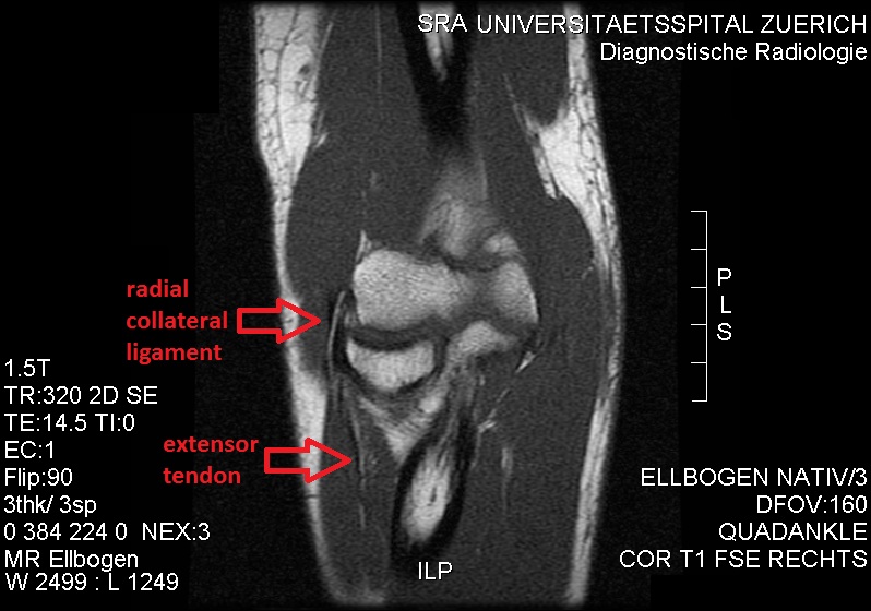
Radial Collateral Ligament (elbow)
The radial collateral ligament (RCL), lateral collateral ligament (LCL), or external lateral ligamentAs opposed to the "internal lateral ligament", better known as the medial or ulnar collateral ligament is a ligament in the elbow on the side of the radius. Structure The composition of the triangular ligamentous structure on the lateral side of the elbow varies widely between individuals, see alsFigure 4/ref> and can be considered either a single ligament, in which case multiple distal attachments are generally mentioned and the annular ligament is described separately, or as several separate ligaments, in which case parts of those ligaments are often described as indistinguishable from each other. In the latter case, the ligaments are collectively referred to as the lateral collateral ligament complex (LCLC), consisting of four ligaments: * the radial collateral ligament roper(RCL), from the lateral epicondyle to the annular ligament deep to the common extensor tendon * the la ... [...More Info...] [...Related Items...] OR: [Wikipedia] [Google] [Baidu] |
Coronoid Process Of The Ulna
The coronoid process of the ulna is a triangular process projecting forward from the anterior proximal portion of the ulna. Structure Its ''base'' is continuous with the body of the bone, and of considerable strength. Anatomy Its ''apex'' is pointed, slightly curved upward, and in flexion of the forearm is received into the coronoid fossa of the humerus. Its ''upper surface'' is smooth, convex, and forms the lower part of the semilunar notch. Its ''antero-inferior'' surface is concave, and marked by a rough impression for the insertion of the brachialis muscle. At the junction of this surface with the front of the body is a rough eminence, the tuberosity of the ulna, which gives insertion to a part of the brachialis; to the lateral border of this tuberosity the oblique cord is attached. Its ''lateral surface'' presents a narrow, oblong, articular depression, the radial notch. Its ''medial surface'', by its prominent, free margin, serves for the attachment of part of the uln ... [...More Info...] [...Related Items...] OR: [Wikipedia] [Google] [Baidu] |
Ulna
The ulna (''pl''. ulnae or ulnas) is a long bone found in the forearm that stretches from the elbow to the smallest finger, and when in anatomical position, is found on the medial side of the forearm. That is, the ulna is on the same side of the forearm as the little finger. It runs parallel to the radius, the other long bone in the forearm. The ulna is usually slightly longer than the radius, but the radius is thicker. Therefore, the radius is considered to be the larger of the two. Structure The ulna is a long bone found in the forearm that stretches from the elbow to the smallest finger, and when in anatomical position, is found on the medial side of the forearm. It is broader close to the elbow, and narrows as it approaches the wrist. Close to the elbow, the ulna has a bony process, the olecranon process, a hook-like structure that fits into the olecranon fossa of the humerus. This prevents hyperextension and forms a hinge joint with the trochlea of the humerus. There is ... [...More Info...] [...Related Items...] OR: [Wikipedia] [Google] [Baidu] |
Humerus
The humerus (; ) is a long bone in the arm that runs from the shoulder to the elbow. It connects the scapula and the two bones of the lower arm, the radius and ulna, and consists of three sections. The humeral upper extremity consists of a rounded head, a narrow neck, and two short processes (tubercles, sometimes called tuberosities). The body is cylindrical in its upper portion, and more prismatic below. The lower extremity consists of 2 epicondyles, 2 processes (trochlea & capitulum), and 3 fossae (radial fossa, coronoid fossa, and olecranon fossa). As well as its true anatomical neck, the constriction below the greater and lesser tubercles of the humerus is referred to as its surgical neck due to its tendency to fracture, thus often becoming the focus of surgeons. Etymology The word "humerus" is derived from la, humerus, umerus meaning upper arm, shoulder, and is linguistically related to Gothic ''ams'' shoulder and Greek ''ōmos''. Structure Upper extremity The upper or pr ... [...More Info...] [...Related Items...] OR: [Wikipedia] [Google] [Baidu] |


