|
Ligament
A ligament is the fibrous connective tissue that connects bones to other bones. It is also known as ''articular ligament'', ''articular larua'', ''fibrous ligament'', or ''true ligament''. Other ligaments in the body include the: * Peritoneal ligament: a fold of peritoneum or other membranes. * Fetal remnant ligament: the remnants of a fetal tubular structure. * Periodontal ligament: a group of fibers that attach the cementum of teeth to the surrounding alveolar bone. Ligaments are similar to tendons and fasciae as they are all made of connective tissue. The differences among them are in the connections that they make: ligaments connect one bone to another bone, tendons connect muscle to bone, and fasciae connect muscles to other muscles. These are all found in the skeletal system of the human body. Ligaments cannot usually be regenerated naturally; however, there are periodontal ligament stem cells located near the periodontal ligament which are involved in the adult ... [...More Info...] [...Related Items...] OR: [Wikipedia] [Google] [Baidu] |
Cruciate Ligament
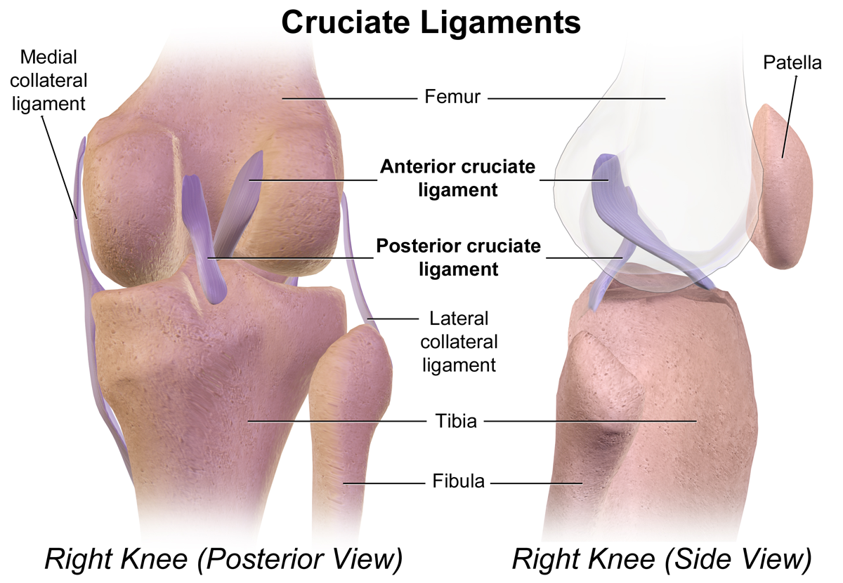
Cruciate ligaments (also cruciform ligaments) are pairs of ligaments arranged like a letter X. They occur in several joints of the body, such as the knee joint and the atlanto-axial joint. In a fashion similar to the cords in a toy Jacob's ladder, the crossed ligaments stabilize the joint while allowing a very large range of motion. Knee Structure Cruciate ligaments occur in the knee of humans and other bipedal animals and the corresponding stifle of quadrupedal animals, and in the neck, fingers, and foot. * The cruciate ligaments of the knee are the anterior cruciate ligament (ACL) and the posterior cruciate ligament (PCL). These ligaments are two strong, rounded bands that extend from the head of the tibia to the intercondyloid notch of the femur. The ACL is lateral and the PCL is medial. They cross each other like the limbs of an X. They are named for their insertion into the tibia: the ACL attaches to the anterior aspect of the intercondylar area, the PCL to the ... [...More Info...] [...Related Items...] OR: [Wikipedia] [Google] [Baidu] |
Periodontal Ligament
The periodontal ligament, commonly abbreviated as the PDL, is a group of specialized connective tissue fibers that essentially attach a tooth to the alveolar bone within which it sits. It inserts into root cementum one side and onto alveolar bone on the other. Structure The PDL consists of principal fibres, loose connective tissue, blast and clast cells, oxytalan fibres and Cell Rest of Malassez. Alveolodental ligament The main principal fiber group is the alveolodental ligament, which consists of five fiber subgroups: alveolar crest, horizontal, oblique, apical, and interradicular on multirooted teeth. Principal fibers other than the alveolodental ligament are the transseptal fibers. All these fibers help the tooth withstand the naturally substantial compressive forces that occur during chewing and remain embedded in the bone. The ends of the principal fibers that are within either cementum or alveolar bone proper are considered Sharpey fibers. * Alveolar crest fibers ( ... [...More Info...] [...Related Items...] OR: [Wikipedia] [Google] [Baidu] |
Peritoneum
The peritoneum is the serous membrane forming the lining of the abdominal cavity or coelom in amniotes and some invertebrates, such as annelids. It covers most of the intra-abdominal (or coelomic) organs, and is composed of a layer of mesothelium supported by a thin layer of connective tissue. This peritoneal lining of the cavity supports many of the abdominal organs and serves as a conduit for their blood vessels, lymphatic vessels, and nerves. The abdominal cavity (the space bounded by the vertebrae, abdominal muscles, diaphragm, and pelvic floor) is different from the intraperitoneal space (located within the abdominal cavity but wrapped in peritoneum). The structures within the intraperitoneal space are called "intraperitoneal" (e.g., the stomach and intestines), the structures in the abdominal cavity that are located behind the intraperitoneal space are called " retroperitoneal" (e.g., the kidneys), and those structures below the intraperitoneal space are called "su ... [...More Info...] [...Related Items...] OR: [Wikipedia] [Google] [Baidu] |
Peritoneal Ligament
Peritoneal ligaments are folds of peritoneum that are used to connect viscera to viscera or the abdominal wall. There are multiple named ligaments that usually are named in accordance with what they are. * Gastrocolic ligament, connects the stomach and the colon. * Splenocolic ligament, connects the spleen and the colon. * Gastrosplenic ligament * Gastrophrenic ligament * Phrenicocolic ligament * Splenorenal ligament * Hepatic ligaments - Ligaments that are associated with the liver ** Coronary ligament ** Left triangular ligament ** Right triangular ligament ** Hepatoduodenal ligament The hepatoduodenal ligament is the portion of the lesser omentum extending between the porta hepatis of the liver and the superior part of the duodenum. Running inside it are the following structures collectively known as the portal triad: * h ... ** Hepatogastric ligament ** Falciform ligament ** Round ligament of liver References {{reflist Abdomen ... [...More Info...] [...Related Items...] OR: [Wikipedia] [Google] [Baidu] |
Dislocation (medicine)
A joint dislocation, also called luxation, occurs when there is an abnormal separation in the joint, where two or more bones meet.Dislocations. Lucile Packard Children’s Hospital at Stanford. Retrieved 3 March 2013 A partial dislocation is referred to as a subluxation. Dislocations are often caused by sudden trauma on the joint like an impact or fall. A joint dislocation can cause damage to the surrounding ligaments, tendons, muscles, and nerves. Dislocations can occur in any major joint (shoulder, knees, etc.) or minor joint (toes, fingers, etc.). The most common joint dislocation is a shoulder dislocation. Treatment for joint dislocation is usually by closed reduction, that is, skilled manipulation to return the bones to their normal position. Reduction should only be performed by trained medical professionals, because it can cause injury to soft tissue and/or the nerves and vascular structures around the dislocation. Symptoms and signs The following symptoms are common wit ... [...More Info...] [...Related Items...] OR: [Wikipedia] [Google] [Baidu] |
Musculoskeletal System
The human musculoskeletal system (also known as the human locomotor system, and previously the activity system) is an organ system that gives humans the ability to move using their muscular and skeletal systems. The musculoskeletal system provides form, support, stability, and movement to the body. It is made up of the bones of the skeleton, muscles, cartilage, tendons, ligaments, joints, and other connective tissue that supports and binds tissues and organs together. The musculoskeletal system's primary functions include supporting the body, allowing motion, and protecting vital organs. The skeletal portion of the system serves as the main storage system for calcium and phosphorus and contains critical components of the hematopoietic system. This system describes how bones are connected to other bones and muscle fibers via connective tissue such as tendons and ligaments. The bones provide stability to the body. Muscles keep bones in place and also play a role in the movem ... [...More Info...] [...Related Items...] OR: [Wikipedia] [Google] [Baidu] |
Cementum
Cementum is a specialized calcified substance covering the root of a tooth. The cementum is the part of the periodontium that attaches the teeth to the alveolar bone by anchoring the periodontal ligament.Illustrated Dental Embryology, Histology, and Anatomy, Bath-Balogh and Fehrenbach, Elsevier, 2011, page 170. Structure The cells of cementum are the entrapped cementoblasts, the cementocytes. Each cementocyte lies in its lacuna, similar to the pattern noted in bone. These lacunae also have canaliculi or canals. Unlike those in bone, however, these canals in cementum do not contain nerves, nor do they radiate outward. Instead, the canals are oriented toward the periodontal ligament and contain cementocytic processes that exist to diffuse nutrients from the ligament because it is vascularized. After the apposition of cementum in layers, the cementoblasts that do not become entrapped in cementum line up along the cemental surface along the length of the outer covering of the per ... [...More Info...] [...Related Items...] OR: [Wikipedia] [Google] [Baidu] |
Connective Tissue
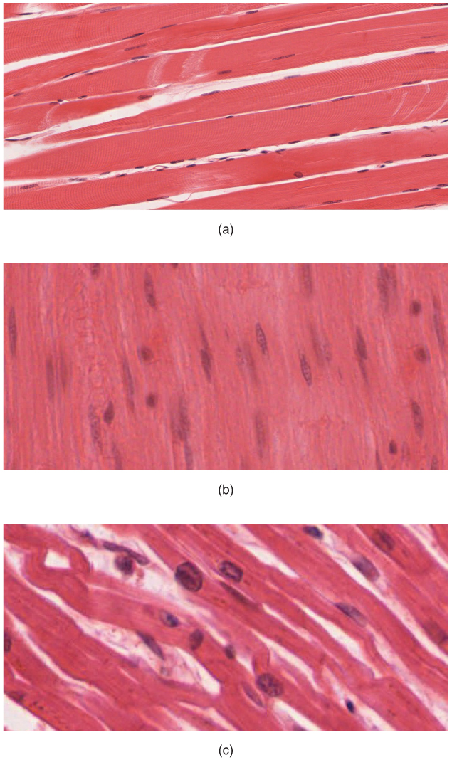
Connective tissue is one of the four primary types of animal tissue, along with epithelial tissue, muscle tissue, and nervous tissue. It develops from the mesenchyme derived from the mesoderm the middle embryonic germ layer. Connective tissue is found in between other tissues everywhere in the body, including the nervous system. The three meninges, membranes that envelop the brain and spinal cord are composed of connective tissue. Most types of connective tissue consists of three main components: elastic and collagen fibers, ground substance, and cells. Blood, and lymph are classed as specialized fluid connective tissues that do not contain fiber. All are immersed in the body water. The cells of connective tissue include fibroblasts, adipocytes, macrophages, mast cells and leucocytes. The term "connective tissue" (in German, ''Bindegewebe'') was introduced in 1830 by Johannes Peter Müller. The tissue was already recognized as a distinct class in the 18th century. ... [...More Info...] [...Related Items...] OR: [Wikipedia] [Google] [Baidu] |
Hypermobility (joints)
Hypermobility, also known as double-jointedness, describes joints that stretch farther than normal. For example, some hypermobile people can bend their thumbs backwards to their wrists, bend their knee joints backwards, put their leg behind the head or perform other contortionist "tricks". It can affect one or more joints throughout the body. Hypermobile joints are common and occur in about 10 to 25% of the population, but in a minority of people, pain and other symptoms are present. This may be a sign of what is known as joint hypermobility syndrome (JMS) or, more recently, hypermobility spectrum disorder (HSD). Hypermobile joints are a feature of genetic connective tissue disorders such as hypermobility spectrum disorder (HSD) or Ehlers–Danlos syndromes (EDS). Until new diagnostic criteria were introduced, hypermobility syndrome was sometimes considered identical to hypermobile Ehlers–Danlos syndrome (hEDS), formerly called EDS Type 3. As no genetic test can distinguish ... [...More Info...] [...Related Items...] OR: [Wikipedia] [Google] [Baidu] |
Connective Tissue
Connective tissue is one of the four primary types of animal tissue, along with epithelial tissue, muscle tissue, and nervous tissue. It develops from the mesenchyme derived from the mesoderm the middle embryonic germ layer. Connective tissue is found in between other tissues everywhere in the body, including the nervous system. The three meninges, membranes that envelop the brain and spinal cord are composed of connective tissue. Most types of connective tissue consists of three main components: elastic and collagen fibers, ground substance, and cells. Blood, and lymph are classed as specialized fluid connective tissues that do not contain fiber. All are immersed in the body water. The cells of connective tissue include fibroblasts, adipocytes, macrophages, mast cells and leucocytes. The term "connective tissue" (in German, ''Bindegewebe'') was introduced in 1830 by Johannes Peter Müller. The tissue was already recognized as a distinct class in the 18th century. ... [...More Info...] [...Related Items...] OR: [Wikipedia] [Google] [Baidu] |
Fetus
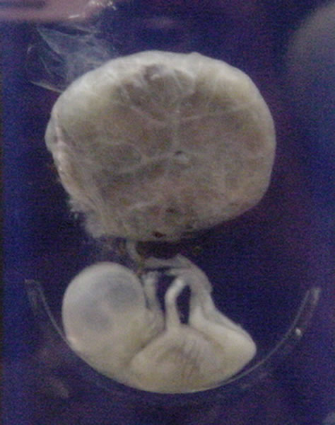
A fetus or foetus (; plural fetuses, feti, foetuses, or foeti) is the unborn offspring that develops from an animal embryo. Following embryonic development the fetal stage of development takes place. In human prenatal development, fetal development begins from the ninth week after fertilization (or eleventh week gestational age) and continues until birth. Prenatal development is a continuum, with no clear defining feature distinguishing an embryo from a fetus. However, a fetus is characterized by the presence of all the major body organs, though they will not yet be fully developed and functional and some not yet situated in their final anatomical location. Etymology The word '' fetus'' (plural '' fetuses'' or '' feti'') is related to the Latin '' fētus'' ("offspring", "bringing forth", "hatching of young") and the Greek "φυτώ" to plant. The word "fetus" was used by Ovid in Metamorphoses, book 1, line 104. The predominant British, Irish, and Commonwealth spelling ... [...More Info...] [...Related Items...] OR: [Wikipedia] [Google] [Baidu] |


