|
Parson Russell Terrier
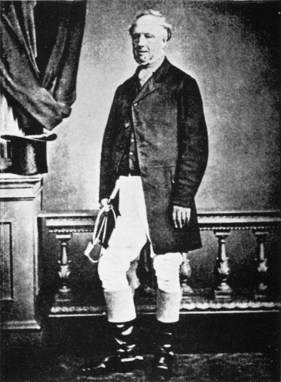
The Parson Russell Terrier is a breed of small white terrier that was the original Fox Terrier of the 18th century. The breed is named after the Reverend Jack Russell, credited with the creation of this type of dog. It is the recognised conformation show variety of the Jack Russell Terrier and was first recognised in 1990 in the United Kingdom as the Parson Jack Russell Terrier. In America, it was first recognised as the Jack Russell Terrier in 1997. The name was changed to its current form in 1999 in the UK and by 2008 all international kennel clubs recognised it under the new name. A mostly white breed with either a smooth, rough or broken coat, it conforms to a narrower range of sizes than the Jack Russell. It is a feisty, energetic terrier, suited to sports and able to get along with children and other animals. It has a range of breed-related health issues, mainly relating to eye disorders. History :''This breed shares a common history with the Jack Russell Terrier until th ... [...More Info...] [...Related Items...] OR: [Wikipedia] [Google] [Baidu] |
England
England is a country that is part of the United Kingdom. It shares land borders with Wales to its west and Scotland to its north. The Irish Sea lies northwest and the Celtic Sea to the southwest. It is separated from continental Europe by the North Sea to the east and the English Channel to the south. The country covers five-eighths of the island of Great Britain, which lies in the North Atlantic, and includes over 100 smaller islands, such as the Isles of Scilly and the Isle of Wight. The area now called England was first inhabited by modern humans during the Upper Paleolithic period, but takes its name from the Angles, a Germanic tribe deriving its name from the Anglia peninsula, who settled during the 5th and 6th centuries. England became a unified state in the 10th century and has had a significant cultural and legal impact on the wider world since the Age of Discovery, which began during the 15th century. The English language, the Anglican Church, and Engli ... [...More Info...] [...Related Items...] OR: [Wikipedia] [Google] [Baidu] |
Breed Standard (dog)
In animal husbandry or animal fancy, a breed standard is a description of the characteristics of a hypothetical or ideal example of a breed. The description may include physical or morphological detail, genetic criteria, or criteria of athletic or productive performance. It may also describe faults or deficiencies that would disqualify an animal from registration or from reproduction. The hypothetical ideal example may be called a "breed type". Breed standards are devised by breed associations or breed clubs, not by individuals, and are written to reflect the use or purpose of the species and breed of the animal. Breed standards help define the ideal animal of a breed and provide goals for breeders in improving stock. In essence a breed standard is a blueprint for an animal fit for the function it was bred - i.e. herding, tracking etc. [...More Info...] [...Related Items...] OR: [Wikipedia] [Google] [Baidu] |
Posterior Vitreous Detachment
A posterior vitreous detachment (PVD) is a condition of the eye in which the vitreous membrane separates from the retina. It refers to the separation of the posterior hyaloid membrane from the retina anywhere posterior to the vitreous base (a 3–4 mm wide attachment to the ora serrata). The condition is common for older adults; over 75% of those over the age of 65 develop it. Although less common among people in their 40s or 50s, the condition is not rare for those individuals. Some research has found that the condition is more common among women. Symptoms When this occurs there is a characteristic pattern of symptoms: * Flashes of light (photopsia) * A sudden dramatic increase in the number of floaters * A ring of floaters or hairs just to the temporal side of the central vision As a posterior vitreous detachment proceeds, adherent vitreous membrane may pull on the retina. While there are no pain fibers in the retina, vitreous traction may stimulate the retina, with ... [...More Info...] [...Related Items...] OR: [Wikipedia] [Google] [Baidu] |
Progressive Retinal Atrophy
Progressive retinal atrophy (PRA) is a group of genetic diseases seen in certain breeds of dogs and, more rarely, cats. Similar to retinitis pigmentosa in humans, it is characterized by the bilateral degeneration of the retina, causing progressive vision loss culminating in blindness. The condition in nearly all breeds is inherited as an autosomal recessive trait, with the exception of the Siberian Husky (inherited as an X chromosome linked trait) and the Bullmastiff (inherited as an autosomal dominant trait). There is no treatment. Types of PRA In general, PRAs are characterised by initial loss of rod photoreceptor cell function followed by that of the cones and for this reason night blindness is the first significant clinical sign for most dogs affected with PRA. As other retinal disorders, PRA can be divided into either dysplastic disease, where the cells develop abnormally, and degenerative, where the cells develop normally but then degenerate during the dog's lifetime. Ge ... [...More Info...] [...Related Items...] OR: [Wikipedia] [Google] [Baidu] |
Corneal Dystrophies In Dogs
Corneal dystrophies are a group of diseases that affect the cornea in dogs. Treatment Corneal dystrophy in dogs usually does not cause any problems and treatment is not required. Suboptimal vision caused by corneal dystrophy usually requires surgical intervention in the form of corneal transplantation. Penetrating keratoplasty is commonly performed for extensive corneal dystrophy. Corneal endothelial dystrophy Corneal endothelial dystrophy is an age-related change that affects the inner layer of the corneal, the endothelium. Leakage of fluid into the cornea causes edema, causing a bluish appearance. This will eventually involve the whole cornea. Bullous keratopathy ( blisters in the cornea) may also form, leading to nonhealing and recurrent corneal ulceration. Hyperosmotic agents are sometimes used topically for treatment, but success with these medications is inconsistent and can cause irritation. Bad cases may require a corneal transplant or thermokeratoplasty, which ... [...More Info...] [...Related Items...] OR: [Wikipedia] [Google] [Baidu] |
Cataracts
A cataract is a cloudy area in the lens of the eye that leads to a decrease in vision. Cataracts often develop slowly and can affect one or both eyes. Symptoms may include faded colors, blurry or double vision, halos around light, trouble with bright lights, and trouble seeing at night. This may result in trouble driving, reading, or recognizing faces. Poor vision caused by cataracts may also result in an increased risk of falling and depression. Cataracts cause 51% of all cases of blindness and 33% of visual impairment worldwide. Cataracts are most commonly due to aging but may also occur due to trauma or radiation exposure, be present from birth, or occur following eye surgery for other problems. Risk factors include diabetes, longstanding use of corticosteroid medication, smoking tobacco, prolonged exposure to sunlight, and alcohol. The underlying mechanism involves accumulation of clumps of protein or yellow-brown pigment in the lens that reduces transmission of li ... [...More Info...] [...Related Items...] OR: [Wikipedia] [Google] [Baidu] |
Anterior Chamber Of Eyeball
The anterior chamber (Optometric Abbreviations#AC, AC) is the aqueous humor-filled space inside the human eye, eye between the iris (anatomy), iris and the cornea's innermost surface, the Corneal endothelium, endothelium. Hyphema, Uveitis, anterior uveitis and glaucoma are three main pathologies in this area. In hyphema, blood fills the anterior chamber as a result of a hemorrhage, most commonly after a blunt eye injury. Anterior uveitis is an inflammatory process affecting the iris (anatomy), iris and ciliary body, with resulting inflammatory signs in the anterior chamber. In glaucoma, blockage of the trabecular meshwork prevents the normal outflow of aqueous humour, resulting in increased intraocular pressure, progressive damage to the optic nerve head, and eventually blindness. The depth of the anterior chamber of the eye varies between 1.5 and 4.0 mm, averaging 3.0 mm. It tends to become shallower at older age and in eyes with Far-sightedness, hypermetropia (far sight ... [...More Info...] [...Related Items...] OR: [Wikipedia] [Google] [Baidu] |
Lens (anatomy)
The lens, or crystalline lens, is a transparent biconvex structure in the eye that, along with the cornea, helps to refract light to be focused on the retina. By changing shape, it functions to change the focal length of the eye so that it can focus on objects at various distances, thus allowing a sharp real image of the object of interest to be formed on the retina. This adjustment of the lens is known as '' accommodation'' (see also below). Accommodation is similar to the focusing of a photographic camera via movement of its lenses. The lens is flatter on its anterior side than on its posterior side. In humans, the refractive power of the lens in its natural environment is approximately 18 dioptres, roughly one-third of the eye's total power. Structure The lens is part of the anterior segment of the human eye. In front of the lens is the iris, which regulates the amount of light entering into the eye. The lens is suspended in place by the suspensory ligament of the lens ... [...More Info...] [...Related Items...] OR: [Wikipedia] [Google] [Baidu] |
Zonular Fibres
The zonule of Zinn () (Zinn's membrane, ciliary zonule) (after Johann Gottfried Zinn) is a ring of fibrous strands forming a zonule (little band) that connects the ciliary body with the crystalline lens of the eye. These fibers are sometimes collectively referred to as the suspensory ligaments of the lens, as they act like suspensory ligaments. Development The ciliary epithelial cells of the eye probably synthesize portions of the zonules. Anatomy The zonule of Zinn is split into two layers: a thin layer, which lines the hyaloid fossa, and a thicker layer, which is a collection of zonular fibers. Together, the fibers are known as the suspensory ligament of the lens. The zonules are about 1–2 μm in diameter. The zonules attach to the lens capsule 2 mm anterior and 1 mm posterior to the equator, and arise of the ciliary epithelium from the pars plana region as well as from the valleys between the ciliary processes in the pars plicata. When colour granules are displaced from th ... [...More Info...] [...Related Items...] OR: [Wikipedia] [Google] [Baidu] |
Lens Luxation
Ectopia lentis is a displacement or malposition of the eye's crystalline lens from its normal location. A partial dislocation of a lens is termed lens subluxation or subluxated lens; a complete dislocation of a lens is termed lens luxation or luxated lens. Ectopia lentis in dogs and cats Although observed in humans and cats, ectopia lentis is most commonly seen in dogs. Ciliary zonules normally hold the lens in place. Abnormal development of these zonules can lead to primary ectopia lentis, usually a bilateral condition. Luxation can also be a secondary condition, caused by trauma, cataract formation (decrease in lens diameter may stretch and break the zonules), or glaucoma (enlargement of the globe stretches the zonules). Steroid administration weakens the zonules and can lead to luxation, as well. Lens luxation in cats can occur secondary to anterior uveitis (inflammation of the inside of the eye). Anterior lens luxation With anterior lens luxation, the lens pushes into the i ... [...More Info...] [...Related Items...] OR: [Wikipedia] [Google] [Baidu] |
Dog Agility
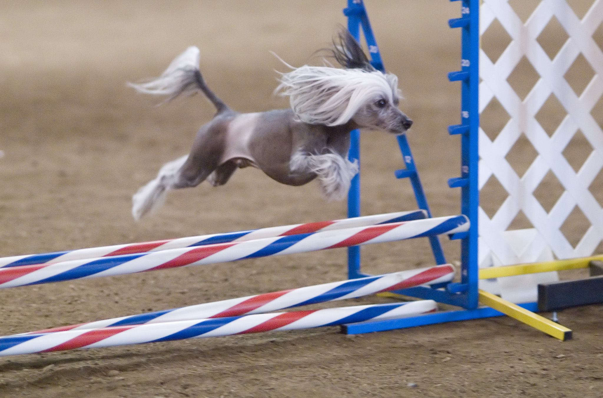
Dog agility is a dog sport in which a handler directs a dog through an obstacle course in a race for both time and accuracy. Dogs run off leash with no food or toys as incentives, and the handler can touch neither dog nor obstacles. The handler's controls are limited to voice, movement, and various body signals, requiring exceptional training of the animal and coordination of the handler. An agility course consists of a set of standard obstacles laid out by a judge in a design of their own choosing in an area of a specified size. The surface may be of grass, dirt, rubber, or special matting. Depending on the type of competition, the obstacles may be marked with numbers indicating the order in which they must be completed. Courses are complicated enough that a dog could not complete them correctly without human direction. In competition, the handler must assess the course, decide on handling strategies, and direct the dog through the course, with precision and speed equally impo ... [...More Info...] [...Related Items...] OR: [Wikipedia] [Google] [Baidu] |
Flyball
Flyball is a dog sport in which teams of dogs race against each other from the start to the finish line, over a line of hurdles, to a box that releases a tennis ball to be caught when the dog presses the spring-loaded pad, then back to their handlers while carrying the ball. Flyball is run in teams of four dogs, as a relay. The course consists of four hurdles placed 10 feet (3 m) apart from each other, with the starting line six feet (1.8 m) from the first hurdle, and the flyball box 15 feet (4.5 m) after the last one, making for a 51-foot (15.5 m) length. The hurdle height is determined by the ulna's length or the smallest dog's shoulder height on the team (depending on the association). For example, under current North American Flyball Association (NAFA) rules, this should be 5 inches (12.7 cm) below the withers height of the smallest dog, to a height of no less than 7 inches (17.8 cm) and no greater than 14 inches (35.6 cm). United Kingdom Flyba ... [...More Info...] [...Related Items...] OR: [Wikipedia] [Google] [Baidu] |


_PHIL_4284_lores.jpg)


