|
Neovascular
Neovascularization is the natural formation of new blood vessels ('' neo-'' + ''vascular'' + '' -ization''), usually in the form of functional microvascular networks, capable of perfusion by red blood cells, that form to serve as collateral circulation in response to local poor perfusion or ischemia. Growth factors that inhibit neovascularization include those that affect endothelial cell division and differentiation. These growth factors often act in a paracrine or autocrine fashion; they include fibroblast growth factor, placental growth factor, insulin-like growth factor, hepatocyte growth factor, and platelet-derived endothelial growth factor. There are three different pathways that comprise neovascularization:(1) vasculogenesis,(2) angiogenesis, and (3) arteriogenesis. Three pathways of neovascularization Vasculogenesis Vasculogenesis is the de novo formation of blood vessels. This primarily occurs in the developing embryo with the development of the first primitive vas ... [...More Info...] [...Related Items...] OR: [Wikipedia] [Google] [Baidu] |
Corneal Neovascularization
Corneal neovascularization (CNV) is the in-growth of new blood vessels from the pericorneal plexus into avascular corneal tissue as a result of oxygen deprivation. Maintaining avascularity of the corneal stroma is an important aspect of corneal pathophysiology as it is required for corneal transparency and optimal vision. A decrease in corneal transparency causes visual acuity deterioration. Corneal tissue is avascular in nature and the presence of vascularization, which can be deep or superficial, is always pathologically related. Corneal neovascularization is a sight-threatening condition that can be caused by inflammation related to infection, chemical injury, autoimmune conditions, immune hypersensitivity, post- corneal transplantation, and traumatic conditions among other ocular pathologies. Common causes of CNV within the cornea include trachoma, corneal ulcers, phylctenular keratoconjunctivitisrosacea keratitis interstitial keratitissclerosing keratitis chemical burns, ... [...More Info...] [...Related Items...] OR: [Wikipedia] [Google] [Baidu] |
Glaucoma
Glaucoma is a group of eye diseases that result in damage to the optic nerve (or retina) and cause vision loss. The most common type is open-angle (wide angle, chronic simple) glaucoma, in which the drainage angle for fluid within the eye remains open, with less common types including closed-angle (narrow angle, acute congestive) glaucoma and normal-tension glaucoma. Open-angle glaucoma develops slowly over time and there is no pain. Peripheral vision may begin to decrease, followed by central vision, resulting in blindness if not treated. Closed-angle glaucoma can present gradually or suddenly. The sudden presentation may involve severe eye pain, blurred vision, mid-dilated pupil, redness of the eye, and nausea. Vision loss from glaucoma, once it has occurred, is permanent. Eyes affected by glaucoma are referred to as being glaucomatous. Risk factors for glaucoma include increasing age, high pressure in the eye, a family history of glaucoma, and use of steroid medication. F ... [...More Info...] [...Related Items...] OR: [Wikipedia] [Google] [Baidu] |
Choroidal Neovascularization
Choroidal neovascularization (CNV) is the creation of new blood vessels in the choroid layer of the eye. Choroidal neovascularization is a common cause of neovascular degenerative maculopathy (i.e. 'wet' macular degeneration) commonly exacerbated by extreme myopia, malignant myopic degeneration, or age-related developments. Causes CNV can occur rapidly in individuals with defects in Bruch's membrane, the innermost layer of the choroid. It is also associated with excessive amounts of vascular endothelial growth factor (VEGF). As well as in wet macular degeneration, CNV can also occur frequently with the rare genetic disease pseudoxanthoma elasticum and rarely with the more common optic disc drusen. CNV has also been associated with extreme myopia or malignant myopic degeneration, where in choroidal neovascularization occurs primarily in the presence of cracks within the retinal (specifically) macular tissue known as lacquer cracks. Symptoms CNV can create a sudden deterioration o ... [...More Info...] [...Related Items...] OR: [Wikipedia] [Google] [Baidu] |
Hypoxia-inducible Factors
Hypoxia-inducible factors (HIFs) are transcription factors that respond to decreases in available oxygen in the cellular environment, or Hypoxia (medical), hypoxia. They are only present in ParaHoxozoa, parahoxozoan animals. Discovery The HIF transcriptional complex was discovered in 1995 by Gregg L. Semenza and postdoctoral fellow Guang Wang. In 2016, William Kaelin Jr., Peter J. Ratcliffe and Gregg L. Semenza were presented the Lasker Award for their work in elucidating the role of HIF-1 in oxygen sensing and its role in surviving low oxygen conditions. In 2019, the same three individuals were jointly awarded the Nobel Prize in Physiology or Medicine for their work in elucidating how HIF senses and adapts cellular response to oxygen availability. Structure Most, if not all, oxygen-breathing species express the conservation (genetics), highly conserved transcriptional complex HIF-1, which is a heterodimer composed of an alpha and a beta subunit, the latter being a const ... [...More Info...] [...Related Items...] OR: [Wikipedia] [Google] [Baidu] |
Rubeosis Iridis
Rubeosis iridis is a medical condition of the iris of the eye in which new abnormal blood vessels (formed by neovascularization) are found on the surface of the iris. Causes This condition is often associated with diabetes in advanced proliferative diabetic retinopathy. Other conditions causing rubeosis iridis include central retinal vein occlusion, ocular ischemic syndrome, and chronic retinal detachment. Pathophysiology It is usually associated with disease processes in the retina, which involve the retina becoming starved of oxygen (ischaemic). The ischemic retina releases a variety of factors, the most important of which is vascular endothelial growth factor (VEGF). These factors stimulate the formation of new blood vessels (angiogenesis). These new vessels do not have the same characteristics as the blood vessels originally formed in the eye. In addition, new blood vessels can form in areas that do not have them. Specifically, new blood vessels can be observed on the iri ... [...More Info...] [...Related Items...] OR: [Wikipedia] [Google] [Baidu] |
Revascularization
In medical and surgical therapy, revascularization is the restoration of perfusion to a body part or organ that has had ischemia. It is typically accomplished by surgical means. Vascular bypass and angioplasty are the two primary means of revascularization. The term derives from the prefix re-, in this case meaning "restoration" and vasculature, which refers to the circulatory structures of an organ. It is often combined with "urgent" to form urgent vascularization. Revascularization involves a thorough analysis and diagnosis and treatment of the existing diseased vasculature of the affected organ, and can be aided by the use of different imaging modalities such as magnetic resonance imaging, PET scan, CT scan, and X-ray fluoroscopy. Applications For coronary artery disease (ischemic heart disease), coronary artery bypass surgery and percutaneous coronary intervention (coronary balloon angioplasty) are the two primary means of revascularization. When those cannot be done, ... [...More Info...] [...Related Items...] OR: [Wikipedia] [Google] [Baidu] |
Blood Vessel
The blood vessels are the components of the circulatory system that transport blood throughout the human body. These vessels transport blood cells, nutrients, and oxygen to the tissues of the body. They also take waste and carbon dioxide away from the tissues. Blood vessels are needed to sustain life, because all of the body's tissues rely on their functionality. There are five types of blood vessels: the arteries, which carry the blood away from the heart; the arterioles; the capillaries, where the exchange of water and chemicals between the blood and the tissues occurs; the venules; and the veins, which carry blood from the capillaries back towards the heart. The word ''vascular'', meaning relating to the blood vessels, is derived from the Latin ''vas'', meaning vessel. Some structures – such as cartilage, the epithelium, and the lens and cornea of the eye – do not contain blood vessels and are labeled ''avascular''. Etymology * artery: late Middle English; from Latin ... [...More Info...] [...Related Items...] OR: [Wikipedia] [Google] [Baidu] |
Inosculation
Inosculation is a natural phenomenon in which trunks, branches or roots of two trees grow together in a manner biologically similar to the artificial process of grafting. The term is derived from the Latin roots ''in'' + '' ōsculārī'', "to kiss into/inward/against" or etymologically and more illustratively "to make a small mouth inward/into/against"; trees having undergone the process are referred to in forestry as gemels, from the Latin word meaning "a pair". It is most common for branches of two trees of the same species to grow together, though inosculation may be noted across related species. The branches first grow separately in proximity to each other until they touch. At this point, the bark on the touching surfaces is gradually abraded away as the trees move in the wind. Once the cambium of two trees touches, they sometimes self-graft and grow together as they expand in diameter. Inosculation customarily results when tree limbs are braided or pleached. The term ''in ... [...More Info...] [...Related Items...] OR: [Wikipedia] [Google] [Baidu] |
Aqueous Humour
The aqueous humour is a transparent water-like fluid similar to plasma, but containing low protein concentrations. It is secreted from the ciliary body, a structure supporting the lens of the eyeball. It fills both the anterior and the posterior chambers of the eye, and is not to be confused with the vitreous humour, which is located in the space between the lens and the retina, also known as the posterior cavity or vitreous chamber. Blood cannot normally enter the eyeball. Structure Composition *Amino acids: transported by ciliary muscles *98% water *Electrolytes ( pH = 7.4 -one source gives 7.1) **Sodium = 142.09 **Potassium = 2.2 - 4.0 **Calcium = 1.8 **Magnesium = 1.1 **Chloride = 131.6 **HCO3- = 20.15 **Phosphate = 0.62 ** Osm = 304 *Ascorbic acid *Glutathione *Immunoglobulins Function *Maintains the intraocular pressure and inflates the globe of the eye. It is this hydrostatic pressure that keeps the eyeball in a roughly spherical shape and keeps the walls of the eyeball ... [...More Info...] [...Related Items...] OR: [Wikipedia] [Google] [Baidu] |
Choroid
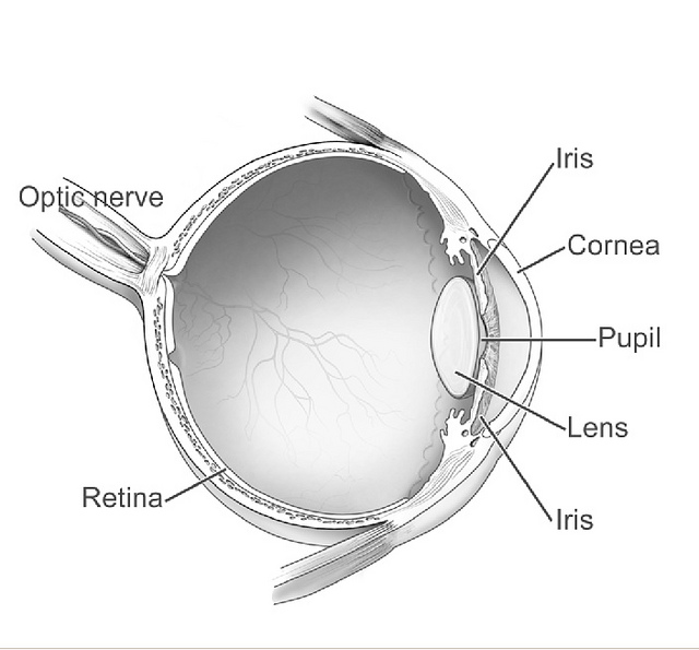
The choroid, also known as the choroidea or choroid coat, is a part of the uvea, the vascular layer of the eye, and contains connective tissues, and lies between the retina and the sclera. The human choroid is thickest at the far extreme rear of the eye (at 0.2 mm), while in the outlying areas it narrows to 0.1 mm. The choroid provides oxygen and nourishment to the outer layers of the retina. Along with the ciliary body and iris, the choroid forms the uveal tract. The structure of the choroid is generally divided into four layers (classified in order of furthest away from the retina to closest): *Haller's layer - outermost layer of the choroid consisting of larger diameter blood vessels; *Sattler's layer - layer of medium diameter blood vessels; * Choriocapillaris - layer of capillaries; and *Bruch's membrane (synonyms: Lamina basalis, Complexus basalis, Lamina vitra) - innermost layer of the choroid. Blood supply There are two circulations of the eye: the retin ... [...More Info...] [...Related Items...] OR: [Wikipedia] [Google] [Baidu] |
Bruch's Membrane
Bruch's membrane is the innermost layer of the choroid of the eye. It is also called the ''vitreous lamina'' or ''Membrane vitriae'', because of its glassy microscopic appearance. It is 2–4 μm thick. Layers Bruch's membrane consists of five layers (from inside to outside): #the basement membrane of the retinal pigment epithelium #the inner collagenous zone #a central band of elastic fibers #the outer collagenous zone #the basement membrane of the choriocapillaris The retinal pigment epithelium transports metabolic waste from the photoreceptors across Bruch's membrane to the choroid. Embryology Bruch's membrane is present by midterm in fetal development as an elastic sheet. Pathology Bruch's membrane thickens with age, slowing the transport of metabolites. This may lead to the formation of drusen in age-related macular degeneration. There is also a buildup of deposits (Basal Linear Deposits or BLinD and Basal Lamellar Deposits BLamD) on and within the membrane, primarily co ... [...More Info...] [...Related Items...] OR: [Wikipedia] [Google] [Baidu] |


