|
Choroid
The choroid, also known as the choroidea or choroid coat, is a part of the uvea, the vascular layer of the eye, and contains connective tissues, and lies between the retina and the sclera. The human choroid is thickest at the far extreme rear of the eye (at 0.2 mm), while in the outlying areas it narrows to 0.1 mm. The choroid provides oxygen and nourishment to the outer layers of the retina. Along with the ciliary body and iris, the choroid forms the uveal tract. The structure of the choroid is generally divided into four layers (classified in order of furthest away from the retina to closest): *Haller's layer - outermost layer of the choroid consisting of larger diameter blood vessels; *Sattler's layer - layer of medium diameter blood vessels; * Choriocapillaris - layer of capillaries; and *Bruch's membrane (synonyms: Lamina basalis, Complexus basalis, Lamina vitra) - innermost layer of the choroid. Blood supply There are two circulations of the eye: the retin ... [...More Info...] [...Related Items...] OR: [Wikipedia] [Google] [Baidu] |
Indocyanine Green Angiography
Indocyanine green angiography (ICGA) is a diagnostic procedure used to examine choroidal blood flow and associated pathology. Indocyanine green (ICG) is a water soluble cyanine dye which shows fluorescence in near-infrared (790–805 nm) range, with peak spectral absorption of 800-810 nm in blood. The near infrared light used in ICGA penetrates ocular pigments such as melanin and xanthophyll, as well as exudates and thin layers of sub-retinal vessels. Age-related macular degeneration is the third main cause of blindness worldwide, and it is the leading cause of blindness in industrialized countries. Indocyanine green angiography is widely used to study choroidal neovascularization in patients with exudative age-related macular degeneration. In nonexudative AMD, ICGA is used in classification of drusen and associated subretinal deposits. Indications Indications for indocyanine green angiography include: * Choroidal neovascularisation (CNV):Indocyanine green angiography is widely us ... [...More Info...] [...Related Items...] OR: [Wikipedia] [Google] [Baidu] |
Tapetum Lucidum
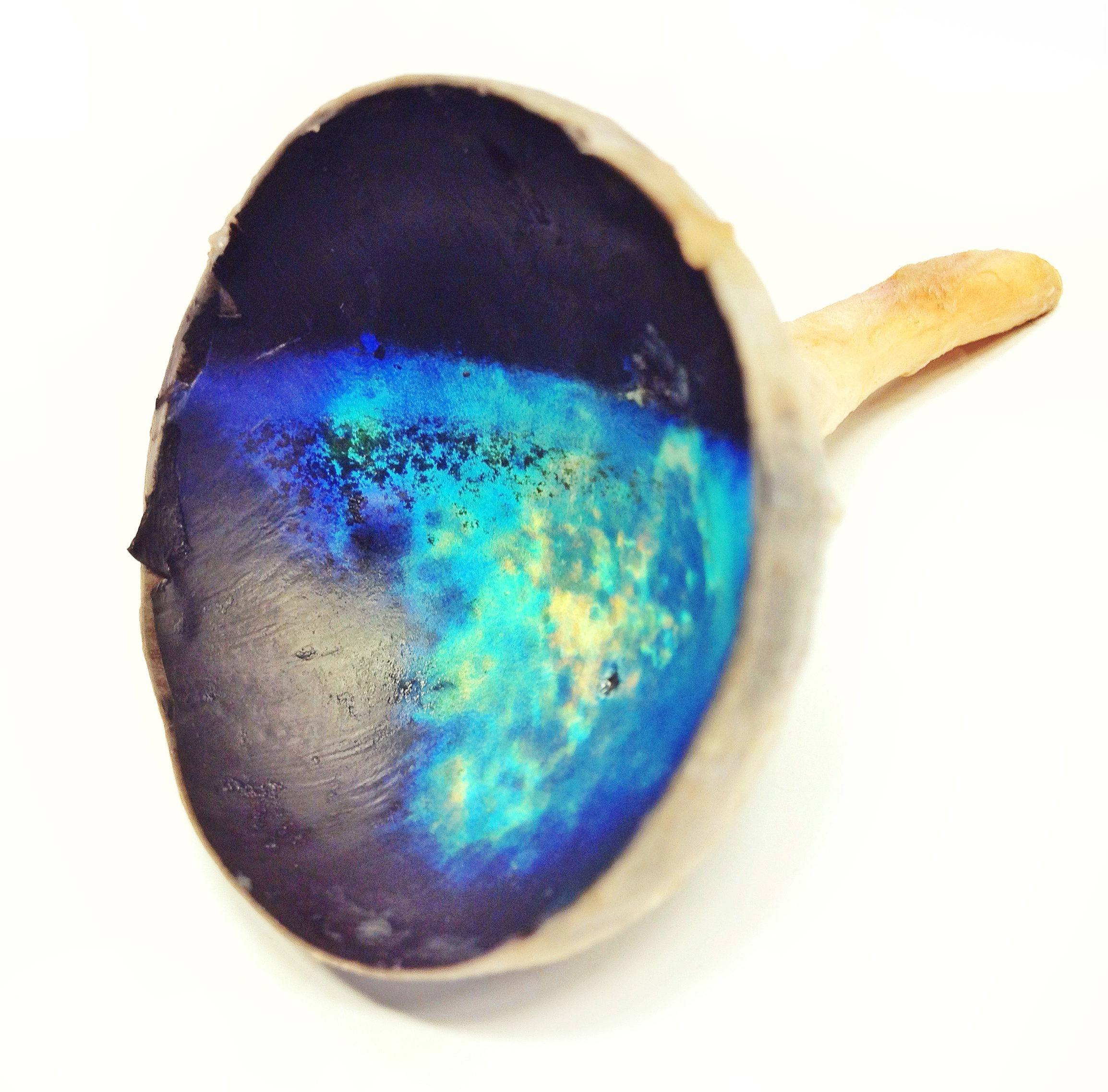
The ''tapetum lucidum'' ( ; ; ) is a layer of tissue in the eye of many vertebrates and some other animals. Lying immediately behind the retina, it is a retroreflector. It reflects visible light back through the retina, increasing the light available to the photoreceptors (although slightly blurring the image). The tapetum lucidum contributes to the superior night vision of some animals. Many of these animals are nocturnal, especially carnivores, while others are deep sea animals. Similar adaptations occur in some species of spiders. Haplorhine primates, including humans, are diurnal and lack a ''tapetum lucidum''. Function and mechanism Presence of a ''tapetum lucidum'' enables animals to see in dimmer light than would otherwise be possible. The ''tapetum lucidum'', which is iridescent, reflects light roughly on the interference principles of thin-film optics, as seen in other iridescent tissues. However, the ''tapetum lucidum'' cells are leucophores, not iridophores. ... [...More Info...] [...Related Items...] OR: [Wikipedia] [Google] [Baidu] |
Sattler's Layer
Sattler's layer, named after Hubert Sattler, an Austrian ophthalmologist, is one of five (or six) layers of medium-diameter blood vessels of the choroid, and a layer of the eye. It is situated between the Bruch's membrane, choriocapillaris below, and the Haller's layer and suprachoroidea above, respectively. The origin seems to be related to a continuous differentiation throughout the growth of the tissue and even further differentiation during adulthood. Measurement methods and clinical impact After excision the choroid collapses partially, histologic preparations also alter the local pressure and fluid content of different sections in the tissue, thus requiring preparations with rubber solution or others that can conserve the vascular status of living tissue. Novel diagnostic methods, especially optical coherence tomography have widened the understanding of the real-time, in vivo status of the different layers. Several papers have shown the relationship between the thick ... [...More Info...] [...Related Items...] OR: [Wikipedia] [Google] [Baidu] |
Retina
The retina (from la, rete "net") is the innermost, light-sensitive layer of tissue of the eye of most vertebrates and some molluscs. The optics of the eye create a focused two-dimensional image of the visual world on the retina, which then processes that image within the retina and sends nerve impulses along the optic nerve to the visual cortex to create visual perception. The retina serves a function which is in many ways analogous to that of the film or image sensor in a camera. The neural retina consists of several layers of neurons interconnected by synapses and is supported by an outer layer of pigmented epithelial cells. The primary light-sensing cells in the retina are the photoreceptor cells, which are of two types: rods and cones. Rods function mainly in dim light and provide monochromatic vision. Cones function in well-lit conditions and are responsible for the perception of colour through the use of a range of opsins, as well as high-acuity vision used for task ... [...More Info...] [...Related Items...] OR: [Wikipedia] [Google] [Baidu] |
Uvea
The uvea (; Lat. ''uva'', "grape"), also called the ''uveal layer'', ''uveal coat'', ''uveal tract'', ''vascular tunic'' or ''vascular layer'' is the pigmented middle of the three concentric layers that make up an eye. History and etymology The originally medieval Latin term comes from the Latin word ''uva'' ("grape") and is a reference to its grape-like appearance (reddish-blue or almost black colour, wrinkled appearance and grape-like size and shape when stripped intact from a cadaveric eye). In fact, it is a partial loan translation of the Ancient Greek term for the choroid, which literally means “covering resembling a grape”. Its use as a technical term for part of the eye is ancient, but it only referred to the choroid in Middle English and before. Structure Regions The uvea is the vascular middle layer of the eye. It is traditionally divided into three areas, from front to back, the: * Iris * Ciliary body * Choroid Function The prime functions of the uveal tra ... [...More Info...] [...Related Items...] OR: [Wikipedia] [Google] [Baidu] |
Short Posterior Ciliary Arteries
The short posterior ciliary arteries, around twenty in number, arise from the medial posterior ciliary artery and lateral posterior ciliary artery, which are branches of the ophthalmic artery as it crosses the optic nerve. Course and target They pass forward around the optic nerve to the posterior part of the eyeball, pierce the sclera around the entrance of the optic nerve, and supply the choroid (up to the equator of the eye) and ciliary processes. Some branches of the short posterior ciliary arteries also supply the optic disc via an anastomotic ring, the circle of Zinn-Haller or ''circle of Zinn'', which is associated with the fibrous extension of the ocular tendons (common tendinous ring (also annulus of Zinn)). Additional images File:Gray880.png, The terminal portion of the optic nerve and its entrance into the eyeball, in horizontal section See also * Long posterior ciliary arteries *Anterior ciliary arteries The anterior ciliary arteries are seven small arteries in ea ... [...More Info...] [...Related Items...] OR: [Wikipedia] [Google] [Baidu] |
Uvea
The uvea (; Lat. ''uva'', "grape"), also called the ''uveal layer'', ''uveal coat'', ''uveal tract'', ''vascular tunic'' or ''vascular layer'' is the pigmented middle of the three concentric layers that make up an eye. History and etymology The originally medieval Latin term comes from the Latin word ''uva'' ("grape") and is a reference to its grape-like appearance (reddish-blue or almost black colour, wrinkled appearance and grape-like size and shape when stripped intact from a cadaveric eye). In fact, it is a partial loan translation of the Ancient Greek term for the choroid, which literally means “covering resembling a grape”. Its use as a technical term for part of the eye is ancient, but it only referred to the choroid in Middle English and before. Structure Regions The uvea is the vascular middle layer of the eye. It is traditionally divided into three areas, from front to back, the: * Iris * Ciliary body * Choroid Function The prime functions of the uveal tra ... [...More Info...] [...Related Items...] OR: [Wikipedia] [Google] [Baidu] |
Laser Doppler Imaging
Laser Doppler imaging (LDI) is an imaging method that uses a laser beam to scan live tissue. When the laser light reaches the tissue, the moving blood cells generate doppler components in the reflected ( backscattered) light. The light that comes back is detected using a photodiode that converts it into an electrical signal. Then the signal is processed to calculate a signal that is proportional to the tissue perfusion in the scanned area. When the process is completed, the signal is processed to generate an image that shows the perfusion on a screen. The laser doppler effect was first used to measure microcirculation by Stern M.D. in 1975. And it is used widely in medicine, some representative research work about it are these: Use in Ophthalmology The eye offers a unique opportunity for the non-invasive exploration of cardiovascular diseases. LDI by digital holography can measure blood flow in the retina and choroid. In particular, the choroid is a highly vascularized tissue s ... [...More Info...] [...Related Items...] OR: [Wikipedia] [Google] [Baidu] |
Macula
The macula (/ˈmakjʊlə/) or macula lutea is an oval-shaped pigmented area in the center of the retina of the human eye and in other animals. The macula in humans has a diameter of around and is subdivided into the umbo, foveola, foveal avascular zone, fovea, parafovea, and perifovea areas. The anatomical macula at a size of is much larger than the clinical macula which, at a size of , corresponds to the anatomical fovea. The macula is responsible for the central, high-resolution, color vision that is possible in good light; and this kind of vision is impaired if the macula is damaged, for example in macular degeneration. The clinical macula is seen when viewed from the pupil, as in ophthalmoscopy or retinal photography. The term macula lutea comes from Latin ''macula'', "spot", and ''lutea'', "yellow". Structure The macula is an oval-shaped pigmented area in the center of the retina of the human eye and other animal eyes. Its center is shifted slightly away from the ... [...More Info...] [...Related Items...] OR: [Wikipedia] [Google] [Baidu] |
Optical Coherence Tomography
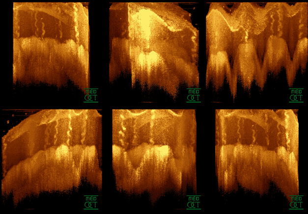
Optical coherence tomography (OCT) is an imaging technique that uses low-coherence light to capture micrometer-resolution, two- and three-dimensional images from within optical scattering media (e.g., biological tissue). It is used for medical imaging and industrial nondestructive testing (NDT). Optical coherence tomography is based on low-coherence interferometry, typically employing near-infrared light. The use of relatively long wavelength light allows it to penetrate into the scattering medium. Confocal microscopy, another optical technique, typically penetrates less deeply into the sample but with higher resolution. Depending on the properties of the light source ( superluminescent diodes, ultrashort pulsed lasers, and supercontinuum lasers have been employed), optical coherence tomography has achieved sub-micrometer resolution (with very wide-spectrum sources emitting over a ~100 nm wavelength range). Optical coherence tomography is one of a class of optical tom ... [...More Info...] [...Related Items...] OR: [Wikipedia] [Google] [Baidu] |
Teleosts
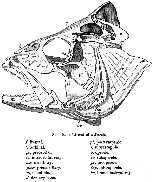
Teleostei (; Greek ''teleios'' "complete" + ''osteon'' "bone"), members of which are known as teleosts ), is, by far, the largest infraclass in the class Actinopterygii, the ray-finned fishes, containing 96% of all extant species of fish. Teleosts are arranged into about 40 orders and 448 families. Over 26,000 species have been described. Teleosts range from giant oarfish measuring or more, and ocean sunfish weighing over , to the minute male anglerfish ''Photocorynus spiniceps'', just long. Including not only torpedo-shaped fish built for speed, teleosts can be flattened vertically or horizontally, be elongated cylinders or take specialised shapes as in anglerfish and seahorses. The difference between teleosts and other bony fish lies mainly in their jaw bones; teleosts have a movable premaxilla and corresponding modifications in the jaw musculature which make it possible for them to protrude their jaws outwards from the mouth. This is of great advantage, enabling them to ... [...More Info...] [...Related Items...] OR: [Wikipedia] [Google] [Baidu] |


