|
Myomesin-2
Myomesin-2, also known as M-protein is a protein that in humans is encoded by the ''MYOM2'' gene. M-protein is expressed in adult cardiac muscle and fast skeletal muscle, and functions to stabilize the three-dimensional arrangement of proteins comprising M-band structures in a sarcomere. Structure Human M-protein is 165.0 kDa and 1465 amino acids in length. ''MYOM2'' is localized to the human chromosome 8p23.3. M-protein belong to the superfamily of cytoskeletal proteins having immunoglobulin/fibronectin repeats; M-protein contains two immunoglobulin C2-type repeats in the N-terminal region, five fibronectin type III repeats in the central region, and an additional four immunoglobulin C2-type repeats in the C-terminal region. M-protein is expressed only in striated muscle, including fast skeletal muscle and cardiac muscle. Function M-protein exhibits a different pattern of expression in cardiac and skeletal muscle, as well as fast- versus slow-skeletal muscle during development ... [...More Info...] [...Related Items...] OR: [Wikipedia] [Google] [Baidu] |
MYOM1
Myomesin-1 is a protein that in humans is encoded by the ''MYOM1'' gene. Myomesin-1 is expressed in muscle cells and functions to stabilize the three-dimensional conformation of the thick filament. Embryonic forms of Myomesin-1 have been detected in dilated cardiomyopathy. Structure Alternatively spliced variants of ''MYOM1'', including EH-myomesin, Skelemin and Myomesin-1 have been identified; with Skelemin having an additional 96 amino acids rich in serine and proline residues. Myomesin-1, like myomesin 2 and titin, is a member of a family of myosin-associated proteins containing structural modules with strong homology to either fibronectin type III (motif I) or immunoglobulin C2 (motif II) domains. Myomesin-1 bears uniqueness within this family in that it has intermediate filament core-like motifs, one near each terminus. Myomesin-1 and Myomesin-2 each have a unique N-terminal region followed by 12 modules of motif I or motif II, in the arrangement II-II-I-I-I-I-I-II ... [...More Info...] [...Related Items...] OR: [Wikipedia] [Google] [Baidu] |
Protein
Proteins are large biomolecules and macromolecules that comprise one or more long chains of amino acid residues. Proteins perform a vast array of functions within organisms, including catalysing metabolic reactions, DNA replication, responding to stimuli, providing structure to cells and organisms, and transporting molecules from one location to another. Proteins differ from one another primarily in their sequence of amino acids, which is dictated by the nucleotide sequence of their genes, and which usually results in protein folding into a specific 3D structure that determines its activity. A linear chain of amino acid residues is called a polypeptide. A protein contains at least one long polypeptide. Short polypeptides, containing less than 20–30 residues, are rarely considered to be proteins and are commonly called peptides. The individual amino acid residues are bonded together by peptide bonds and adjacent amino acid residues. The sequence of amino acid residue ... [...More Info...] [...Related Items...] OR: [Wikipedia] [Google] [Baidu] |
Cardiac Hypertrophy
Ventricular hypertrophy (VH) is thickening of the walls of a ventricle (lower chamber) of the heart. Although left ventricular hypertrophy (LVH) is more common, right ventricular hypertrophy (RVH), as well as concurrent hypertrophy of both ventricles can also occur. Ventricular hypertrophy can result from a variety of conditions, both adaptive and maladaptive. For example, it occurs in what is regarded as a physiologic, adaptive process in pregnancy in response to increased blood volume; but can also occur as a consequence of ventricular remodeling following a heart attack. Importantly, pathologic and physiologic remodeling engage different cellular pathways in the heart and result in different gross cardiac phenotypes. Presentation In individuals with eccentric hypertrophy there may be little or no indication that hypertrophy has occurred as it is generally a healthy response to increased demands on the heart. Conversely, concentric hypertrophy can make itself known in a variety ... [...More Info...] [...Related Items...] OR: [Wikipedia] [Google] [Baidu] |
Titin
Titin (contraction for Titan protein) (also called connectin) is a protein that in humans is encoded by the ''TTN'' gene. Titin is a giant protein, greater than 1 µm in length, that functions as a molecular spring that is responsible for the passive elasticity of muscle. It comprises 244 individually folded protein domains connected by unstructured peptide sequences. These domains unfold when the protein is stretched and refold when the tension is removed. Titin is important in the contraction of striated muscle tissues. It connects the Z line to the M line in the sarcomere. The protein contributes to force transmission at the Z line and resting tension in the I band region. It limits the range of motion of the sarcomere in tension, thus contributing to the passive stiffness of muscle. Variations in the sequence of titin between different types of striated muscle (cardiac or skeletal) have been correlated with differences in the mechanical properties of these muscles. ... [...More Info...] [...Related Items...] OR: [Wikipedia] [Google] [Baidu] |
Reperfusion Injury
Reperfusion injury, sometimes called ischemia-reperfusion injury (IRI) or reoxygenation injury, is the tissue damage caused when blood supply returns to tissue ('' re-'' + '' perfusion'') after a period of ischemia or lack of oxygen (anoxia or hypoxia). The absence of oxygen and nutrients from blood during the ischemic period creates a condition in which the restoration of circulation results in inflammation and oxidative damage through the induction of oxidative stress rather than (or along with) restoration of normal function. Reperfusion injury is distinct from cerebral hyperperfusion syndrome (sometimes called "Reperfusion syndrome"), a state of abnormal cerebral vasodilation. Mechanisms Reperfusion of ischemic tissues is often associated with microvascular injury, particularly due to increased permeability of capillaries and arterioles that lead to an increase of diffusion and fluid filtration across the tissues. Activated endothelial cells produce more reactive oxygen sp ... [...More Info...] [...Related Items...] OR: [Wikipedia] [Google] [Baidu] |
Broiler Chicken
A broiler is any chicken (''Gallus gallus domesticus'') that is bred and raised specifically for meat production. Most commercial broilers reach slaughter weight between four and six weeks of age, although slower growing breeds reach slaughter weight at approximately 14 weeks of age. Typical broilers have white feathers and yellowish skin. Broiler or sometimes broiler-fryer is also used sometimes to refer specifically to younger chickens under , as compared with the larger roasters. Due to extensive breeding selection for rapid early growth and the husbandry used to sustain this, broilers are susceptible to several welfare concerns, particularly skeletal malformation and dysfunction, skin and eye lesions and congestive heart conditions. Management of ventilation, housing, stocking density and in-house procedures must be evaluated regularly to support good welfare of the flock. The breeding stock (broiler-breeders) do grow to maturity but also have their own welfare concerns re ... [...More Info...] [...Related Items...] OR: [Wikipedia] [Google] [Baidu] |
Ascites
Ascites is the abnormal build-up of fluid in the abdomen. Technically, it is more than 25 ml of fluid in the peritoneal cavity, although volumes greater than one liter may occur. Symptoms may include increased abdominal size, increased weight, abdominal discomfort, and shortness of breath. Complications can include spontaneous bacterial peritonitis. In the developed world, the most common cause is liver cirrhosis. Other causes include cancer, heart failure, tuberculosis, pancreatitis, and blockage of the hepatic vein. In cirrhosis, the underlying mechanism involves high blood pressure in the portal system and dysfunction of blood vessels. Diagnosis is typically based on an examination together with ultrasound or a CT scan. Testing the fluid can help in determining the underlying cause. Treatment often involves a low-salt diet, medication such as diuretics, and draining the fluid. A transjugular intrahepatic portosystemic shunt (TIPS) may be placed but is associated with co ... [...More Info...] [...Related Items...] OR: [Wikipedia] [Google] [Baidu] |
Myocardium
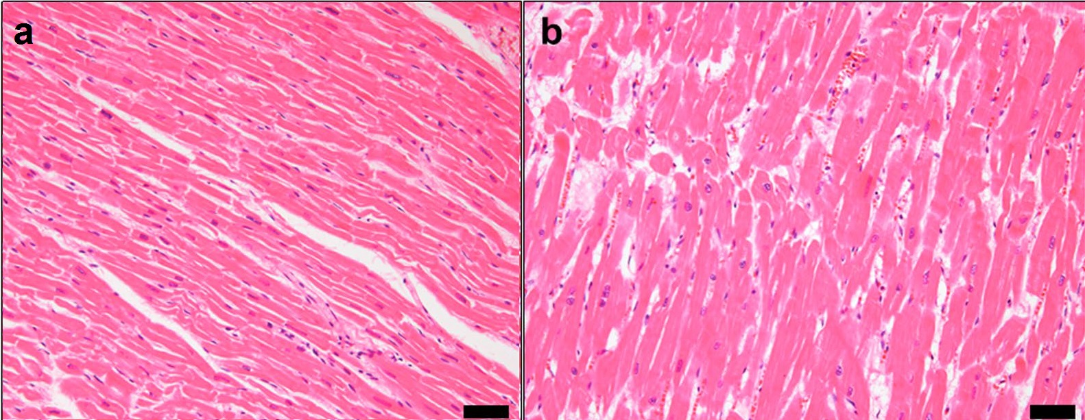
Cardiac muscle (also called heart muscle, myocardium, cardiomyocytes and cardiac myocytes) is one of three types of vertebrate muscle tissues, with the other two being skeletal muscle and smooth muscle. It is an involuntary, striated muscle that constitutes the main tissue of the wall of the heart. The cardiac muscle (myocardium) forms a thick middle layer between the outer layer of the heart wall (the pericardium) and the inner layer (the endocardium), with blood supplied via the coronary circulation. It is composed of individual cardiac muscle cells joined by intercalated discs, and encased by collagen fibers and other substances that form the extracellular matrix. Cardiac muscle contracts in a similar manner to skeletal muscle, although with some important differences. Electrical stimulation in the form of a cardiac action potential triggers the release of calcium from the cell's internal calcium store, the sarcoplasmic reticulum. The rise in calcium causes the cell's m ... [...More Info...] [...Related Items...] OR: [Wikipedia] [Google] [Baidu] |
Ventricle (heart)
A ventricle is one of two large chambers toward the bottom of the heart that collect and expel blood towards the peripheral beds within the body and lungs. The blood pumped by a ventricle is supplied by an atrium, an adjacent chamber in the upper heart that is smaller than a ventricle. Interventricular means between the ventricles (for example the interventricular septum), while intraventricular means within one ventricle (for example an intraventricular block). In a four-chambered heart, such as that in humans, there are two ventricles that operate in a double circulatory system: the right ventricle pumps blood into the pulmonary circulation to the lungs, and the left ventricle pumps blood into the systemic circulation through the aorta. Structure Ventricles have thicker walls than atria and generate higher blood pressures. The physiological load on the ventricles requiring pumping of blood throughout the body and lungs is much greater than the pressure generated by the atria ... [...More Info...] [...Related Items...] OR: [Wikipedia] [Google] [Baidu] |
Matrix Metalloproteinase 2
Gelatinase A, also known as MMP2 (, ''72-kDa gelatinase'', ''matrix metalloproteinase 2'', ''type IV collagenase'', ''3/4 collagenase'', ''matrix metalloproteinase 5'', ''72 kDa gelatinase type A'', ''collagenase IV'', ''collagenase type IV'', ''MMP 2'', ''type IV collagen metalloproteinase'', ''type IV collagenase/gelatinase'') is an enzyme. This enzyme catalyses the following chemical reaction : Cleavage of gelatin type I and collagen types IV, V, VII, X. Cleaves the collagen-like sequence Pro-Gln-Gly-Ile-Ala-Gly-Gln This secreted endopeptidase belongs to the peptidase A protease (also called a peptidase, proteinase, or proteolytic enzyme) is an enzyme that catalyzes (increases reaction rate or "speeds up") proteolysis, breaking down proteins into smaller polypeptides or single amino acids, and spurring the fo ... family M10. References External links * {{Portal bar, Biology, border=no EC 3.4.24 ... [...More Info...] [...Related Items...] OR: [Wikipedia] [Google] [Baidu] |


