|
Middle Cervical Ganglion
The middle cervical ganglion is the smallest of the three cervical ganglia, and is occasionally absent. It is placed opposite the sixth cervical vertebra, usually in front of, or close to, the inferior thyroid artery. It sends gray rami communicantes to the fifth and sixth cervical nerves, and gives off the middle cardiac nerve. It is probably formed by the coalescence of two ganglia corresponding to the fifth and sixth cervical nerves. Branches # Gray Rami Communicantes to the anterior rami of the fifth and sixth cervical nerves. # Thyroid Branches which pass along the inferior thyroid artery to the thyroid gland. # The middle cardiac branch, which descends in the neck and ends in the cardiac plexus in the thorax See also * Middle cardiac nerve The middle cardiac nerve (''great cardiac nerve''), the largest of the three cardiac nerves, arises from the middle cervical ganglion, or from the trunk between the middle and inferior ganglia. On the right side it descends behind the ... [...More Info...] [...Related Items...] OR: [Wikipedia] [Google] [Baidu] |
Thyroid
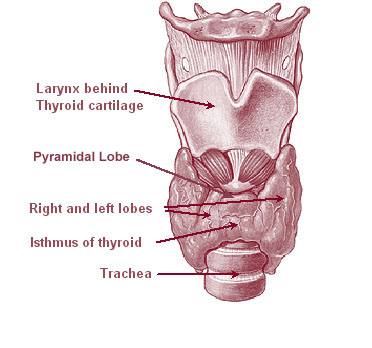
The thyroid, or thyroid gland, is an endocrine gland in vertebrates. In humans it is in the neck and consists of two connected lobes. The lower two thirds of the lobes are connected by a thin band of tissue called the thyroid isthmus. The thyroid is located at the front of the neck, below the Adam's apple. Microscopically, the functional unit of the thyroid gland is the spherical thyroid follicle, lined with follicular cells (thyrocytes), and occasional parafollicular cells that surround a lumen containing colloid. The thyroid gland secretes three hormones: the two thyroid hormones triiodothyronine (T3) and thyroxine (T4)and a peptide hormone, calcitonin. The thyroid hormones influence the metabolic rate and protein synthesis, and in children, growth and development. Calcitonin plays a role in calcium homeostasis. Secretion of the two thyroid hormones is regulated by thyroid-stimulating hormone (TSH), which is secreted from the anterior pituitary gland. TSH is regula ... [...More Info...] [...Related Items...] OR: [Wikipedia] [Google] [Baidu] |
Middle Cardiac Nerve
The middle cardiac nerve (''great cardiac nerve''), the largest of the three cardiac nerves, arises from the middle cervical ganglion, or from the trunk between the middle and inferior ganglia. On the right side it descends behind the common carotid artery, and at the root of the neck runs either in front of or behind the subclavian artery; it then descends on the trachea, receives a few filaments from the recurrent nerve, and joins the right half of the deep part of the cardiac plexus. In the neck, it communicates with the superior cardiac and recurrent nerves. On the left side, the middle cardiac nerve enters the chest between the left carotid and the left subclavian artery, and joins the left half of the deep part of the cardiac plexus. See also * Middle cervical ganglion The middle cervical ganglion is the smallest of the three cervical ganglia, and is occasionally absent. It is placed opposite the sixth cervical vertebra, usually in front of, or close to, the inferior t ... [...More Info...] [...Related Items...] OR: [Wikipedia] [Google] [Baidu] |
Cervical Ganglia
The cervical ganglia are paravertebral ganglia of the sympathetic nervous system. Preganglionic nerves from the thoracic spinal cord enter into the cervical ganglions and synapse with its postganglionic fibers or nerves. The cervical ganglion has three paravertebral ganglia: * superior cervical ganglion (largest) - adjacent to C2 & C3; postganglionic axon projects to target: (heart, head, neck) via "hitchhiking" on the carotid arteries * middle cervical ganglion (smallest) - adjacent to C6; target: heart, neck * inferior cervical ganglion. The inferior ganglion may be fused with the first thoracic ganglion to form a single structure, the stellate ganglion. - adjacent to C7; target: heart, lower neck, arm, posterior cranial arteries Nerves emerging from cervical sympathetic ganglia contribute to the cardiac plexus The cardiac plexus is a plexus of nerves situated at the base of the heart that innervates the heart. Structure The cardiac plexus is divided into a superficial p ... [...More Info...] [...Related Items...] OR: [Wikipedia] [Google] [Baidu] |
Cervical Vertebra
In tetrapods, cervical vertebrae (singular: vertebra) are the vertebrae of the neck, immediately below the skull. Truncal vertebrae (divided into thoracic and lumbar vertebrae in mammals) lie caudal (toward the tail) of cervical vertebrae. In sauropsid species, the cervical vertebrae bear cervical ribs. In lizards and saurischian dinosaurs, the cervical ribs are large; in birds, they are small and completely fused to the vertebrae. The vertebral transverse processes of mammals are homologous to the cervical ribs of other amniotes. Most mammals have seven cervical vertebrae, with the only three known exceptions being the manatee with six, the two-toed sloth with five or six, and the three-toed sloth with nine. In humans, cervical vertebrae are the smallest of the true vertebrae and can be readily distinguished from those of the thoracic or lumbar regions by the presence of a foramen (hole) in each transverse process, through which the vertebral artery, vertebral veins, and inferio ... [...More Info...] [...Related Items...] OR: [Wikipedia] [Google] [Baidu] |
Inferior Thyroid Artery
The inferior thyroid artery is an artery in the neck. It arises from the thyrocervical trunk and passes upward, in front of the vertebral artery and longus colli muscle. It then turns medially behind the carotid sheath and its contents, and also behind the sympathetic trunk, the middle cervical ganglion resting upon the vessel. Reaching the lower border of the thyroid gland it divides into two branches, which supply the postero-inferior parts of the gland, and anastomose with the superior thyroid artery, and with the corresponding artery of the opposite side. Structure The branches of the inferior thyroid artery are the inferior laryngeal, the oesophageal, the tracheal, the ascending cervical and the pharyngeal arteries. The inferior laryngeal artery climbs the trachea to the back part of the larynx under cover of the inferior pharyngeal constrictor muscle. It is accompanied by the recurrent nerve, and supplies the muscles and mucous membrane of this part, anastomosing with th ... [...More Info...] [...Related Items...] OR: [Wikipedia] [Google] [Baidu] |
Gray Rami Communicantes
Each spinal nerve receives a branch called a gray ramus communicans (plural rami communicantes) from the adjacent paravertebral ganglion of the sympathetic trunk. The gray rami communicantes contain postganglionic nerve fibers of the sympathetic nervous system and are composed of largely unmyelinated neurons. This is in contrast to the white rami communicantes, in which heavily myelinated neurons give the rami their white appearance. Function Preganglionic sympathetic fibres from the intermediolateral nucleus in the lateral grey column of the spinal cord are carried in the white ramus communicans to the paravertebral ganglia of the sympathetic trunk. Once the preganglionic nerve has traversed a white ramus communicans, it can do one of three things. * The preganglionic neuron can synapse with a postganglionic sympathetic neuron in the sympathetic paravertebral ganglion at that level. From here, the postganglionic sympathetic neuron can travel back out the grey ramus communicans of ... [...More Info...] [...Related Items...] OR: [Wikipedia] [Google] [Baidu] |
Ganglia
A ganglion is a group of neuron cell bodies in the peripheral nervous system. In the somatic nervous system this includes dorsal root ganglia and trigeminal ganglia among a few others. In the autonomic nervous system there are both sympathetic and parasympathetic ganglia which contain the cell bodies of postganglionic sympathetic and parasympathetic neurons respectively. A pseudoganglion looks like a ganglion, but only has nerve fibers and has no nerve cell bodies. Structure Ganglia are primarily made up of somata and dendritic structures which are bundled or connected. Ganglia often interconnect with other ganglia to form a complex system of ganglia known as a plexus. Ganglia provide relay points and intermediary connections between different neurological structures in the body, such as the peripheral and central nervous systems. Among vertebrates there are three major groups of ganglia: *Dorsal root ganglia (also known as the spinal ganglia) contain the cell bodies of se ... [...More Info...] [...Related Items...] OR: [Wikipedia] [Google] [Baidu] |
Cervical Nerves
A spinal nerve is a mixed nerve, which carries motor, sensory, and autonomic signals between the spinal cord and the body. In the human body there are 31 pairs of spinal nerves, one on each side of the vertebral column. These are grouped into the corresponding cervical, thoracic, lumbar, sacral and coccygeal regions of the spine. There are eight pairs of cervical nerves, twelve pairs of thoracic nerves, five pairs of lumbar nerves, five pairs of sacral nerves, and one pair of coccygeal nerves. The spinal nerves are part of the peripheral nervous system. Structure Each spinal nerve is a mixed nerve, formed from the combination of nerve fibers from its dorsal and ventral roots. The dorsal root is the afferent sensory root and carries sensory information to the brain. The ventral root is the efferent motor root and carries motor information from the brain. The spinal nerve emerges from the spinal column through an opening (intervertebral foramen) between adjacent vertebrae. Th ... [...More Info...] [...Related Items...] OR: [Wikipedia] [Google] [Baidu] |
Middle Cardiac Nerve
The middle cardiac nerve (''great cardiac nerve''), the largest of the three cardiac nerves, arises from the middle cervical ganglion, or from the trunk between the middle and inferior ganglia. On the right side it descends behind the common carotid artery, and at the root of the neck runs either in front of or behind the subclavian artery; it then descends on the trachea, receives a few filaments from the recurrent nerve, and joins the right half of the deep part of the cardiac plexus. In the neck, it communicates with the superior cardiac and recurrent nerves. On the left side, the middle cardiac nerve enters the chest between the left carotid and the left subclavian artery, and joins the left half of the deep part of the cardiac plexus. See also * Middle cervical ganglion The middle cervical ganglion is the smallest of the three cervical ganglia, and is occasionally absent. It is placed opposite the sixth cervical vertebra, usually in front of, or close to, the inferior t ... [...More Info...] [...Related Items...] OR: [Wikipedia] [Google] [Baidu] |


