|
Middle Cardiac Nerve
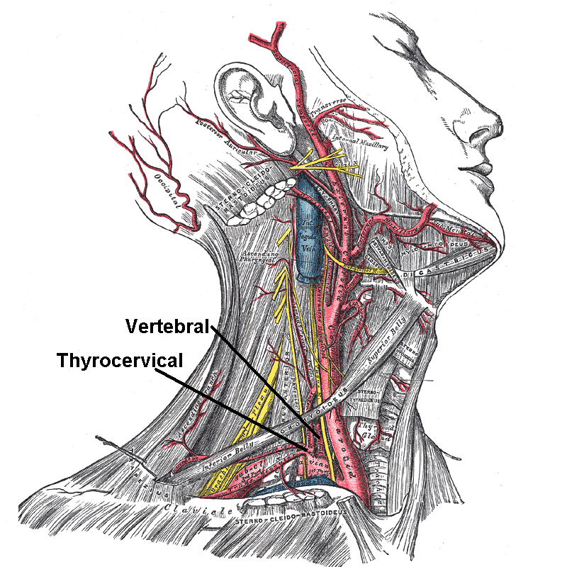
The middle cardiac nerve (''great cardiac nerve''), the largest of the three cardiac nerves, arises from the middle cervical ganglion, or from the trunk between the middle and inferior ganglia. On the right side it descends behind the common carotid artery, and at the root of the neck runs either in front of or behind the subclavian artery; it then descends on the trachea, receives a few filaments from the recurrent nerve, and joins the right half of the deep part of the cardiac plexus. In the neck, it communicates with the superior cardiac and recurrent nerves. On the left side, the middle cardiac nerve enters the chest between the left carotid and the left subclavian artery, and joins the left half of the deep part of the cardiac plexus. See also * Middle cervical ganglion The middle cervical ganglion is the smallest of the three cervical ganglia, and is occasionally absent. It is placed opposite the sixth cervical vertebra, usually in front of, or close to, the inferior t ... [...More Info...] [...Related Items...] OR: [Wikipedia] [Google] [Baidu] |
Middle Cervical Ganglion
The middle cervical ganglion is the smallest of the three cervical ganglia, and is occasionally absent. It is placed opposite the sixth cervical vertebra, usually in front of, or close to, the inferior thyroid artery. It sends gray rami communicantes to the fifth and sixth cervical nerves, and gives off the middle cardiac nerve. It is probably formed by the coalescence of two ganglia corresponding to the fifth and sixth cervical nerves. Branches # Gray Rami Communicantes to the anterior rami of the fifth and sixth cervical nerves. # Thyroid Branches which pass along the inferior thyroid artery to the thyroid gland. # The middle cardiac branch, which descends in the neck and ends in the cardiac plexus in the thorax See also * Middle cardiac nerve The middle cardiac nerve (''great cardiac nerve''), the largest of the three cardiac nerves, arises from the middle cervical ganglion, or from the trunk between the middle and inferior ganglia. On the right side it descends behind the ... [...More Info...] [...Related Items...] OR: [Wikipedia] [Google] [Baidu] |
Cardiac Nerves
The cardiac nerves are autonomic nerves which supply the heart. They include: * Superior cardiac nerve (nervus cardiacus cervicalis superior) * Middle cardiac nerve (nervus cardiacus cervicalis medius) * Inferior cardiac nerve The inferior cardiac nerve arises from either the inferior cervical or the first thoracic ganglion. It descends behind the subclavian artery and along the front of the trachea, to join the deep part of the cardiac plexus. It communicates free ... (nervus cardiacus inferior) Anatomy The nerves go down to the root of the neck with these following association: Posterior: "prevertebral fascia overlying anterolateral surface of vertebral bodies" Superior: "common carotid artery" Inferior: "subclavian artery" Laterally: "sympathetic trunk" References Cardiology {{Neuroanatomy-stub ... [...More Info...] [...Related Items...] OR: [Wikipedia] [Google] [Baidu] |
Common Carotid Artery
In anatomy, the left and right common carotid arteries (carotids) ( in Merriam-Webster Online Dictionary '.) are that supply the head and neck with ; they divide in the neck to form the and |
Subclavian Artery
In human anatomy, the subclavian arteries are paired major arteries of the upper thorax, below the clavicle. They receive blood from the aortic arch. The left subclavian artery supplies blood to the left arm and the right subclavian artery supplies blood to the right arm, with some branches supplying the head and thorax. On the left side of the body, the subclavian comes directly off the aortic arch, while on the right side it arises from the relatively short brachiocephalic artery when it bifurcates into the subclavian and the right common carotid artery. The usual branches of the subclavian on both sides of the body are the vertebral artery, the internal thoracic artery, the thyrocervical trunk, the costocervical trunk and the dorsal scapular artery, which may branch off the transverse cervical artery, which is a branch of the thyrocervical trunk. The subclavian becomes the axillary artery at the lateral border of the first rib. Structure From its origin, the subclavian artery t ... [...More Info...] [...Related Items...] OR: [Wikipedia] [Google] [Baidu] |
Recurrent Nerve
The recurrent laryngeal nerve (RLN) is a branch of the vagus nerve ( cranial nerve X) that supplies all the intrinsic muscles of the larynx, with the exception of the cricothyroid muscles. There are two recurrent laryngeal nerves, right and left. The right and left nerves are not symmetrical, with the left nerve looping under the aortic arch, and the right nerve looping under the right subclavian artery then traveling upwards. They both travel alongside the trachea. Additionally, the nerves are among the few nerves that follow a ''recurrent'' course, moving in the opposite direction to the nerve they branch from, a fact from which they gain their name. The recurrent laryngeal nerves supply sensation to the larynx below the vocal cords, give cardiac branches to the deep cardiac plexus, and branch to the trachea, esophagus and the inferior constrictor muscles. The posterior cricoarytenoid muscles, the only muscles that can open the vocal folds, are innervated by this nerve. ... [...More Info...] [...Related Items...] OR: [Wikipedia] [Google] [Baidu] |
Cardiac Plexus
The cardiac plexus is a plexus of nerves situated at the base of the heart that innervates the heart. Structure The cardiac plexus is divided into a superficial part, which lies in the concavity of the aortic arch, and a deep part, between the aortic arch and the trachea. The two parts are, however, closely connected. The sympathetic component of the cardiac plexus comes from cardiac nerves, which originate from the sympathetic trunk. The parasympathetic component of the cardiac plexus originates from the cardiac branches of the vagus nerve. Superficial part The superficial part of the cardiac plexus lies beneath the arch of the aorta, in front of the right pulmonary artery. It is formed by the superior cervical cardiac branch of the left sympathetic trunk and the inferior cardiac branch of the left vagus nerve. A small ganglion, the ''cardiac ganglion of Wrisberg'', is occasionally found connected with these nerves at their point of junction. This ganglion, when present, is si ... [...More Info...] [...Related Items...] OR: [Wikipedia] [Google] [Baidu] |
Carotid
In anatomy, the left and right common carotid arteries (carotids) ( in Merriam-Webster Online Dictionary '.) are that supply the head and neck with ; they divide in the neck to form the and |
Middle Cervical Ganglion
The middle cervical ganglion is the smallest of the three cervical ganglia, and is occasionally absent. It is placed opposite the sixth cervical vertebra, usually in front of, or close to, the inferior thyroid artery. It sends gray rami communicantes to the fifth and sixth cervical nerves, and gives off the middle cardiac nerve. It is probably formed by the coalescence of two ganglia corresponding to the fifth and sixth cervical nerves. Branches # Gray Rami Communicantes to the anterior rami of the fifth and sixth cervical nerves. # Thyroid Branches which pass along the inferior thyroid artery to the thyroid gland. # The middle cardiac branch, which descends in the neck and ends in the cardiac plexus in the thorax See also * Middle cardiac nerve The middle cardiac nerve (''great cardiac nerve''), the largest of the three cardiac nerves, arises from the middle cervical ganglion, or from the trunk between the middle and inferior ganglia. On the right side it descends behind the ... [...More Info...] [...Related Items...] OR: [Wikipedia] [Google] [Baidu] |


