|
Metachromasy
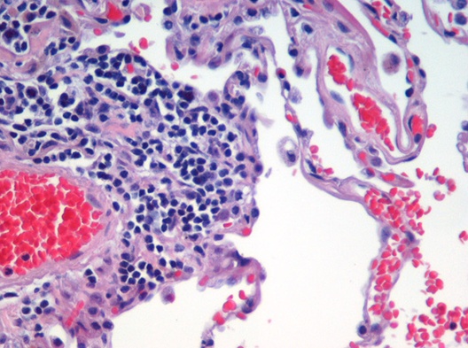
Metachromasia (var. metachromasy) is a characteristical change in the color of staining carried out in biological tissues, exhibited by certain dyes when they bind to particular substances present in these tissues, called chromotropes. For example, toluidine blue becomes dark blue (with a colour range from blue-red dependent on glycosaminoglycan content) when bound to cartilage. Other widely used metachromatic stains are the haematological Giemsa and May-Grunwald stains that also contain thiazine dyes. The white cell nucleus stains purple, basophil granules intense magenta, whilst the cytoplasms (of mononuclear cells) stains blue. The absence of color change in staining is named orthochromasia. The underlying mechanism for metachromasia requires the presence of polyanions within the tissue. When these tissues are stained with a concentrated basic dye solution, such as toluidine blue, the bound dye molecules are close enough to form dimeric and polymeric aggregates. The light a ... [...More Info...] [...Related Items...] OR: [Wikipedia] [Google] [Baidu] |
Lucien Lison
Lucien Alphonse Joseph Lison (1908–1984) was a Belgian/Brazilian physician and biomedical scientist, considered the "father of histochemistry".Ronan O’RahillyThree and one-half centuries of histology ''Irish Journal of Medical Science (1926-1967)'', 33(6): 288-292, June, 1958 Lison was born in Trazegnies, Belgium. He studied medicine at the Universite Libre de Bruxelles, graduating in 1931. Deciding for a career in experimental biological research, Lison started to work in histology, developing a number of new techniques for dyeing specific substances present in a slice of tissue. Before the advent of radiolabeling, this was the only group of techniques which could infer function based on biochemical activity and it represented a great promise not only for basic science, such as physiology and pharmacology, but for pathology and laboratory diagnosis of diseases, as well. He developed the Lison-Dunn stain, a technique using leuco patent blue V and hydrogen peroxidase ... [...More Info...] [...Related Items...] OR: [Wikipedia] [Google] [Baidu] |
Color
Color (American English) or colour (British English) is the visual perceptual property deriving from the spectrum of light interacting with the photoreceptor cells of the eyes. Color categories and physical specifications of color are associated with objects or materials based on their physical properties such as light absorption, reflection, or emission spectra. By defining a color space, colors can be identified numerically by their coordinates. Because perception of color stems from the varying spectral sensitivity of different types of cone cells in the retina to different parts of the spectrum, colors may be defined and quantified by the degree to which they stimulate these cells. These physical or physiological quantifications of color, however, do not fully explain the psychophysical perception of color appearance. Color science includes the perception of color by the eye and brain, the origin of color in materials, color theory in art, and the physics of ele ... [...More Info...] [...Related Items...] OR: [Wikipedia] [Google] [Baidu] |
Staining
Staining is a technique used to enhance contrast in samples, generally at the microscopic level. Stains and dyes are frequently used in histology (microscopic study of biological tissues), in cytology (microscopic study of cells), and in the medical fields of histopathology, hematology, and cytopathology that focus on the study and diagnoses of diseases at the microscopic level. Stains may be used to define biological tissues (highlighting, for example, muscle fibers or connective tissue), cell populations (classifying different blood cells), or organelles within individual cells. In biochemistry, it involves adding a class-specific ( DNA, proteins, lipids, carbohydrates) dye to a substrate to qualify or quantify the presence of a specific compound. Staining and fluorescent tagging can serve similar purposes. Biological staining is also used to mark cells in flow cytometry, and to flag proteins or nucleic acids in gel electrophoresis. Light microscopes are used for ... [...More Info...] [...Related Items...] OR: [Wikipedia] [Google] [Baidu] |
Tissue (biology)
In biology, tissue is a biological organizational level between cells and a complete organ. A tissue is an ensemble of similar cells and their extracellular matrix from the same origin that together carry out a specific function. Organs are then formed by the functional grouping together of multiple tissues. The English word "tissue" derives from the French word "tissu", the past participle of the verb tisser, "to weave". The study of tissues is known as histology or, in connection with disease, as histopathology. Xavier Bichat is considered as the "Father of Histology". Plant histology is studied in both plant anatomy and physiology. The classical tools for studying tissues are the paraffin block in which tissue is embedded and then sectioned, the histological stain, and the optical microscope. Developments in electron microscopy, immunofluorescence, and the use of frozen tissue-sections have enhanced the detail that can be observed in tissues. With these tools, th ... [...More Info...] [...Related Items...] OR: [Wikipedia] [Google] [Baidu] |
Toluidine Blue
Toluidine blue, also known as TBO or tolonium chloride (INN) is a blue cationic (basic) dye used in histology (as the toluidine blue stain) and sometimes clinically. Test for lignin Toluidine blue solution is used in testing for lignin, a complex organic molecule that bonds to cellulose fibres and strengthens and hardens the cell walls in plants. A positive toluidine blue test causes the solution to turn from blue to pink. A similar test can be performed with phloroglucinol- HCl solution, which turns red. Histological uses Toluidine blue is a basic thiazine metachromatic dye with high affinity for acidic tissue components. It stains nucleic acids blue and polysaccharides purple and also increases the sharpness of histology slide images. It is especially useful today for staining chromosomes in plant or animal tissues, as a replacement for Aceto-orcein stain. Toluidine blue is often used to identify mast cells, by virtue of the heparin in their cytoplasmic granules. It is ... [...More Info...] [...Related Items...] OR: [Wikipedia] [Google] [Baidu] |
Cartilage
Cartilage is a resilient and smooth type of connective tissue. In tetrapods, it covers and protects the ends of long bones at the joints as articular cartilage, and is a structural component of many body parts including the rib cage, the neck and the bronchial tubes, and the intervertebral discs. In other taxa, such as chondrichthyans, but also in cyclostomes, it may constitute a much greater proportion of the skeleton. It is not as hard and rigid as bone, but it is much stiffer and much less flexible than muscle. The matrix of cartilage is made up of glycosaminoglycans, proteoglycans, collagen fibers and, sometimes, elastin. Because of its rigidity, cartilage often serves the purpose of holding tubes open in the body. Examples include the rings of the trachea, such as the cricoid cartilage and carina. Cartilage is composed of specialized cells called chondrocytes that produce a large amount of collagenous extracellular matrix, abundant ground substance that is rich in p ... [...More Info...] [...Related Items...] OR: [Wikipedia] [Google] [Baidu] |
Orthochromasia
In chemistry, orthochromasia is the property of a dye or stain to not change color on binding to a target, as opposed to metachromatic stains, which change color. The word is derived from the Greek '' orthos'' (correct, upright), and chromatic (color). Toluidine blue is an example of a partially orthochromatic dye, as it stains nucleic acids by its orthochromatic color (blue), but stains mast cell granules in its metachromatic color (red). In spectral terms, orthochromasia refers to maintaining the position of spectral peaks, while metachromasia refers to a shift in wavelength, becoming either shorter or longer. In photography, an orthochromatic light spectrum is one devoid of red light. In biology, orthochromatic refers to the greyish staining because of acidophilic and basophilic mixture in the cell. Orthochromatic photography Orthochromatic photography refers to a photographic emulsion that is sensitive to only blue and green light, and thus can be processed with a ... [...More Info...] [...Related Items...] OR: [Wikipedia] [Google] [Baidu] |
Polyanions
Polyelectrolytes are polymers whose repeating units bear an electrolyte group. Polycations and polyanions are polyelectrolytes. These groups dissociate in aqueous solutions (water), making the polymers charged. Polyelectrolyte properties are thus similar to both electrolytes (salts) and polymers (high molecular weight compounds) and are sometimes called polysalts. Like salts, their solutions are electrically conductive. Like polymers, their solutions are often viscous. Charged molecular chains, commonly present in soft matter systems, play a fundamental role in determining structure, stability and the interactions of various molecular assemblies. Theoretical approaches to describing their statistical properties differ profoundly from those of their electrically neutral counterparts, while technological and industrial fields exploit their unique properties. Many biological molecules are polyelectrolytes. For instance, polypeptides, glycosaminoglycans, and DNA are polyelectrolyt ... [...More Info...] [...Related Items...] OR: [Wikipedia] [Google] [Baidu] |
Victor André Cornil
Victor André Cornil, also André-Victor Cornil (17 June 1837 – 13 April 1908) was a French pathologist, histologist and politician born in Cusset, Allier. Biography He studied medicine in Paris, earning his doctorate in 1864. In 1869 he became '' professeur agrègé'' to the Paris faculty, and in 1884 a member of the Académie Nationale de Médecine. Cornil was elected a member of the Royal Swedish Academy of Sciences in 1902. Cornil specialized in pathological anatomy, and made important contributions in the fields of bacteriology, histology and microscopic anatomy. In 1863 Cornil demonstrated histological evidence that supported Guillaume Duchenne's hypothesis regarding the cause of paralysis in poliomyelitis. With Austrian anatomist Richard Heschl (1824-1881) and Rudolph Jürgens of Berlin, he was among the first to use methyl violet as an histological stain for detection of amyloid. In 1864 he was the first physician to describe chronic childhood arthritis ... [...More Info...] [...Related Items...] OR: [Wikipedia] [Google] [Baidu] |
Louis-Antoine Ranvier
Louis-Antoine Ranvier (2 October 1835 – 22 March 1922) was a French physician, pathologist, anatomist and histologist, who discovered the nodes of Ranvier, regularly spaced discontinuities of the myelin sheath, occurring at varying intervals along the length of a nerve fiber. Career Ranvier was born and studied medicine at Lyon, graduating in 1865 from the Ecole Préparatoire de Médecine et de Pharmacie. He moved to Paris after receiving the internship of Parisian hospitals. Here he founded a small private research laboratory on Rue Christine along with fellow intern Victor André Cornil, and together they later offered a course in histology to medical students which involved the careful examination of tissues under a microscope. Their course was unique in the time as microscopy had not been viewed favourably in medicine especially by Henri Ducrotay de Blainville (1777-1850) and Auguste Comte (1798-1857). Their histology course material became an influential textbook on histop ... [...More Info...] [...Related Items...] OR: [Wikipedia] [Google] [Baidu] |
Paul Ehrlich
Paul Ehrlich (; 14 March 1854 – 20 August 1915) was a Nobel Prize-winning German physician and scientist who worked in the fields of hematology, immunology, and antimicrobial chemotherapy. Among his foremost achievements were finding a cure for syphilis in 1909 and inventing the precursor technique to Gram staining bacteria. The methods he developed for staining tissue made it possible to distinguish between different types of blood cells, which led to the ability to diagnose numerous blood diseases. His laboratory discovered arsphenamine (Salvarsan), the first effective medicinal treatment for syphilis, thereby initiating and also naming the concept of chemotherapy. Ehrlich popularized the concept of a magic bullet. He also made a decisive contribution to the development of an antiserum to combat diphtheria and conceived a method for standardizing therapeutic serums. In 1908, he received the Nobel Prize in Physiology or Medicine for his contributions to immunology. H ... [...More Info...] [...Related Items...] OR: [Wikipedia] [Google] [Baidu] |


.jpg)


