|
List Of Materials Analysis Methods
This is a list of analysis methods used in materials science. Analysis methods are listed by their acronym, if one exists. Symbols * μSR – see muon spin spectroscopy * χ – see magnetic susceptibility A * AAS – Atomic absorption spectroscopy * AED – Auger electron diffraction * AES – Auger electron spectroscopy * AFM – Atomic force microscopy * AFS – Atomic fluorescence spectroscopy * Analytical ultracentrifugation * APFIM – Atom probe field ion microscopy * APS – Appearance potential spectroscopy * ARPES – Angle resolved photoemission spectroscopy * ARUPS – Angle resolved ultraviolet photoemission spectroscopy * ATR – Attenuated total reflectance B * BET – BET surface area measurement (BET from Brunauer, Emmett, Teller) * BiFC – Bimolecular fluorescence complementation * BKD – Backscatter Kikuchi diffraction, see EBSD * BRET – Bioluminescence resonance energy transfer * BSED – Back scattered electron diffraction, see EBSD C * C ... [...More Info...] [...Related Items...] OR: [Wikipedia] [Google] [Baidu] |
Coaxial Impact Collision Ion Scattering Spectroscopy
In geometry, coaxial means that several three-dimensional linear or planar forms share a common axis. The two-dimensional analog is ''concentric''. Common examples: A coaxial cable is a three-dimensional linear structure. It has a wire conductor in the centre (D), a circumferential outer conductor (B), and an insulating medium called the dielectric (C) separating these two conductors. The outer conductor is usually sheathed in a protective PVC outer jacket (A). All these have a common axis. The dimension and material of the conductors and insulation determine the cable's characteristic impedance and attenuation at various frequencies. Coaxial rotors are a three-dimensional planar structure: a pair of helicopter rotors (wings) mounted one above the other on concentric shafts, with the same axis of rotation (but turning in opposite directions). In loudspeaker design, coaxial speakers A coaxial loudspeaker is a loudspeaker system in which the individual driver units radiate so ... [...More Info...] [...Related Items...] OR: [Wikipedia] [Google] [Baidu] |
Dielectric Thermal Analysis
Dielectric thermal analysis (DETA), or dielectric analysis (DEA), is a materials science technique similar to dynamic mechanical analysis except that an oscillating electrical field is used instead of a mechanical force. For investigation of the curing behavior of thermosetting resin systems, composite materials, adhesives and paints, Dielectric Analysis (DEA) can be used in accordance with ASTM E 2038 or E 2039. The great advantage of DEA is that it can be employed not only on a laboratory scale, but also in process. Measuring principle In a typical test, the sample is placed in contact with two electrodes (the dielectric sensor) and a sinusoidal voltage (the excitation) is applied to one electrode. The resulting sinusoidal current (the response) is measured at the second electrode. The response signal is attenuated in amplitude and shifted in phase in relation to the mobility of the ions and alignment of the dipoles. Dipoles in the material will attempt to align with the ele ... [...More Info...] [...Related Items...] OR: [Wikipedia] [Google] [Baidu] |
Cyclic Voltammetry
Cyclic voltammetry (CV) is a type of potentiodynamic electrochemical measurement. In a cyclic voltammetry experiment, the working electrode potential is ramped linearly versus time. Unlike in linear sweep voltammetry, after the set potential is reached in a CV experiment, the working electrode's potential is ramped in the opposite direction to return to the initial potential. These cycles of ramps in potential may be repeated as many times as needed. The current at the working electrode is plotted versus the applied voltage (that is, the working electrode's potential) to give the cyclic voltammogram trace. Cyclic voltammetry is generally used to study the electrochemical properties of an analyte in solution or of a molecule that is adsorbed onto the electrode. Experimental method In cyclic voltammetry (CV), the electrode potential ramps linearly versus time in cyclical phases (Figure 2). The rate of voltage change over time during each of these phases is known as the experim ... [...More Info...] [...Related Items...] OR: [Wikipedia] [Google] [Baidu] |
Cryo-scanning Electron Microscopy
Scanning electron cryomicroscopy (CryoSEM) is a form of electron microscopy where a hydrated but cryogenically fixed sample is imaged on a scanning electron microscope's cold stage in a cryogenic chamber. The cooling is usually achieved with liquid nitrogen. CryoSEM of biological samples with a high moisture content can be done faster with fewer sample preparation steps than conventional SEM. In addition, the dehydration processes needed to prepare a biological sample for a conventional SEM chamber create numerous distortions in the tissue leading to structural artifacts during imaging. See also * Electron microscopy * Electron cryomicroscopy * Transmission electron cryomicroscopy Transmission electron cryomicroscopy (CryoTEM), commonly known as cryo-EM, is a form of cryogenic electron microscopy, more specifically a type of transmission electron microscopy (TEM) where the sample is studied at cryogenic temperatures (genera ... References Electron microscopy Scientific tec ... [...More Info...] [...Related Items...] OR: [Wikipedia] [Google] [Baidu] |
Cryo-electron Microscopy
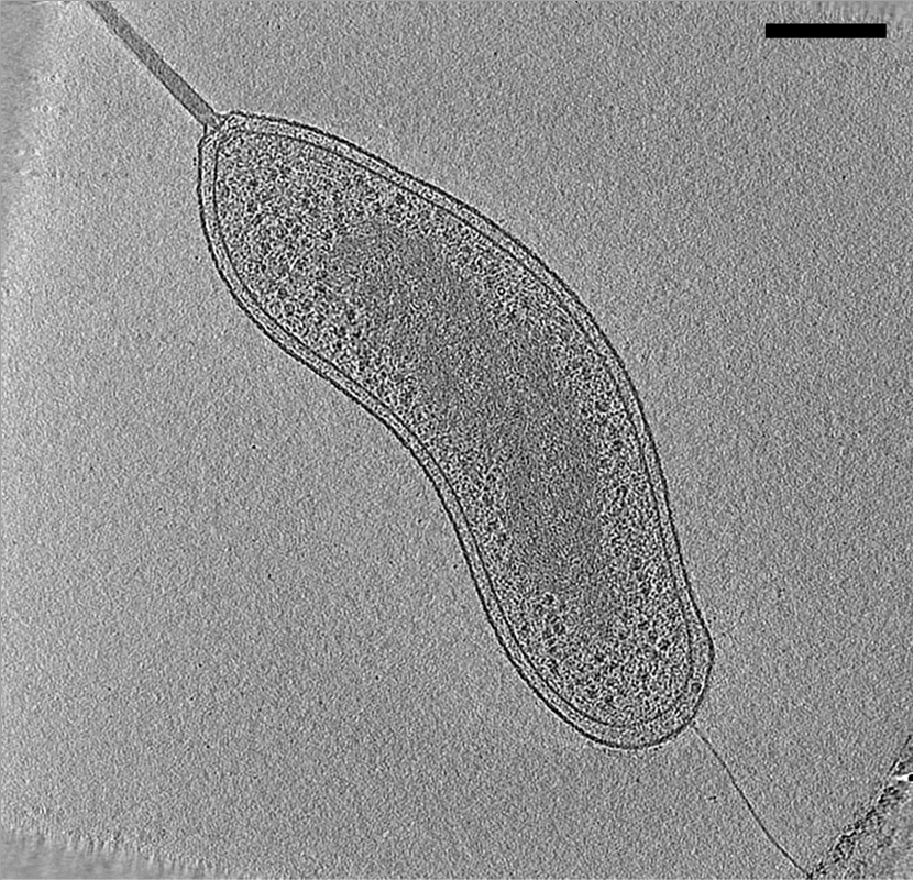
Cryogenic electron microscopy (cryo-EM) is a cryomicroscopy technique applied on samples cooled to cryogenic temperatures. For biological specimens, the structure is preserved by embedding in an environment of vitreous ice. An aqueous sample solution is applied to a grid-mesh and plunge-frozen in liquid ethane or a mixture of liquid ethane and propane. While development of the technique began in the 1970s, recent advances in detector technology and software algorithms have allowed for the determination of biomolecular structures at near-atomic resolution. This has attracted wide attention to the approach as an alternative to X-ray crystallography or NMR spectroscopy for macromolecular structure determination without the need for crystallization. In 2017, the Nobel Prize in Chemistry was awarded to Jacques Dubochet, Joachim Frank, and Richard Henderson "for developing cryo-electron microscopy for the high-resolution structure determination of biomolecules in solution." ''Nature ... [...More Info...] [...Related Items...] OR: [Wikipedia] [Google] [Baidu] |
Correlation Spectroscopy
Two-dimensional nuclear magnetic resonance spectroscopy (2D NMR) is a set of nuclear magnetic resonance spectroscopy (NMR) methods which give data plotted in a space defined by two frequency axes rather than one. Types of 2D NMR include correlation spectroscopy (COSY), J-spectroscopy, exchange spectroscopy (EXSY), and nuclear Overhauser effect spectroscopy (NOESY). Two-dimensional NMR spectra provide more information about a molecule than one-dimensional NMR spectra and are especially useful in determining the structure of a molecule, particularly for molecules that are too complicated to work with using one-dimensional NMR. The first two-dimensional experiment, COSY, was proposed by Jean Jeener, a professor at the Université Libre de Bruxelles, in 1971. This experiment was later implemented by Walter P. Aue, Enrico Bartholdi and Richard R. Ernst, who published their work in 1976. Fundamental concepts Each experiment consists of a sequence of radio frequency (RF) pulses with ... [...More Info...] [...Related Items...] OR: [Wikipedia] [Google] [Baidu] |
Confocal Laser Scanning Microscopy
Confocal microscopy, most frequently confocal laser scanning microscopy (CLSM) or laser confocal scanning microscopy (LCSM), is an optical imaging technique for increasing optical resolution and contrast of a micrograph by means of using a spatial pinhole to block out-of-focus light in image formation. Capturing multiple two-dimensional images at different depths in a sample enables the reconstruction of three-dimensional structures (a process known as optical sectioning) within an object. This technique is used extensively in the scientific and industrial communities and typical applications are in life sciences, semiconductor inspection and materials science. Light travels through the sample under a conventional microscope as far into the specimen as it can penetrate, while a confocal microscope only focuses a smaller beam of light at one narrow depth level at a time. The CLSM achieves a controlled and highly limited depth of field. Basic concept The principle of ... [...More Info...] [...Related Items...] OR: [Wikipedia] [Google] [Baidu] |
Cathodoluminescence
Cathodoluminescence is an optical and electromagnetic phenomenon in which electrons impacting on a luminescent material such as a phosphor, cause the emission of photons which may have wavelengths in the visible spectrum. A familiar example is the generation of light by an electron beam scanning the phosphor-coated inner surface of the screen of a television that uses a cathode ray tube. Cathodoluminescence is the inverse of the photoelectric effect, in which electron emission is induced by irradiation with photons. Origin Luminescence in a semiconductor results when an electron in the conduction band recombines with a hole in the valence band. The difference energy (band gap) of this transition can be emitted in form of a photon. The energy (color) of the photon, and the probability that a photon and not a phonon will be emitted, depends on the material, its purity, and the presence of defects. First, the electron has to be excited from the valence band into the conduction b ... [...More Info...] [...Related Items...] OR: [Wikipedia] [Google] [Baidu] |
Cryo-electron Tomography
Electron cryotomography (CryoET) is an imaging technique used to produce high-resolution (~1–4 nm) three-dimensional views of samples, often (but not limited to) biological macromolecules and cells. CryoET is a specialized application of transmission electron cryomicroscopy (CryoTEM) in which samples are imaged as they are tilted, resulting in a series of 2D images that can be combined to produce a 3D reconstruction, similar to a CT scan of the human body. In contrast to other electron tomography techniques, samples are imaged under cryogenic conditions (< −150 °C). For cellular material, the structure is immobilized in non-crystalline, vitreous ice, allowing them to be imaged without dehydration or chemical fixation, which would otherwise disrupt or distort biological structures. Description of technique [...More Info...] [...Related Items...] OR: [Wikipedia] [Google] [Baidu] |
Capillary Electrophoresis
Capillary electrophoresis (CE) is a family of electrokinetic separation methods performed in submillimeter diameter capillaries and in micro- and nanofluidic channels. Very often, CE refers to capillary zone electrophoresis (CZE), but other electrophoretic techniques including capillary gel electrophoresis (CGE), capillary isoelectric focusing (CIEF), capillary isotachophoresis and micellar electrokinetic chromatography (MEKC) belong also to this class of methods. In CE methods, analytes migrate through electrolyte solutions under the influence of an electric field. Analytes can be separated according to ionic mobility and/or partitioning into an alternate phase via non-covalent interactions. Additionally, analytes may be concentrated or "focused" by means of gradients in conductivity and pH. Instrumentation The instrumentation needed to perform capillary electrophoresis is relatively simple. A basic schematic of a capillary electrophoresis system is shown in ''figure ... [...More Info...] [...Related Items...] OR: [Wikipedia] [Google] [Baidu] |
Coherent Diffraction Imaging
Coherent diffractive imaging (CDI) is a "lensless" technique for 2D or 3D reconstruction of the image of nanoscale structures such as nanotubes, nanocrystals, porous nanocrystalline layers, defects, potentially proteins, and more. In CDI, a highly coherent beam of X-rays, electrons or other wavelike particle or photon is incident on an object. The beam scattered by the object produces a diffraction pattern downstream which is then collected by a detector. This recorded pattern is then used to reconstruct an image via an iterative feedback algorithm. Effectively, the objective lens in a typical microscope is replaced with software to convert from the reciprocal space diffraction pattern into a real space image. The advantage in using no lenses is that the final image is aberration–free and so resolution is only diffraction and dose limited (dependent on wavelength, aperture size and exposure). Applying a simple inverse Fourier transform to information with only intensities i ... [...More Info...] [...Related Items...] OR: [Wikipedia] [Google] [Baidu] |


.png)

