Cryo-electron Tomography on:
[Wikipedia]
[Google]
[Amazon]
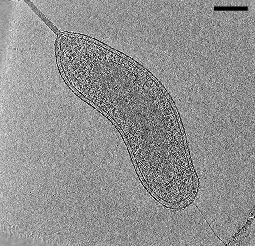
Electron cryotomography (CryoET) is an imaging technique used to produce high-resolution (~1–4 nm) three-dimensional views of samples, often (but not limited to) biological
 In
In
Getting started in cryo-EM course (Caltech)
Cell biology Electron microscopy techniques
macromolecule
A macromolecule is a very large molecule important to biophysical processes, such as a protein or nucleic acid. It is composed of thousands of covalently bonded atoms. Many macromolecules are polymers of smaller molecules called monomers. The ...
s and cells
Cell most often refers to:
* Cell (biology), the functional basic unit of life
Cell may also refer to:
Locations
* Monastic cell, a small room, hut, or cave in which a religious recluse lives, alternatively the small precursor of a monastery w ...
. CryoET is a specialized application of transmission electron cryomicroscopy (CryoTEM) in which samples are imaged as they are tilted, resulting in a series of 2D images that can be combined to produce a 3D reconstruction, similar to a CT scan
A computed tomography scan (CT scan; formerly called computed axial tomography scan or CAT scan) is a medical imaging technique used to obtain detailed internal images of the body. The personnel that perform CT scans are called radiographers ...
of the human body. In contrast to other electron tomography techniques, samples are imaged under cryogenic
In physics, cryogenics is the production and behaviour of materials at very low temperatures.
The 13th IIR International Congress of Refrigeration (held in Washington DC in 1971) endorsed a universal definition of “cryogenics” and “cr ...
conditions (< −150 °C). For cellular material, the structure is immobilized in non-crystalline, vitreous ice, allowing them to be imaged without dehydration or chemical fixation, which would otherwise disrupt or distort biological structures.
Description of technique
 In
In electron microscopy
An electron microscope is a microscope that uses a beam of accelerated electrons as a source of illumination. As the wavelength of an electron can be up to 100,000 times shorter than that of visible light photons, electron microscopes have a hi ...
(EM), samples are imaged in a high vacuum. Such a vacuum is incompatible with biological samples such as cells; the water would boil off, and the difference in pressure would explode the cell. In room-temperature EM techniques, samples are therefore prepared by fixation and dehydration. Another approach to stabilize biological samples, however, is to freeze them (electron cryomicroscopy
Cryogenic electron microscopy (cryo-EM) is a cryomicroscopy technique applied on samples cooled to cryogenic temperatures. For biological specimens, the structure is preserved by embedding in an environment of vitreous ice. An aqueous sample sol ...
). As in other electron cryomicroscopy techniques, samples for CryoET (typically small cells such as Bacteria, Archaea
Archaea ( ; singular archaeon ) is a domain of single-celled organisms. These microorganisms lack cell nuclei and are therefore prokaryotes. Archaea were initially classified as bacteria, receiving the name archaebacteria (in the Archaebac ...
, or viruses) are prepared in standard aqueous media and applied to an EM grid. The grid is then plunged into a cryogen (typically liquid ethane) so efficiently such that water molecules
A molecule is a group of two or more atoms held together by attractive forces known as chemical bonds; depending on context, the term may or may not include ions which satisfy this criterion. In quantum physics, organic chemistry, and bioche ...
do not have time to rearrange into a crystalline lattice. The resulting water state is called "vitreous ice" and preserves native cellular structures, such as lipid membranes, that would normally be destroyed by freezing. Plunge-frozen samples are subsequently stored and imaged at liquid-nitrogen
Liquid nitrogen—LN2—is nitrogen in a liquid state at low temperature. Liquid nitrogen has a boiling point of about . It is produced industrially by fractional distillation of liquid air. It is a colorless, low viscosity liquid that is wide ...
temperatures so that the water never warms enough to crystallize.
Samples are imaged in a transmission electron microscope (TEM). As in other electron tomography techniques, the sample is tilted to different angles relative to the electron beam (typically every 1 or 2 degrees from about −60° to +60°), and an image is acquired at each angle. This tilt-series of images can then be computationally reconstructed into a three-dimensional view of the object of interest. This is called a tomogram, or tomographic reconstruction.
Applications
In transmission electron microscopy (TEM), because electrons interact strongly with matter, resolution is limited by the thickness of the sample. Also, the thickness of the sample increases as the sample is tilted, and thicker samples can then completely block the electron beam, making the image dark or completely black. Therefore, for CryoET, samples should be less than ~500 nm thick to achieve "macromolecular" resolution (~4 nm). For this reason, most ECT studies have focused on purified macromolecular complexes, viruses, or small cells such as those of many species of Bacteria and Archaea. Cryotomography was used to understand encapsulation of 12 nm size protein cage nanoparticles inside 60 nm sized virus-like nanoparticles. Larger cells, and even tissues, can be prepared for CryoET by thinning, either by cryo-sectioning or by focused ion beam (FIB) milling. In cryo-sectioning, frozen blocks of cells or tissue are sectioned into thin samples with a cryo-microtome
A microtome (from the Greek ''mikros'', meaning "small", and ''temnein'', meaning "to cut") is a cutting tool used to produce extremely thin slices of material known as ''sections''. Important in science, microtomes are used in microscopy, allow ...
. In FIB-milling, plunge-frozen samples are exposed to a focused beam of ions, typically gallium, that precisely whittle away material from the top and bottom of a sample, leaving a thin lamella suitable for ECT imaging.
The strong interaction of electrons with matter also results in an anisotropic resolution effect. As the sample is tilted during imaging, the electron beam interacts with a relatively greater cross-sectional area at higher tilt angles. In practice, tilt angles greater than approximately 60–70° do not yield much information and are therefore not used. This results in a "missing wedge" of information in the final tomogram that decreases resolution parallel to the electron beam.
For structures that are present in multiple copies in one or multiple tomograms, higher resolution (even ≤1 nm) can be obtained by subtomogram averaging. Similar to single particle analysis, subtomogram averaging computationally combines images of identical objects to increase the signal-to-noise ratio
Signal-to-noise ratio (SNR or S/N) is a measure used in science and engineering that compares the level of a desired signal to the level of background noise. SNR is defined as the ratio of signal power to the noise power, often expressed in deci ...
.
A major obstacle in CryoET is identifying structures of interest within complicated cellular environments. One solution is to apply correlated cryo- fluorescence light microscopy, and even super-resolution light microscopy
Super-resolution microscopy is a series of techniques in optical microscopy that allow such images to have resolutions higher than those imposed by the diffraction limit, which is due to the diffraction of light. Super-resolution imaging tech ...
(e.g. cryo-PALM), and CryoET. In these techniques, a sample containing a fluorescently-tagged protein of interest is plunge-frozen and first imaged in a light microscope equipped with a special stage to allow the sample to be kept at sub-crystallization temperatures (< −150 °C). The location of the fluorescent signal is identified and the sample is transferred to the CryoTEM, where the same location is then imaged at high resolution by CryoET.
See also
*Electron microscopy
An electron microscope is a microscope that uses a beam of accelerated electrons as a source of illumination. As the wavelength of an electron can be up to 100,000 times shorter than that of visible light photons, electron microscopes have a hi ...
* Electron tomography
* Transmission electron cryomicroscopy
* Transmission electron microscopy
References
{{ReflistExternal links
Getting started in cryo-EM course (Caltech)
Cell biology Electron microscopy techniques