|
Laminitis
Laminitis is a disease that affects the feet of ungulates and is found mostly in horses and cattle. Clinical signs include foot tenderness progressing to inability to walk, increased digital pulses, and increased temperature in the hooves. Severe cases with outwardly visible clinical signs are known by the colloquial term ''founder'', and progression of the disease will lead to perforation of the coffin bone through the sole of the hoof or being unable to stand up, requiring euthanasia. Laminae The bones of the hoof are suspended within the axial hooves of ungulates by layers of modified skin cells, known as laminae or lamellae, which act as shock absorbers during locomotion. In horses, there are about 550–600 pairs of primary epidermal laminae, each with 150–200 secondary laminae projection from their surface. These interdigitate with equivalent structures on the surface of the coffin bone (PIII, P3, the third phalanx, pedal bone, or distal phalanx), known as dermal lamina ... [...More Info...] [...Related Items...] OR: [Wikipedia] [Google] [Baidu] |
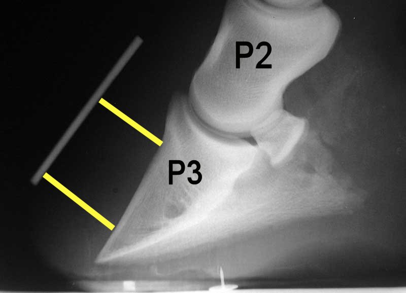
Laminitis Radiograph With Annotation
Laminitis is a disease that affects the feet of ungulates and is found mostly in horses and cattle. Clinical signs include foot tenderness progressing to inability to walk, increased digital pulses, and increased temperature in the hooves. Severe cases with outwardly visible clinical signs are known by the colloquial term ''founder'', and progression of the disease will lead to perforation of the coffin bone through the sole of the hoof or being unable to stand up, requiring euthanasia. Laminae The bones of the hoof are suspended within the axial hooves of ungulates by layers of modified skin cells, known as laminae or lamellae, which act as shock absorbers during locomotion. In horses, there are about 550–600 pairs of primary epidermal laminae, each with 150–200 secondary laminae projection from their surface. These interdigitate with equivalent structures on the surface of the coffin bone (PIII, P3, the third phalanx, pedal bone, or distal phalanx), known as dermal lamina ... [...More Info...] [...Related Items...] OR: [Wikipedia] [Google] [Baidu] |
Laminitis Embedding Hc Biovision
Laminitis is a disease that affects the feet of ungulates and is found mostly in horses and cattle. Clinical signs include foot tenderness progressing to inability to walk, increased digital pulses, and increased temperature in the hooves. Severe cases with outwardly visible clinical signs are known by the colloquial term ''founder'', and progression of the disease will lead to perforation of the coffin bone through the sole of the hoof or being unable to stand up, requiring euthanasia. Laminae The bones of the hoof are suspended within the axial hooves of ungulates by layers of modified skin cells, known as laminae or lamellae, which act as shock absorbers during locomotion. In horses, there are about 550–600 pairs of primary epidermal laminae, each with 150–200 secondary laminae projection from their surface. These interdigitate with equivalent structures on the surface of the coffin bone (PIII, P3, the third phalanx, pedal bone, or distal phalanx), known as dermal lamina ... [...More Info...] [...Related Items...] OR: [Wikipedia] [Google] [Baidu] |
Equine Metabolic Syndrome
Equine metabolic syndrome (EMS) is an endocrinopathy affecting horses and ponies. It is of primary concern due to its link to obesity, insulin dysregulation, and subsequent laminitis. There are some similarities in clinical signs between EMS and pituitary pars intermedia dysfunction, also known as PPID or Cushing's disease, and some equines may develop both, but they are not the same condition, having different causes and different treatment. Pathogenesis The cells of adipose (fat) tissue synthesizes hormones known as adipokines. In humans, dysfunction of adipose tissue, even in cases without obesity, has been associated with the development of insulin resistance, hypertension, systemic inflammation, and increased risk of blood clots (thrombosis). The inflammation produced by these hormones are thought to inflame adipose tissue, leading to the production of more adipokines and perpetuation of the cycle, and a constant low-level, pro-inflammatory state. Although it is suspected th ... [...More Info...] [...Related Items...] OR: [Wikipedia] [Google] [Baidu] |
Horse Hoof
A horse hoof is the lower extremity of each leg of a horse, the part that makes contact with the ground and carries the weight of the animal. It is both hard and flexible. It is a complex structure surrounding the distal phalanx of the 3rd digit (digit III of the basic pentadactyl limb of vertebrates, evolved into a single weight-bearing digit in horses) of each of the four limbs, which is covered by soft tissue and keratinised (cornified) matter. Anatomy The hoof is made up of two parts. The outer part, called the hoof capsule, is composed of various cornified specialized structures. The inner, living part of the hoof, is made up of soft tissues and bone. The cornified material of the hoof capsule differ in structure and properties. Dorsally, it covers, protects, and supports P3 (also known as the coffin bone, pedal bone, or PIII). Palmarly/plantarly, it covers and protects specialised soft tissues, such as tendons, ligaments, fibro-fatty and/or fibrocartilaginous tissues, an ... [...More Info...] [...Related Items...] OR: [Wikipedia] [Google] [Baidu] |
Pituitary Pars Intermedia Dysfunction
Pituitary pars intermedia dysfunction (PPID), or equine Cushing's disease, is an endocrine disease affecting the pituitary gland of horses. It is most commonly seen in older animals, and is classically associated with the formation of a long, wavy coat (hirsutism) and chronic laminitis. Pathophysiology Unlike the human and canine forms of Cushing's disease, which most commonly affect the ''pars distalis'' region of the pituitary gland, equine Cushing's disease is a result of hyperplasia or adenoma formation in the pars intermedia. This adenoma then secretes excessive amounts of normal products, leading to clinical signs. Dopaminergic control of the pars intermedia The pituitary gland consists of three parts: the ''pars nervosa'', the ''pars intermedia'', and the ''pars distalis''. The most critical structure to PPID, the ''pars intermedia'', is regulated by the hypothalamus. The neurons of the hypothalamus innervate cells known as melanotropes within the ''pars intermedia'', relea ... [...More Info...] [...Related Items...] OR: [Wikipedia] [Google] [Baidu] |
Black Walnut
''Juglans nigra'', the eastern American black walnut, is a species of deciduous tree in the walnut family, Juglandaceae, native to North America. It grows mostly in riparian zones, from southern Ontario, west to southeast South Dakota, south to Georgia, northern Florida and southwest to central Texas. Wild trees in the upper Ottawa Valley may be an isolated native population or may have derived from planted trees. Black walnut is an important tree commercially, as the wood is a deep brown color and easily worked. Walnut seeds ( nuts) are cultivated for their distinctive and desirable taste. Walnut trees are grown both for lumber and food, and many cultivars have been developed for improved quality wood or nuts. Black walnut is susceptible to thousand cankers disease, which provoked a decline of walnut trees in some regions. Black walnut is anecdotally known for being allelopathic, which means that it releases chemicals from its roots and other tissues that may harm other orga ... [...More Info...] [...Related Items...] OR: [Wikipedia] [Google] [Baidu] |
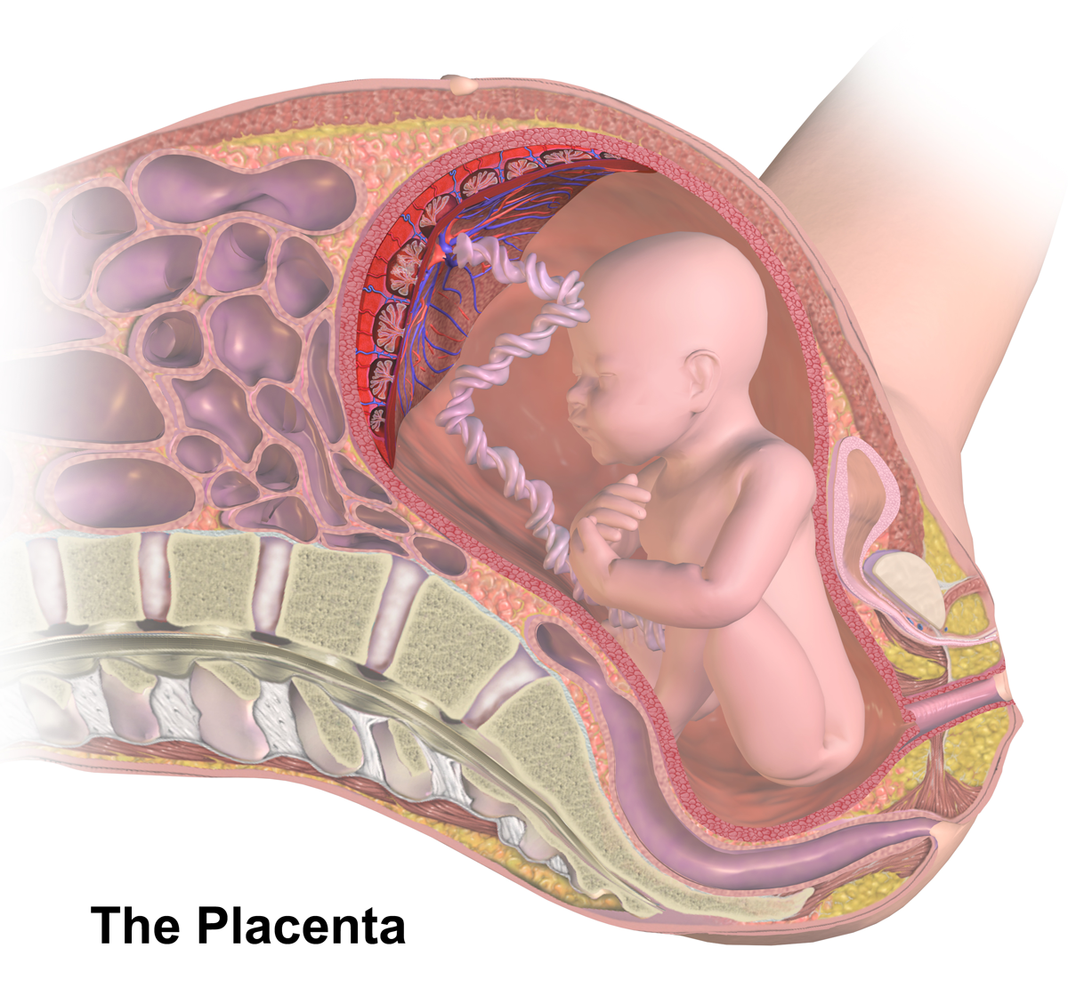
Placenta
The placenta is a temporary embryonic and later fetal organ that begins developing from the blastocyst shortly after implantation. It plays critical roles in facilitating nutrient, gas and waste exchange between the physically separate maternal and fetal circulations, and is an important endocrine organ, producing hormones that regulate both maternal and fetal physiology during pregnancy. The placenta connects to the fetus via the umbilical cord, and on the opposite aspect to the maternal uterus in a species-dependent manner. In humans, a thin layer of maternal decidual (endometrial) tissue comes away with the placenta when it is expelled from the uterus following birth (sometimes incorrectly referred to as the 'maternal part' of the placenta). Placentas are a defining characteristic of placental mammals, but are also found in marsupials and some non-mammals with varying levels of development. Mammalian placentas probably first evolved about 150 million to 200 million years ... [...More Info...] [...Related Items...] OR: [Wikipedia] [Google] [Baidu] |
Tissue Inhibitor Of Metalloproteinase
Tissue inhibitors of metalloproteinases (TIMPs) are specific endogenous protease inhibitors to the matrix metalloproteinases. There are four TIMPs; ''TIMP1'', ''TIMP2'', ''TIMP3'' and ''TIMP4''. TIMP3 has been observed progressively downregulated in Human papillomavirus-positive neoplastic keratinocytes derived from uterine cervical preneoplastic lesions at different levels of malignancy. For this reason, TIMP3 is likely to be associated with tumorigenesis and may be a potential prognostic marker for uterine cervical preneoplastic lesions progression. Overall, all MMPs are inhibited by TIMPs once they are activated but the gelatinases (MMP-2 and MMP-9 Matrix metallopeptidase 9 (MMP-9), also known as 92 kDa type IV collagenase, 92 kDa gelatinase or gelatinase B (GELB), is a matrixin, a class of enzymes that belong to the zinc-metalloproteinases family involved in the degradation of the extracel ...) can form complexes with TIMPs when the enzymes are in the latent form. Th ... [...More Info...] [...Related Items...] OR: [Wikipedia] [Google] [Baidu] |
Metalloproteinase
A metalloproteinase, or metalloprotease, is any protease enzyme whose catalytic mechanism involves a metal. An example is ADAM12 which plays a significant role in the fusion of muscle cells during embryo development, in a process known as myogenesis. Most metalloproteases require zinc, but some use cobalt. The metal ion is coordinated to the protein via three ligands. The ligands coordinating the metal ion can vary with histidine, glutamate, aspartate, lysine, and arginine. The fourth coordination position is taken up by a labile water molecule. Treatment with chelating agents such as EDTA leads to complete inactivation. EDTA is a metal chelator that removes zinc, which is essential for activity. They are also inhibited by the chelator orthophenanthroline. Classification There are two subgroups of metalloproteinases: * Exopeptidases, metalloexopeptidases ( EC number: 3.4.17). * Endopeptidases, metalloendopeptidases (3.4.24). Well known metalloendopeptidases include ADAM pr ... [...More Info...] [...Related Items...] OR: [Wikipedia] [Google] [Baidu] |
Matrix Metalloproteinase Matrix metalloproteinases (MMPs), also known as matrix metallopeptidases or matrixins, are metalloproteinases that are calcium-dependent zinc-containing endopeptidases; other family members are adamalysins, serralysins, and astacins. The MMPs belong to a larger family of proteases known as the metzincin superfamily. Collectively, these enzymes are capable of degrading all kinds of extracellular matrix proteins, but also can process a number of bioactive molecules. They are known to be involved in the cleavage of cell surface receptors, the release of apoptotic ligands (such as the FAS ligand), and chemokine/cytokine inactivation. MMPs are also thought to play a major role in cell behaviors such as cell proliferation, migration (adhesion/dispersion), differentiation, angiogenesis, apoptosis, and host defense. They were first described in vertebrates (1962), including humans, but have since been found in invertebrates and plants. They are distinguished from other endopeptida ... [...More Info...] [...Related Items...] OR: [Wikipedia] [Google] |


