|
LBBB
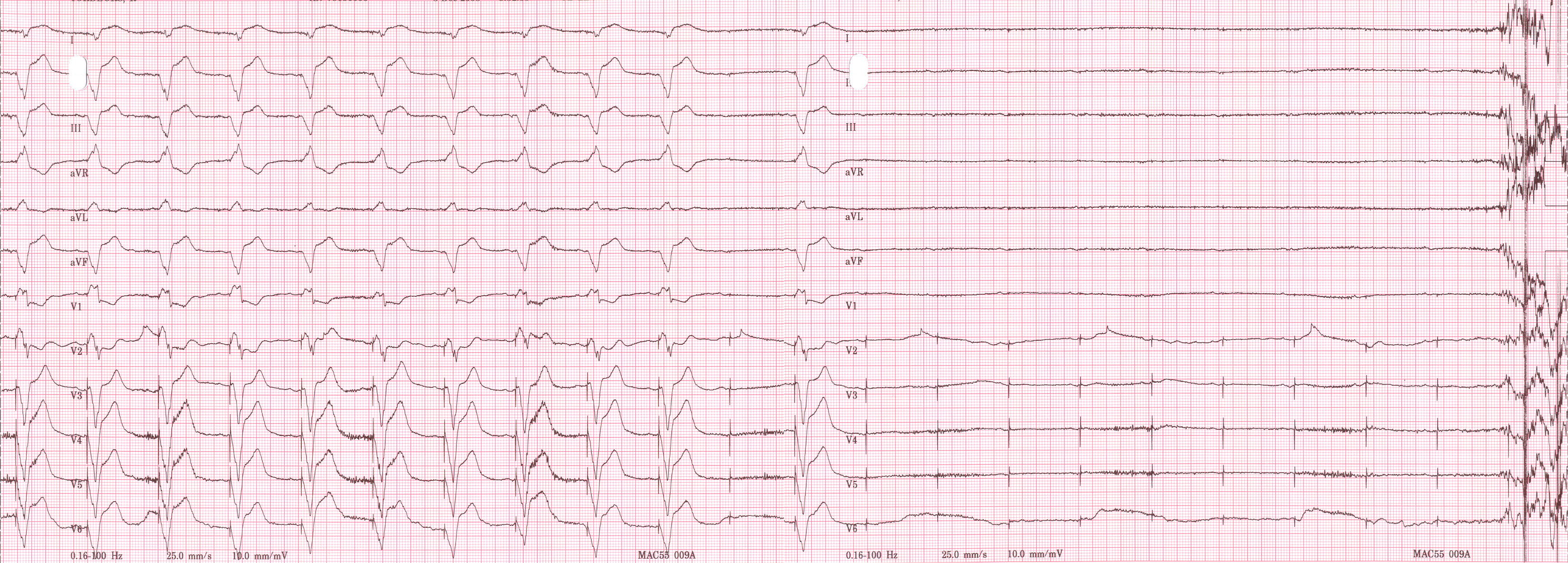
Left bundle branch block (LBBB) is a conduction abnormality in the heart that can be seen on an electrocardiogram (ECG). In this condition, activation of the left ventricle of the heart is delayed, which causes the left ventricle to contract later than the right ventricle. Causes Among the causes of LBBB are: * Aortic stenosis * Dilated cardiomyopathy * Acute myocardial infarction * Extensive coronary artery disease * Primary disease of the cardiac electrical conduction system * Long standing hypertension leading to aortic root dilatation and subsequent aortic regurgitation * Lyme disease Mechanisms Slow or absent conduction through the left bundle branch means that it takes longer than normal for the left ventricle to fully depolarise. This can be due to a damaged bundle branch that is completely unable to conduct, but may represent intact conduction that is slower than normal. LBBB may be fixed, present at all times, but may be intermittent for example occurring on ... [...More Info...] [...Related Items...] OR: [Wikipedia] [Google] [Baidu] |
LBBB2009
Left bundle branch block (LBBB) is a conduction abnormality in the heart that can be seen on an electrocardiogram (ECG). In this condition, activation of the left ventricle of the heart is delayed, which causes the left ventricle to contract later than the right ventricle. Causes Among the causes of LBBB are: * Aortic stenosis * Dilated cardiomyopathy * Acute myocardial infarction * Extensive coronary artery disease * Primary disease of the cardiac electrical conduction system * Long standing hypertension leading to aortic root dilatation and subsequent aortic regurgitation * Lyme disease Mechanisms Slow or absent conduction through the left bundle branch means that it takes longer than normal for the left ventricle to fully depolarise. This can be due to a damaged bundle branch that is completely unable to conduct, but may represent intact conduction that is slower than normal. LBBB may be fixed, present at all times, but may be intermittent for example occurring only durin ... [...More Info...] [...Related Items...] OR: [Wikipedia] [Google] [Baidu] |
Electrocardiography
Electrocardiography is the process of producing an electrocardiogram (ECG or EKG), a recording of the heart's electrical activity. It is an electrogram of the heart which is a graph of voltage versus time of the electrical activity of the heart using electrodes placed on the skin. These electrodes detect the small electrical changes that are a consequence of cardiac muscle depolarization followed by repolarization during each cardiac cycle (heartbeat). Changes in the normal ECG pattern occur in numerous cardiac abnormalities, including cardiac rhythm disturbances (such as atrial fibrillation and ventricular tachycardia), inadequate coronary artery blood flow (such as myocardial ischemia and myocardial infarction), and electrolyte disturbances (such as hypokalemia and hyperkalemia). Traditionally, "ECG" usually means a 12-lead ECG taken while lying down as discussed below. However, other devices can record the electrical activity of the heart such as a Holter monitor but also s ... [...More Info...] [...Related Items...] OR: [Wikipedia] [Google] [Baidu] |
Bundle Branch Block
A bundle branch block is a defect in one the bundle branches in the electrical conduction system of the heart. Anatomy and physiology The heart's electrical activity begins in the sinoatrial node (the heart's natural pacemaker), which is situated on the upper right atrium. The impulse travels next through the left and right atria and summates at the atrioventricular node. From the AV node the electrical impulse travels down the bundle of His and divides into the right and left bundle branches. The right bundle branch contains one fascicle. The left bundle branch subdivides into two fascicles: the left anterior fascicle, and the left posterior fascicle. Other sources divide the left bundle branch into three fascicles: the left anterior, the left posterior, and the left septal fascicle. The thicker left posterior fascicle bifurcates, with one fascicle being in the septal aspect. Ultimately, the fascicles divide into millions of Purkinje fibres, which in turn interdigitate with ind ... [...More Info...] [...Related Items...] OR: [Wikipedia] [Google] [Baidu] |
Dilated Cardiomyopathy
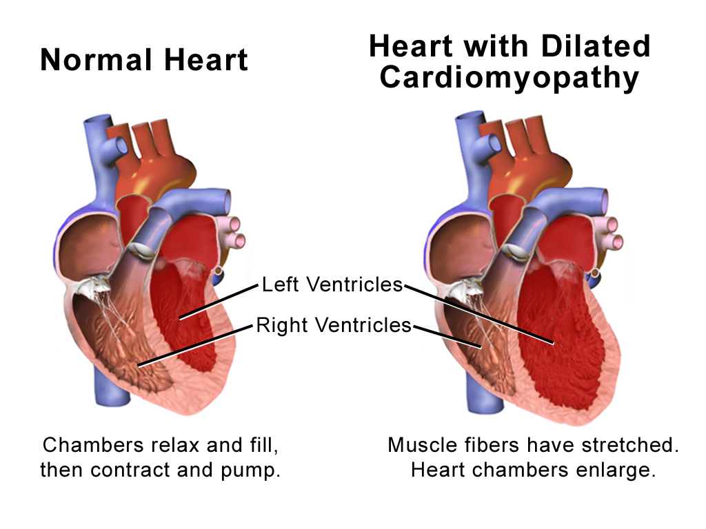
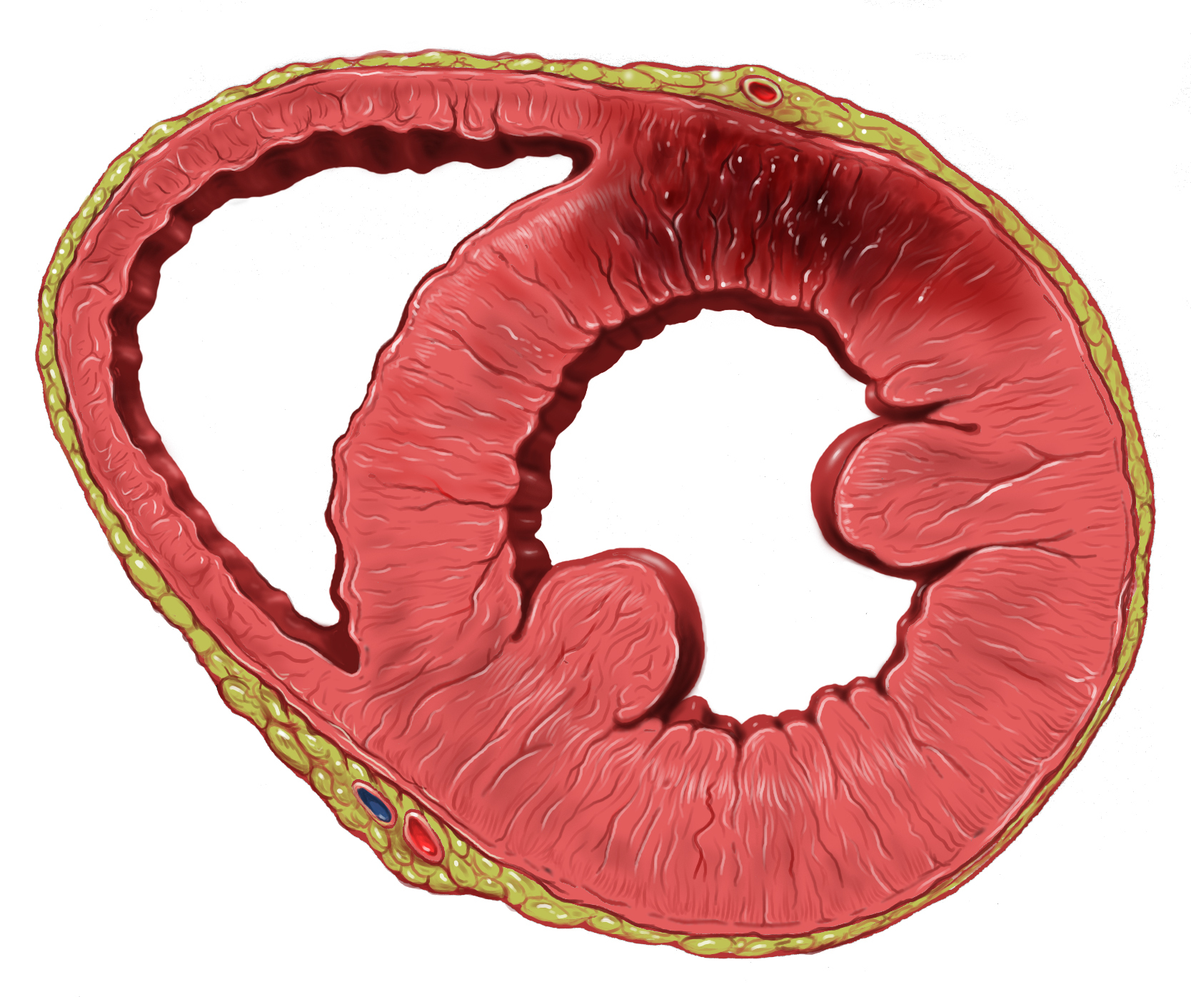
Dilated cardiomyopathy (DCM) is a condition in which the heart becomes enlarged and cannot pump blood effectively. Symptoms vary from none to feeling tired, leg swelling, and shortness of breath. It may also result in chest pain or fainting. Complications can include heart failure, heart valve disease, or an irregular heartbeat. Causes include genetics, alcohol, cocaine, certain toxins, complications of pregnancy, and certain infections. Coronary artery disease and high blood pressure may play a role, but are not the primary cause. In many cases the cause remains unclear. It is a type of cardiomyopathy, a group of diseases that primarily affects the heart muscle. The diagnosis may be supported by an electrocardiogram, chest X-ray, or echocardiogram. In those with heart failure, treatment may include medications in the ACE inhibitor, beta blocker, and diuretic families. A low salt diet may also be helpful. In those with certain types of irregular heartbeat, blood thinners or ... [...More Info...] [...Related Items...] OR: [Wikipedia] [Google] [Baidu] |
Left Anterior Fascicular Block
Left anterior fascicular block (LAFB) is an abnormal condition of the left ventricle of the heart, related to, but distinguished from, left bundle branch block (LBBB). It is caused by only the left anterior fascicle – one half of the left bundle branch being defective. It is manifested on the ECG by left axis deviation. It is much more common than left posterior fascicular block. Mechanism Normal activation of the left ventricle (LV) proceeds down the left bundle branch, which consist of three fascicles, the left anterior fascicle, the left posterior fascicle, and the septal fascicle. The posterior fascicle supplies the posterior and inferoposterior walls of the LV, the anterior fascicle supplies the upper and anterior parts of the LV and the septal fascicle supplies the septal wall with innervation. LAFB — which is also known as left anterior hemiblock (LAHB) — occurs when a cardiac impulse spreads first through the left posterior fascicle, causing a delay in activation of ... [...More Info...] [...Related Items...] OR: [Wikipedia] [Google] [Baidu] |
QRS Complex
The QRS complex is the combination of three of the graphical deflections seen on a typical electrocardiogram (ECG or EKG). It is usually the central and most visually obvious part of the tracing. It corresponds to the depolarization of the right and left ventricles of the heart and contraction of the large ventricular muscles. In adults, the QRS complex normally lasts ; in children it may be shorter. The Q, R, and S waves occur in rapid succession, do not all appear in all leads, and reflect a single event and thus are usually considered together. A Q wave is any downward deflection immediately following the P wave. An R wave follows as an upward deflection, and the S wave is any downward deflection after the R wave. The T wave follows the S wave, and in some cases, an additional U wave follows the T wave. To measure the QRS interval start at the end of the PR interval (or beginning of the Q wave) to the end of the S wave. Normally this interval is 0.08 to 0.10 seconds. When ... [...More Info...] [...Related Items...] OR: [Wikipedia] [Google] [Baidu] |
Artificial Cardiac Pacemaker
An artificial cardiac pacemaker (or artificial pacemaker, so as not to be confused with the natural cardiac pacemaker) or pacemaker is a medical device that generates electrical impulses delivered by electrodes to the chambers of the heart either the upper atria, or lower ventricles to cause the targeted chambers to contract and pump blood. By doing so, the pacemaker regulates the function of the electrical conduction system of the heart. The primary purpose of a pacemaker is to maintain an adequate heart rate, either because the heart's natural pacemaker is not fast enough, or because there is a block in the heart's electrical conduction system. Modern pacemakers are externally programmable and allow a cardiologist, particularly a cardiac electrophysiologist, to select the optimal pacing modes for individual patients. Most pacemakers are on demand, in which the stimulation of the heart is based on the dynamic demand of the circulatory system. Others send out a fixed rate ... [...More Info...] [...Related Items...] OR: [Wikipedia] [Google] [Baidu] |
Pacemaker
An artificial cardiac pacemaker (or artificial pacemaker, so as not to be confused with the natural cardiac pacemaker) or pacemaker is a medical device that generates electrical impulses delivered by electrodes to the chambers of the heart either the upper atria, or lower ventricles to cause the targeted chambers to contract and pump blood. By doing so, the pacemaker regulates the function of the electrical conduction system of the heart. The primary purpose of a pacemaker is to maintain an adequate heart rate, either because the heart's natural pacemaker is not fast enough, or because there is a block in the heart's electrical conduction system. Modern pacemakers are externally programmable and allow a cardiologist, particularly a cardiac electrophysiologist, to select the optimal pacing modes for individual patients. Most pacemakers are on demand, in which the stimulation of the heart is based on the dynamic demand of the circulatory system. Others send out a fixed rate of ... [...More Info...] [...Related Items...] OR: [Wikipedia] [Google] [Baidu] |
Syncope (medicine)
Syncope, commonly known as fainting, or passing out, is a loss of consciousness and muscle strength characterized by a fast onset, short duration, and spontaneous recovery. It is caused by a decrease in blood flow to the brain, typically from low blood pressure. There are sometimes symptoms before the loss of consciousness such as lightheadedness, sweating, pale skin, blurred vision, nausea, vomiting, or feeling warm. Syncope may also be associated with a short episode of muscle twitching. Psychiatric causes can also be determined when a patient experiences fear, anxiety, or panic; particularly before a stressful event usually medical in nature. When consciousness and muscle strength are not completely lost, it is called presyncope. It is recommended that presyncope be treated the same as syncope. Causes range from non-serious to potentially fatal. There are three broad categories of causes: heart or blood vessel related; reflex, also known as neurally mediated; and orthos ... [...More Info...] [...Related Items...] OR: [Wikipedia] [Google] [Baidu] |
Southern Illinois University School Of Medicine
Southern Illinois University School of Medicine is a medical school located in Springfield, the capital of the U.S. state of Illinois. It is part of the Southern Illinois University system, which includes a campus in Edwardsville as well as the flagship in Carbondale. The medical school was founded in 1970 and achieved full accreditation in 1972. It was founded to relieve a chronic shortage of physicians in downstate Illinois. Notability SIU was once the only medical school in Illinois with its main campus outside of Chicago or its suburbs until the Carle Illinois College of Medicine was formed in 2018 (although the University of Illinois at Chicago does maintain satellites in Peoria and Rockford). SIU was early to incorporate problem-based learning (PBL) into their curricula (see below) and "standardized patients" for medical student testing purposes. Being able to interact with standardized patients now comprises the majority of one of the four major exams that all US ... [...More Info...] [...Related Items...] OR: [Wikipedia] [Google] [Baidu] |
Myocardial Infarction
A myocardial infarction (MI), commonly known as a heart attack, occurs when blood flow decreases or stops to the coronary artery of the heart, causing damage to the heart muscle. The most common symptom is chest pain or discomfort which may travel into the shoulder, arm, back, neck or jaw. Often it occurs in the center or left side of the chest and lasts for more than a few minutes. The discomfort may occasionally feel like heartburn. Other symptoms may include shortness of breath, nausea, feeling faint, a cold sweat or feeling tired. About 30% of people have atypical symptoms. Women more often present without chest pain and instead have neck pain, arm pain or feel tired. Among those over 75 years old, about 5% have had an MI with little or no history of symptoms. An MI may cause heart failure, an irregular heartbeat, cardiogenic shock or cardiac arrest. Most MIs occur due to coronary artery disease. Risk factors include high blood pressure, smoking, diabetes, ... [...More Info...] [...Related Items...] OR: [Wikipedia] [Google] [Baidu] |


