|
Khdrbs3
KH domain-containing, RNA-binding, signal transduction-associated protein 3 is a protein that in humans is encoded by the ''KHDRBS3'' gene. Interactions KHDRBS3 has been shown to interact with SIAH1. KHDRBS3 interacts with splicing protein Sam68 and oncogene metadherin in prostate cancer cells. Clinical significance KHDRBS3 (T-STAR) expression has been shown to be increased in prostate cancer tissue compared to the surrounding benign tissue. Expression of KHDRBS3 correlates with mpMRI signal measured through Likert score a system similar to PI-RADS. While still under debate, mpMRI signal correlates with higher Gleason grade and tumour size, in addition to histopathological features associated with clinically aggressive prostate cancer. Expression of KHDRBS3 was increased in the failing human myocardium of heart failure patients, here KHDRBS3 protein interacted with several important mRNAs coding for sarcomere components, such as actin gamma 1 (''ACTG1''), myosin light ch ... [...More Info...] [...Related Items...] OR: [Wikipedia] [Google] [Baidu] |
Titin
Titin (contraction for Titan protein) (also called connectin) is a protein that in humans is encoded by the ''TTN'' gene. Titin is a giant protein, greater than 1 µm in length, that functions as a molecular spring that is responsible for the passive elasticity of muscle. It comprises 244 individually folded protein domains connected by unstructured peptide sequences. These domains unfold when the protein is stretched and refold when the tension is removed. Titin is important in the contraction of striated muscle tissues. It connects the Z line to the M line in the sarcomere. The protein contributes to force transmission at the Z line and resting tension in the I band region. It limits the range of motion of the sarcomere in tension, thus contributing to the passive stiffness of muscle. Variations in the sequence of titin between different types of striated muscle (cardiac or skeletal) have been correlated with differences in the mechanical properties of these muscles. ... [...More Info...] [...Related Items...] OR: [Wikipedia] [Google] [Baidu] |
SIAH1
E3 ubiquitin-protein ligase SIAH1 is an enzyme that in humans is encoded by the ''SIAH1'' gene. Function This gene encodes for a polypeptide structure that is a member of the seven in absentia homolog (SIAH) family. The protein is an E3 ligase and is involved in ubiquitination and proteasome-mediated degradation of specific proteins. The activity of this ubiquitin ligase has been implicated in the development of certain forms of Parkinson's disease, the regulation of the cellular response to hypoxia and induction of apoptosis. Alternative splicing results in several additional transcript variants, some encoding different isoforms and others that have not been fully characterized. Interactions SIAH1 has been shown to interact with: * APC, * BAG1, * CACYBP, * KHDRBS3, * KIF22, * NUMB, * PEG10, * PEG3 * POU2AF1, * RBBP8, and * TRIB3 Tribbles homolog 3 is a protein that in humans is encoded by the ''TRIB3'' gene. Function The protein encoded by this gene is a p ... [...More Info...] [...Related Items...] OR: [Wikipedia] [Google] [Baidu] |
Protein
Proteins are large biomolecules and macromolecules that comprise one or more long chains of amino acid residues. Proteins perform a vast array of functions within organisms, including catalysing metabolic reactions, DNA replication, responding to stimuli, providing structure to cells and organisms, and transporting molecules from one location to another. Proteins differ from one another primarily in their sequence of amino acids, which is dictated by the nucleotide sequence of their genes, and which usually results in protein folding into a specific 3D structure that determines its activity. A linear chain of amino acid residues is called a polypeptide. A protein contains at least one long polypeptide. Short polypeptides, containing less than 20–30 residues, are rarely considered to be proteins and are commonly called peptides. The individual amino acid residues are bonded together by peptide bonds and adjacent amino acid residues. The sequence of amino acid residue ... [...More Info...] [...Related Items...] OR: [Wikipedia] [Google] [Baidu] |
Ryanodine Receptor 2
Ryanodine receptor 2 (RYR2) is one of a class of ryanodine receptors and a protein found primarily in cardiac muscle. In humans, it is encoded by the ''RYR2'' gene. In the process of cardiac calcium-induced calcium release, RYR2 is the major mediator for sarcoplasmic release of stored calcium ions. Structure The channel is composed of RYR2 homotetramers and FK506-binding proteins found in a 1:4 stoichiometric ratio. Calcium channel function is affected by the specific type of FK506 isomer interacting with the RYR2 protein, due to binding differences and other factors. Function The RYR2 protein functions as the major component of a calcium channel located in the sarcoplasmic reticulum that supplies ions to the cardiac muscle during systole. To enable cardiac muscle contraction, calcium influx through voltage-gated L-type calcium channels in the plasma membrane allows calcium ions to bind to RYR2 located on the sarcoplasmic reticulum. This bindi ... [...More Info...] [...Related Items...] OR: [Wikipedia] [Google] [Baidu] |
Sarcomere
A sarcomere (Greek σάρξ ''sarx'' "flesh", μέρος ''meros'' "part") is the smallest functional unit of striated muscle tissue. It is the repeating unit between two Z-lines. Skeletal muscles are composed of tubular muscle cells (called muscle fibers or myofibers) which are formed during embryonic myogenesis. Muscle fibers contain numerous tubular myofibrils. Myofibrils are composed of repeating sections of sarcomeres, which appear under the microscope as alternating dark and light bands. Sarcomeres are composed of long, fibrous proteins as filaments that slide past each other when a muscle contracts or relaxes. The costamere is a different component that connects the sarcomere to the sarcolemma. Two of the important proteins are myosin, which forms the thick filament, and actin, which forms the thin filament. Myosin has a long, fibrous tail and a globular head, which binds to actin. The myosin head also binds to ATP, which is the source of energy for muscle movement. Myos ... [...More Info...] [...Related Items...] OR: [Wikipedia] [Google] [Baidu] |
RNA Splicing
RNA splicing is a process in molecular biology where a newly-made precursor messenger RNA (pre-mRNA) transcript is transformed into a mature messenger RNA (mRNA). It works by removing all the introns (non-coding regions of RNA) and ''splicing'' back together exons (coding regions). For nuclear-encoded genes, splicing occurs in the nucleus either during or immediately after transcription. For those eukaryotic genes that contain introns, splicing is usually needed to create an mRNA molecule that can be translated into protein. For many eukaryotic introns, splicing occurs in a series of reactions which are catalyzed by the spliceosome, a complex of small nuclear ribonucleoproteins (snRNPs). There exist self-splicing introns, that is, ribozymes that can catalyze their own excision from their parent RNA molecule. The process of transcription, splicing and translation is called gene expression, the central dogma of molecular biology. Splicing pathways Several methods of RNA splici ... [...More Info...] [...Related Items...] OR: [Wikipedia] [Google] [Baidu] |
Metribolone
Metribolone (developmental code R1881, also known as methyltrienolone) is a synthetic compound, synthetic and oral administration, orally active anabolic–androgenic steroid (AAS) and a 17α-alkylated anabolic steroid, 17α-alkylated nandrolone (19-nortestosterone) chemical derivative, derivative which was never marketed for medical use but has been widely used in scientific research as a hot ligand in androgen receptor (AR) ligand binding assays (LBAs) and as a photoaffinity labeling, photoaffinity label for the AR. More precisely, metribolone is the 17α-methylated derivative of trenbolone. It was investigated briefly for the treatment of advanced breast cancer in women in the late 1960s and early 1970s, but was found to produce signs of severe hepatotoxicity at very low dosages, and its development was subsequently discontinued. Medical uses Metribolone was never approved for medical use, a situation unlikely to change given its liver toxicity even at low doses. It was studied ... [...More Info...] [...Related Items...] OR: [Wikipedia] [Google] [Baidu] |
TPM2
β-Tropomyosin, also known as tropomyosin beta chain is a protein that in humans is encoded by the ''TPM2'' gene. β-tropomyosin is striated muscle-specific coiled coil dimer that functions to stabilize actin filaments and regulate muscle contraction. Structure β-tropomyosin is roughly 32 kDa in molecular weight (284 amino acids), but multiple splice variants exist. Tropomysin is a flexible protein homodimer or heterodimer composed of two alpha-helical chains, which adopt a bent coiled coil conformation to wrap around the seven actin molecules in a functional unit of muscle. It is polymerized end to end along the two grooves of actin filaments and provides stability to the filaments. Tropomyosin dimers are composed of varying combinations of tropomyosin isoforms; human striated muscles express protein from the ''TPM1'' (α-tropoomyosin), ''TPM2'' (β-tropomyosin) and ''TPM3'' (γ-tropomyosin) genes, with α-tropomyosin being the predominant isoform in striated muscle. Fast skel ... [...More Info...] [...Related Items...] OR: [Wikipedia] [Google] [Baidu] |
TPM1
Tropomyosin alpha-1 chain is a protein that in humans is encoded by the ''TPM1'' gene. This gene is a member of the tropomyosin (Tm) family of highly conserved, widely distributed actin-binding proteins involved in the contractile system of striated and smooth muscles and the cytoskeleton of non-muscle cells. Structure Tm is a 32.7 kDa protein composed of 284 amino acids. Tm is a flexible protein homodimer or heterodimer composed of two alpha-helical chains, which adopt a bent coiled coil conformation to wrap around the seven actin molecules in a functional unit of muscle. It is polymerized end to end along the two grooves of actin filaments and provides stability to the filaments. Human striated muscles express protein from the ''TPM1'' (α-Tm), ''TPM2'' (β-Tm) and '' TPM3'' (γ-Tm) genes, with α-Tm being the predominant isoform in striated muscle. In human cardiac muscle the ratio of α-Tm to β-Tm is roughly 5:1. Function Tm functions in association with the troponin complex ... [...More Info...] [...Related Items...] OR: [Wikipedia] [Google] [Baidu] |
TNNT2
Cardiac muscle troponin T (cTnT) is a protein that in humans is encoded by the ''TNNT2'' gene. Cardiac TnT is the tropomyosin-binding subunit of the troponin complex, which is located on the thin filament of striated muscles and regulates muscle contraction in response to alterations in intracellular calcium ion concentration. The TNNT2 gene is located at 1q32 in the human chromosomal genome, encoding the cardiac muscle isoform of troponin T (cTnT). Human cTnT is an ~36-kDa protein consisting of 297 amino acids including the first methionine with an isoelectric point (pI) of 4.88. It is the tropomyosin- binding and thin filament anchoring subunit of the troponin complex in cardiac muscle cells. TNNT2 gene is expressed in vertebrate cardiac muscles and embryonic skeletal muscles. Structure Cardiac TnT is a 35.9 kDa protein composed of 298 amino acids. Cardiac TnT is the largest of the three troponin subunits (cTnT, troponin I (TnI), troponin C (TnC)) on the actin thin filament o ... [...More Info...] [...Related Items...] OR: [Wikipedia] [Google] [Baidu] |
TNNI3
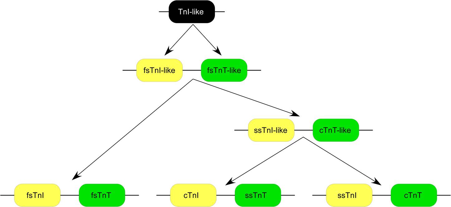
Troponin I, cardiac muscle is a protein that in humans is encoded by the ''TNNI3'' gene. It is a tissue-specific subtype of troponin I, which in turn is a part of the troponin complex image:Troponin Ribbon Diagram.png, 400px, Ribbon representation of the human cardiac troponin core complex (52 kDa core) in the calcium-saturated form. Blue = troponin C; green = troponin I; magenta = troponin T.; ; rendered with PyMOL Troponin, .... The ''TNNI3'' gene encoding cardiac troponin I (cTnI) is located at 19q13.4 in the human chromosomal genome. Human cTnI is a 24 kDa protein consisting of 210 amino acids with isoelectric point (pI) of 9.87. cTnI is exclusively expressed in adult cardiac muscle. Gene evolution left, Figure 1: A phylogenetic tree is derived from alignment of amino acid sequences. cTnI has diverged from the skeletal muscle isoforms of TnI (slow TnI and fast TnI) mainly with a unique N-terminal extension. The amino acid sequence of cTnI is strongly conserved among ma ... [...More Info...] [...Related Items...] OR: [Wikipedia] [Google] [Baidu] |
ACTG1
Actin, cytoplasmic 2, or gamma-actin is a protein that in humans is encoded by the ''ACTG1'' gene. Gamma-actin is widely expressed in cellular cytoskeletons of many tissues; in adult striated muscle cells, gamma-actin is localized to Z-discs and costamere structures, which are responsible for force transduction and transmission in muscle cells. Mutations in ''ACTG1'' have been associated with nonsyndromic hearing loss and Baraitser-Winter syndrome, as well as susceptibility of adolescent patients to vincristine toxicity. Structure Human gamma-actin is 41.8 kDa in molecular weight and 375 amino acids in length. Actins are highly conserved proteins that are involved in various types of cell motility, and maintenance of the cytoskeleton. In vertebrates, three main groups of actin paralogs, alpha, beta, and gamma, have been identified. The alpha actins are found in muscle tissues and are a major constituent of the sarcomere contractile apparatus. The beta and gamma actins co-exist ... [...More Info...] [...Related Items...] OR: [Wikipedia] [Google] [Baidu] |


