|
Tpm1
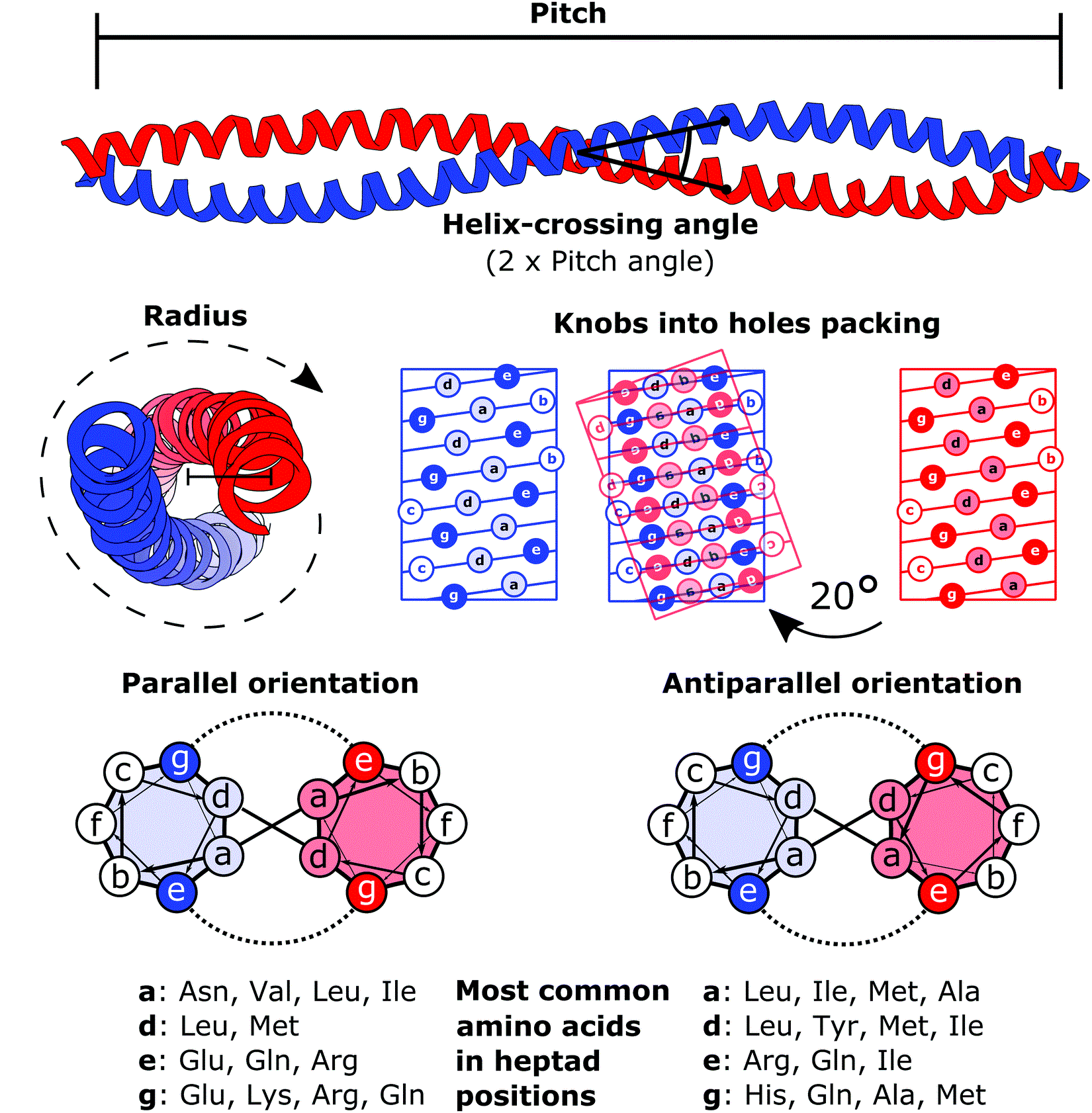
Tropomyosin alpha-1 chain is a protein that in humans is encoded by the ''TPM1'' gene. This gene is a member of the tropomyosin (Tm) family of highly conserved, widely distributed actin-binding proteins involved in the contractile system of striated and smooth muscles and the cytoskeleton of non-muscle cells. Structure Tm is a 32.7 kDa protein composed of 284 amino acids. Tm is a flexible protein homodimer or heterodimer composed of two alpha-helical chains, which adopt a bent coiled coil conformation to wrap around the seven actin molecules in a functional unit of muscle. It is polymerized end to end along the two grooves of actin filaments and provides stability to the filaments. Human striated muscles express protein from the ''TPM1'' (α-Tm), ''TPM2'' (β-Tm) and ''TPM3'' (γ-Tm) genes, with α-Tm being the predominant isoform in striated muscle. In human cardiac muscle the ratio of α-Tm to β-Tm is roughly 5:1. Function Tm functions in association with the troponin co ... [...More Info...] [...Related Items...] OR: [Wikipedia] [Google] [Baidu] |
Protein
Proteins are large biomolecules and macromolecules that comprise one or more long chains of amino acid residue (biochemistry), residues. Proteins perform a vast array of functions within organisms, including Enzyme catalysis, catalysing metabolic reactions, DNA replication, Cell signaling, responding to stimuli, providing Cytoskeleton, structure to cells and Fibrous protein, organisms, and Intracellular transport, transporting molecules from one location to another. Proteins differ from one another primarily in their sequence of amino acids, which is dictated by the Nucleic acid sequence, nucleotide sequence of their genes, and which usually results in protein folding into a specific Protein structure, 3D structure that determines its activity. A linear chain of amino acid residues is called a polypeptide. A protein contains at least one long polypeptide. Short polypeptides, containing less than 20–30 residues, are rarely considered to be proteins and are commonly called pep ... [...More Info...] [...Related Items...] OR: [Wikipedia] [Google] [Baidu] |
Gene
In biology, the word gene has two meanings. The Mendelian gene is a basic unit of heredity. The molecular gene is a sequence of nucleotides in DNA that is transcribed to produce a functional RNA. There are two types of molecular genes: protein-coding genes and non-coding genes. During gene expression (the synthesis of Gene product, RNA or protein from a gene), DNA is first transcription (biology), copied into RNA. RNA can be non-coding RNA, directly functional or be the intermediate protein biosynthesis, template for the synthesis of a protein. The transmission of genes to an organism's offspring, is the basis of the inheritance of phenotypic traits from one generation to the next. These genes make up different DNA sequences, together called a genotype, that is specific to every given individual, within the gene pool of the population (biology), population of a given species. The genotype, along with environmental and developmental factors, ultimately determines the phenotype ... [...More Info...] [...Related Items...] OR: [Wikipedia] [Google] [Baidu] |
Alpha Helix
An alpha helix (or α-helix) is a sequence of amino acids in a protein that are twisted into a coil (a helix). The alpha helix is the most common structural arrangement in the Protein secondary structure, secondary structure of proteins. It is also the most extreme type of local structure, and it is the local structure that is most easily predicted from a sequence of amino acids. The alpha helix has a right-handed helix conformation in which every backbone amino, N−H group hydrogen bonds to the backbone carbonyl, C=O group of the amino acid that is four residue (biochemistry), residues earlier in the protein sequence. Other names The alpha helix is also commonly called a: * Pauling–Corey–Branson α-helix (from the names of three scientists who described its structure) * 3.613-helix because there are 3.6 amino acids in one ring, with 13 atoms being involved in the ring formed by the hydrogen bond (starting with amidic hydrogen and ending with carbonyl oxygen) Discovery ... [...More Info...] [...Related Items...] OR: [Wikipedia] [Google] [Baidu] |
Coiled Coil
A coiled coil is a structural motif in proteins in which two to seven alpha-helices are coiled together like the strands of a rope. ( Dimers and trimers are the most common types.) They have been found in roughly 5-10% of proteins and have a variety of functions. They are one of the most widespread motifs found in protein-protein interactions. To aid protein study, several tools have been developed to predict coiled-coils in protein structures. Many coiled coil-type proteins are involved in important biological functions, such as the regulation of gene expression — e.g., transcription factors. Notable examples are the oncoproteins c-Fos and c-Jun, as well as the muscle protein tropomyosin. Discovery The possibility of coiled coils for α-keratin was initially somewhat controversial. Linus Pauling and Francis Crick independently came to the conclusion that this was possible at about the same time. In the summer of 1952, Pauling visited the laboratory in England where C ... [...More Info...] [...Related Items...] OR: [Wikipedia] [Google] [Baidu] |
Actin
Actin is a family of globular multi-functional proteins that form microfilaments in the cytoskeleton, and the thin filaments in muscle fibrils. It is found in essentially all eukaryotic cells, where it may be present at a concentration of over 100 μM; its mass is roughly 42 kDa, with a diameter of 4 to 7 nm. An actin protein is the monomeric subunit of two types of filaments in cells: microfilaments, one of the three major components of the cytoskeleton, and thin filaments, part of the contractile apparatus in muscle cells. It can be present as either a free monomer called G-actin (globular) or as part of a linear polymer microfilament called F-actin (filamentous), both of which are essential for such important cellular functions as the mobility and contraction of cells during cell division. Actin participates in many important cellular processes, including muscle contraction, cell motility, cell division and cytokinesis, vesicle and organelle mov ... [...More Info...] [...Related Items...] OR: [Wikipedia] [Google] [Baidu] |
TPM2
β-Tropomyosin, also known as tropomyosin beta chain is a protein that in humans is encoded by the ''TPM2'' gene. β-tropomyosin is striated muscle-specific coiled coil dimer that functions to stabilize actin filaments and regulate muscle contraction. Structure β-tropomyosin is roughly 32 kDa in molecular weight (284 amino acids), but multiple splice variants exist. Tropomysin is a flexible protein homodimer or heterodimer composed of two alpha-helical chains, which adopt a bent coiled coil conformation to wrap around the seven actin molecules in a functional unit of muscle. It is polymerized end to end along the two grooves of actin filaments and provides stability to the filaments. Tropomyosin dimers are composed of varying combinations of tropomyosin isoforms; human striated muscles express protein from the ''TPM1'' (α-tropoomyosin), ''TPM2'' (β-tropomyosin) and ''TPM3'' (γ-tropomyosin) genes, with α-tropomyosin being the predominant isoform in striated muscle. Fast s ... [...More Info...] [...Related Items...] OR: [Wikipedia] [Google] [Baidu] |
TPM3
Tropomyosin alpha-3 chain is a protein that in humans is encoded by the ''TPM3'' gene In biology, the word gene has two meanings. The Mendelian gene is a basic unit of heredity. The molecular gene is a sequence of nucleotides in DNA that is transcribed to produce a functional RNA. There are two types of molecular genes: protei .... This gene encodes a member of the tropomyosin family of actin-binding proteins involved in the contractile system of striated and smooth muscles and the cytoskeleton of non-muscle cells. Tropomyosins are dimers of coiled-coil proteins that polymerize end-to-end along the major groove in most actin filaments. They provide stability to the filaments and regulate access of other actin-binding proteins. In muscle cells, they regulate muscle contraction by controlling the binding of myosin heads to the actin filament. Mutations in this gene result in autosomal dominant nemaline myopathy, and oncogenes formed by chromosomal translocations involving this ... [...More Info...] [...Related Items...] OR: [Wikipedia] [Google] [Baidu] |
ACTC1
ACTC1 encodes cardiac muscle alpha actin. This isoform differs from the alpha actin that is expressed in skeletal muscle, ACTA1. Alpha cardiac actin is the major protein of the thin filament in cardiac sarcomeres, which are responsible for muscle contraction and generation of force to support the pump function of the heart. Structure Cardiac alpha actin is a 42.0 kDa protein composed of 377 amino acids. Cardiac alpha actin is a filamentous protein extending from a complex mesh with cardiac alpha-actinin (ACTN2) at Z-lines towards the center of the sarcomere. Polymerization of globular actin (G-actin) leads to a structural filament (F-actin) in the form of a two-stranded helix. Each actin can bind to four others. The atomic structure of monomeric actin was solved by Kabsch et al., and closely thereafter this same group published the structure of the actin filament. Actins are highly conserved proteins; the alpha actins are found in muscle tissues and are a major constituent of ... [...More Info...] [...Related Items...] OR: [Wikipedia] [Google] [Baidu] |
MYH7
Myosin-7 is a protein that in humans is encoded by the ''MYH7'' gene. It is the myosin heavy chain beta (MHC-β) isoform (slow twitch) expressed primarily in the heart, but also in skeletal muscles (type I fibers). This isoform is distinct from the fast isoform of cardiac myosin heavy chain, MYH6, referred to as MHC-α. MHC-β is the major protein comprising the thick filament that forms the sarcomeres in cardiac muscle and plays a major role in cardiac muscle contraction. Structure MHC-β is a 223 kDa protein composed of 1935 amino acids. MHC-β is a hexameric, asymmetric motor forming the bulk of the thick filament in cardiac muscle. MHC-β is composed of N-terminal globular heads (20 nm) that project laterally, and alpha helical tails (130 nm) that dimerize and multimerize into a coiled-coil motif to form the light meromyosin (LMM), thick filament rod. The 9 nm alpha-helical neck region of each MHC-β head non-covalently binds two light chains, essential ... [...More Info...] [...Related Items...] OR: [Wikipedia] [Google] [Baidu] |
TNNT2
Cardiac muscle troponin T (cTnT) is a protein that in humans is encoded by the ''TNNT2'' gene. Cardiac TnT is the tropomyosin-binding subunit of the troponin complex, which is located on the thin filament of striated muscles and regulates muscle contraction in response to alterations in intracellular calcium ion concentration. The TNNT2 gene is located at 1q32 in the human chromosomal genome, encoding the cardiac muscle isoform of troponin T (cTnT). Human cTnT is an ~36-kDa protein consisting of 297 amino acids including the first methionine with an isoelectric point (pI) of 4.88. It is the tropomyosin- binding and thin filament anchoring subunit of the troponin complex in cardiac muscle cells. TNNT2 gene is expressed in vertebrate cardiac muscles and embryonic skeletal muscles. Structure Cardiac TnT is a 35.9 kDa protein composed of 298 amino acids. Cardiac TnT is the largest of the three troponin subunits (cTnT, TNNI3, troponin I (TnI), TNNC1, troponin C (TnC)) on the ACTC1 ... [...More Info...] [...Related Items...] OR: [Wikipedia] [Google] [Baidu] |
Hypertrophic Cardiomyopathy
Hypertrophic cardiomyopathy (HCM, or HOCM when obstructive) is a condition in which muscle tissues of the heart become thickened without an obvious cause. The parts of the heart most commonly affected are the interventricular septum and the ventricles. This results in the heart being less able to pump blood effectively and also may cause electrical conduction problems. Specifically, within the bundle branches that conduct impulses through the interventricular septum and into the Purkinje fibers, as these are responsible for the depolarization of contractile cells of both ventricles. People who have HCM may have a range of symptoms. People may be asymptomatic, or may have fatigue, leg swelling, and shortness of breath. It may also result in chest pain or fainting. Symptoms may be worse when the person is dehydrated. Complications may include heart failure, an irregular heartbeat, and sudden cardiac death. HCM is most commonly inherited in an autosomal dominant pattern. I ... [...More Info...] [...Related Items...] OR: [Wikipedia] [Google] [Baidu] |
Dilated Cardiomyopathy
Dilated cardiomyopathy (DCM) is a condition in which the heart becomes enlarged and cannot pump blood effectively. Symptoms vary from none to feeling tired, leg swelling, and shortness of breath. It may also result in chest pain or fainting. Complications can include heart failure, heart valve disease, or an irregular heartbeat. Causes include genetics, alcohol, cocaine, certain toxins, complications of pregnancy, and certain infections. Coronary artery disease and high blood pressure may play a role, but are not the primary cause. In many cases the cause remains unclear. It is a type of cardiomyopathy, a group of diseases that primarily affects the heart muscle. The diagnosis may be supported by an electrocardiogram, chest X-ray, or echocardiogram. In those with heart failure, treatment may include medications in the ACE inhibitor, beta blocker, and diuretic families. A low salt diet may also be helpful. In those with certain types of irregular heartbeat, blood thinner ... [...More Info...] [...Related Items...] OR: [Wikipedia] [Google] [Baidu] |


