|
Isovolumetric Contraction
In cardiac physiology, isometric contraction is an event occurring in early systole during which the ventricles contract with no corresponding volume change ( isometrically). This short-lasting portion of the cardiac cycle takes place while all heart valves are closed. Description In a healthy young adult, blood enters the atria and flows to the ventricles via the opened atrioventricular valves (tricuspid and mitral valves). Atrial contraction rapidly follows, actively pumping about 30% of the returning blood. As diastole ends, the ventricles begin depolarizing and, while ventricular pressure starts to rise owing to contraction, the atrioventricular valves close in order to prevent backflow to the atria. At this stage, which corresponds to the R peak or the QRS complex seen on an ECG, the semilunar valves (aortic and pulmonary valves) are also closed. The net result is that, while contraction causes ventricular pressures to rise sharply, there is no overall change in volume ... [...More Info...] [...Related Items...] OR: [Wikipedia] [Google] [Baidu] |
Wiggers Diagram
A Wiggers diagram, named after its developer, Carl Wiggers, is a unique diagram that has been used in teaching cardiac physiology for more than a century. In the Wiggers diagram, the X-axis is used to plot time subdivided into the cardiac phases, while the Y-axis typically contains the following on a single grid: * Blood pressure ** Aortic pressure ** Ventricular pressure ** Atrial pressure * Ventricular volume * Electrocardiogram * Arterial flow (optional) * Heart sounds (optional) The Wiggers diagram clearly illustrates the coordinated variation of these values as the heart beats, assisting one in understanding the entire cardiac cycle. Events Note that during isovolumetric/isovolumic contraction and relaxation, all the heart valves are closed; at no time are all the heart valves open. *S3 and S4 heart sounds are associated with pathologies and are not routinely heard. Additional images File:Heart systole.svg, Ventricular systole File:Heart diasystole.svg, Cardiac dias ... [...More Info...] [...Related Items...] OR: [Wikipedia] [Google] [Baidu] |
Depolarization
In biology, depolarization or hypopolarization is a change within a cell, during which the cell undergoes a shift in electric charge distribution, resulting in less negative charge inside the cell compared to the outside. Depolarization is essential to the function of many cells, communication between cells, and the overall physiology of an organism. Most cells in higher organisms maintain an internal environment that is negatively charged relative to the cell's exterior. This difference in charge is called the cell's membrane potential. In the process of depolarization, the negative internal charge of the cell temporarily becomes more positive (less negative). This shift from a negative to a more positive membrane potential occurs during several processes, including an action potential. During an action potential, the depolarization is so large that the potential difference across the cell membrane briefly reverses polarity, with the inside of the cell becoming positively char ... [...More Info...] [...Related Items...] OR: [Wikipedia] [Google] [Baidu] |
Blood Pressure
Blood pressure (BP) is the pressure of circulating blood against the walls of blood vessels. Most of this pressure results from the heart pumping blood through the circulatory system. When used without qualification, the term "blood pressure" refers to the pressure in the large arteries. Blood pressure is usually expressed in terms of the systolic pressure (maximum pressure during one heartbeat) over diastolic pressure (minimum pressure between two heartbeats) in the cardiac cycle. It is measured in millimeters of mercury ( mmHg) above the surrounding atmospheric pressure. Blood pressure is one of the vital signs—together with respiratory rate, heart rate, oxygen saturation, and body temperature—that healthcare professionals use in evaluating a patient's health. Normal resting blood pressure, in an adult is approximately systolic over diastolic, denoted as "120/80 mmHg". Globally, the average blood pressure, age standardized, has remained about the same since 1 ... [...More Info...] [...Related Items...] OR: [Wikipedia] [Google] [Baidu] |
Cardiac Cycle
The cardiac cycle is the performance of the human heart from the beginning of one heartbeat to the beginning of the next. It consists of two periods: one during which the heart muscle relaxes and refills with blood, called diastole, following a period of robust contraction and pumping of blood, called systole. After emptying, the heart immediately relaxes and expands to receive another influx of blood returning from the lungs and other systems of the body, before again contracting to pump blood to the lungs and those systems. A normally performing heart must be fully expanded before it can efficiently pump again. Assuming a healthy heart and a typical rate of 70 to 75 beats per minute, each cardiac cycle, or heartbeat, takes about 0.8 second to complete the cycle. There are two atrial and two ventricle chambers of the heart; they are paired as the left heart and the right heart—that is, the left atrium with the left ventricle, the right atrium with the right ventricle—and t ... [...More Info...] [...Related Items...] OR: [Wikipedia] [Google] [Baidu] |
Isovolumic Relaxation Time
Isovolumic relaxation time (IVRT) is an interval in the cardiac cycle, from the aortic component of the second heart sound, that is, closure of the aortic valve, to onset of filling by opening of the mitral valve."Inductance cardiography (thoracocardiography): A novel, noninvasive technique for monitoring left ventricular filling". ''Journal of Critical Care'', Volume 14, Issue 4, Pages 177–185 It can be used as an indicator of diastolic dysfunction. It can be measured by simultaneous Doppler echocardiography and M-mode sonography, or better still, by simultaneous phonocardiogram and transmitral Doppler.anaesthetist.com >Diastolic dysfunctionLara Hopley and Jo van Schalkwyk. Date of last update: 2009-06-05 Prolonged IVRT indicates poor myocardial relaxation. A normal IVRT is about 70 ± 12 ms, and approximately 10 ms longer in people over forty years. In abnormal relaxation, IVRT is usually in excess of 110 ms. With restrictive ventricular filling, it is usua ... [...More Info...] [...Related Items...] OR: [Wikipedia] [Google] [Baidu] |
Ancient Greek
Ancient Greek includes the forms of the Greek language used in ancient Greece and the ancient world from around 1500 BC to 300 BC. It is often roughly divided into the following periods: Mycenaean Greek (), Dark Ages (), the Archaic period (), and the Classical period (). Ancient Greek was the language of Homer and of fifth-century Athenian historians, playwrights, and philosophers. It has contributed many words to English vocabulary and has been a standard subject of study in educational institutions of the Western world since the Renaissance. This article primarily contains information about the Epic and Classical periods of the language. From the Hellenistic period (), Ancient Greek was followed by Koine Greek, which is regarded as a separate historical stage, although its earliest form closely resembles Attic Greek and its latest form approaches Medieval Greek. There were several regional dialects of Ancient Greek, of which Attic Greek developed into Koine. Dia ... [...More Info...] [...Related Items...] OR: [Wikipedia] [Google] [Baidu] |
Pulmonary Artery
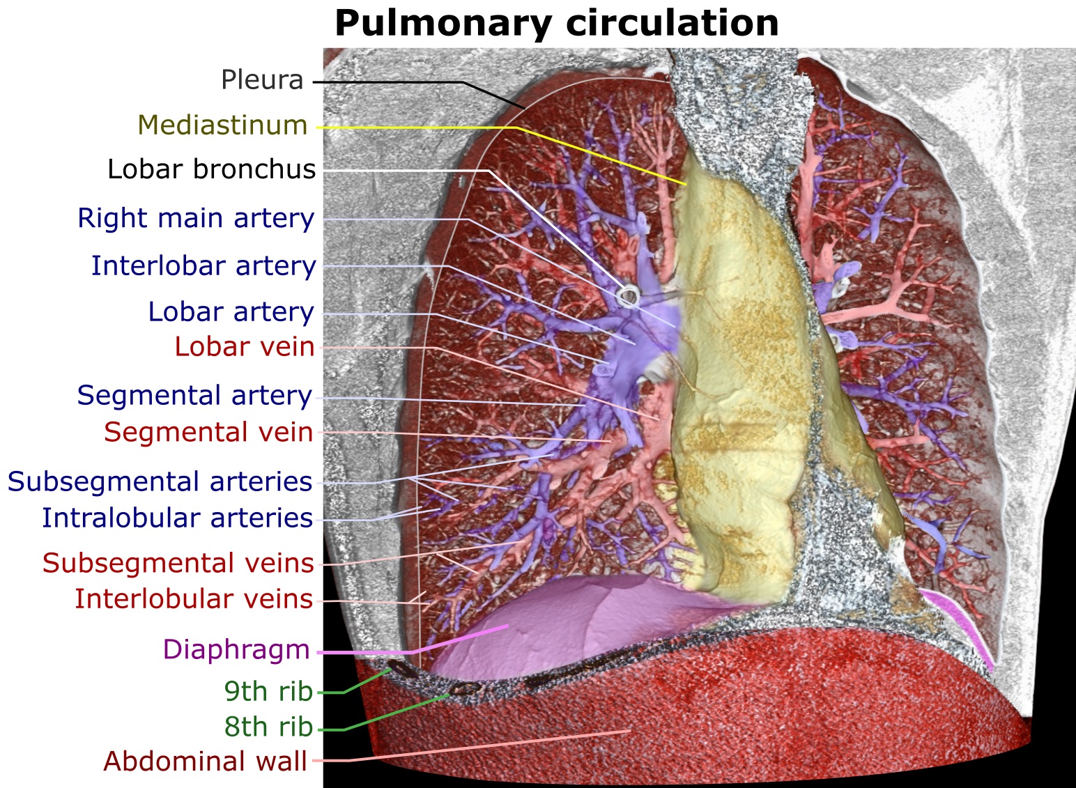
A pulmonary artery is an artery in the pulmonary circulation that carries deoxygenated blood from the right side of the heart to the lungs. The largest pulmonary artery is the ''main pulmonary artery'' or ''pulmonary trunk'' from the heart, and the smallest ones are the arterioles, which lead to the capillaries that surround the pulmonary alveoli. Structure The pulmonary arteries are blood vessels that carry systemic venous blood from the right ventricle of the heart to the microcirculation of the lungs. Unlike in other organs where arteries supply oxygenated blood, the blood carried by the pulmonary arteries is deoxygenated, as it is venous blood returning to the heart. The main pulmonary arteries emerge from the right side of the heart, and then split into smaller arteries that progressively divide and become arterioles, eventually narrowing into the capillary microcirculation of the lungs where gas exchange occurs. Pulmonary trunk In order of blood flow, the pulmonary art ... [...More Info...] [...Related Items...] OR: [Wikipedia] [Google] [Baidu] |
Aorta
The aorta ( ) is the main and largest artery in the human body, originating from the left ventricle of the heart and extending down to the abdomen, where it splits into two smaller arteries (the common iliac arteries). The aorta distributes oxygenated blood to all parts of the body through the systemic circulation. Structure Sections In anatomical sources, the aorta is usually divided into sections. One way of classifying a part of the aorta is by anatomical compartment, where the thoracic aorta (or thoracic portion of the aorta) runs from the heart to the diaphragm. The aorta then continues downward as the abdominal aorta (or abdominal portion of the aorta) from the diaphragm to the aortic bifurcation. Another system divides the aorta with respect to its course and the direction of blood flow. In this system, the aorta starts as the ascending aorta, travels superiorly from the heart, and then makes a hairpin turn known as the aortic arch. Following the aortic arch ... [...More Info...] [...Related Items...] OR: [Wikipedia] [Google] [Baidu] |
Pulmonary Valve
The pulmonary valve (sometimes referred to as the pulmonic valve) is a valve of the heart that lies between the right ventricle and the pulmonary artery and has three cusps. It is one of the four valves of the heart and one of the two semilunar valves, the other being the aortic valve. Similar to the aortic valve, the pulmonary valve opens in ventricular systole, when the pressure in the right ventricle rises above the pressure in the pulmonary artery. At the end of ventricular systole, when the pressure in the right ventricle falls rapidly, the pressure in the pulmonary artery will close the pulmonary valve. The closure of the pulmonary valve contributes the P2 component of the second heart sound (S2). The right heart is a low-pressure system, so the P2 component of the second heart sound is usually softer than the A2 component of the second heart sound. However, it is physiologically normal in some young people to hear both components separated during inhalation. Description * ... [...More Info...] [...Related Items...] OR: [Wikipedia] [Google] [Baidu] |
Aortic Valve
The aortic valve is a valve in the heart of humans and most other animals, located between the left ventricle and the aorta. It is one of the four valves of the heart and one of the two semilunar valves, the other being the pulmonary valve. The aortic valve normally has three cusps or leaflets, although in 1–2% of the population it is found to congenitally have two leaflets. The aortic valve is the last structure in the heart the blood travels through before stopping the flow through the systemic circulation. Structure The aortic valve normally has three cusps however there is some discrepancy in their naming. They may be called the left coronary, right coronary and non-coronary cusp. Some sources also advocate they be named as a left, right and posterior cusp. Anatomists have traditionally named them the left posterior (origin of left coronary), anterior (origin of the right coronary) and right posterior. The three cusps, when the valve is closed, contain a sinus called an a ... [...More Info...] [...Related Items...] OR: [Wikipedia] [Google] [Baidu] |
Electrocardiography
Electrocardiography is the process of producing an electrocardiogram (ECG or EKG), a recording of the heart's electrical activity. It is an electrogram of the heart which is a graph of voltage versus time of the electrical activity of the heart using electrodes placed on the skin. These electrodes detect the small electrical changes that are a consequence of cardiac muscle depolarization followed by repolarization during each cardiac cycle (heartbeat). Changes in the normal ECG pattern occur in numerous cardiac abnormalities, including cardiac rhythm disturbances (such as atrial fibrillation and ventricular tachycardia), inadequate coronary artery blood flow (such as myocardial ischemia and myocardial infarction), and electrolyte disturbances (such as hypokalemia and hyperkalemia). Traditionally, "ECG" usually means a 12-lead ECG taken while lying down as discussed below. However, other devices can record the electrical activity of the heart such as a Holter monitor but also s ... [...More Info...] [...Related Items...] OR: [Wikipedia] [Google] [Baidu] |
Backflow
Backflow is a term in plumbing for an unwanted flow of water in the reverse direction. It can be a serious health risk for the contamination of potable water supplies with foul water. In the most obvious case, a toilet flush cistern and its water supply must be isolated from the toilet bowl. For this reason, building codes mandate a series of measures and backflow prevention devices to prevent backflow. Causes Backflow occurs for one of two reasons, either ''back pressure'' or ''back siphonage''. ''Back pressure'' is the result of a higher pressure in the system than in its supply, i.e. the system pressure has been ''increased'' by some means. This may occur in unvented heating systems, where thermal expansion increases the pressure. ''Back siphonage'' is the result of supply pressure being ''lowered'' below that of the system. This may occur when a supply is interrupted or drained down. Risk of contamination The precise measures required to prevent backflow depend on t ... [...More Info...] [...Related Items...] OR: [Wikipedia] [Google] [Baidu] |


