|
Pulmonary Artery
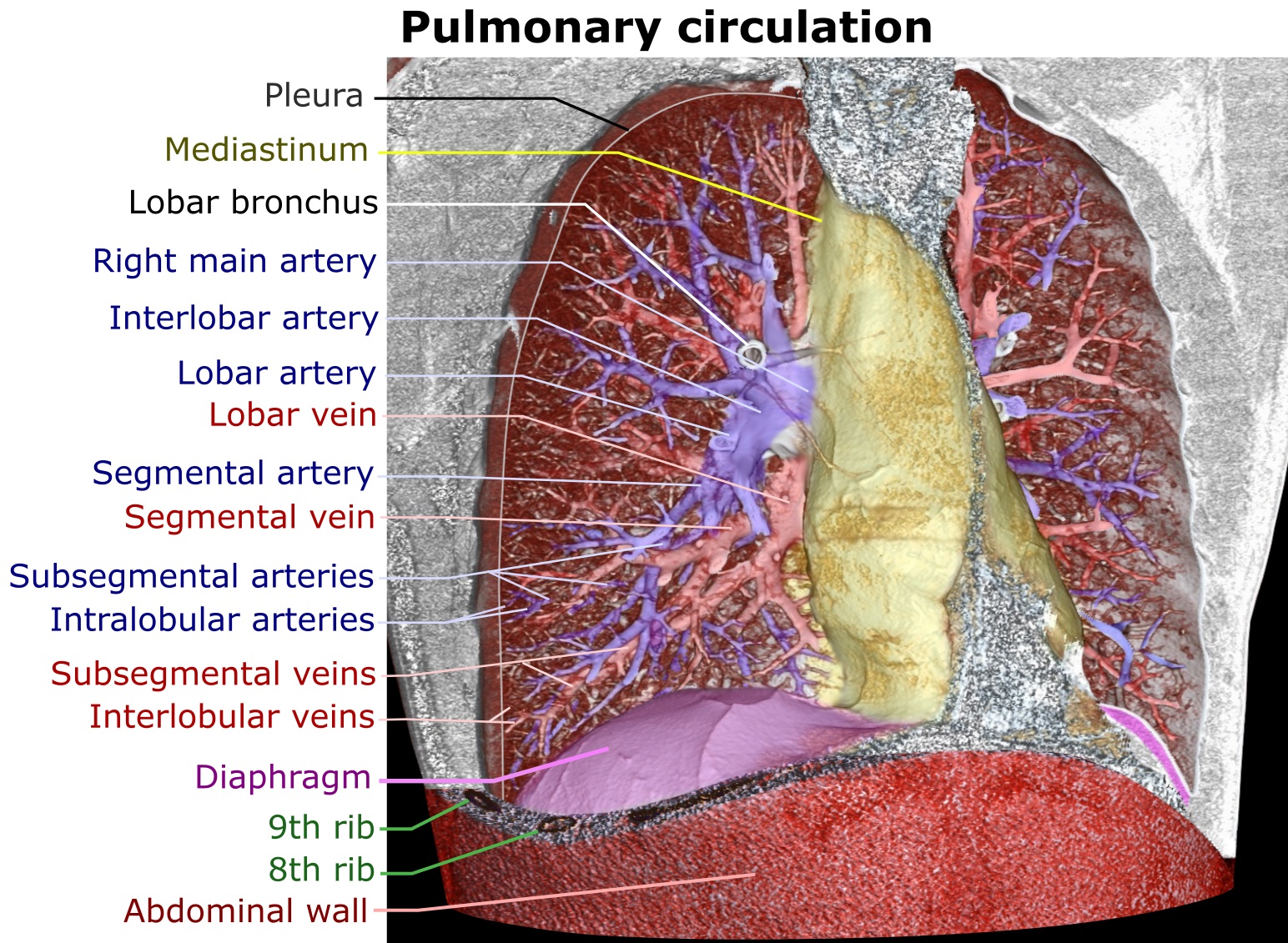
A pulmonary artery is an artery in the pulmonary circulation that carries deoxygenated blood from the right side of the heart to the lungs. The largest pulmonary artery is the ''main pulmonary artery'' or ''pulmonary trunk'' from the heart, and the smallest ones are the arterioles, which lead to the capillaries that surround the pulmonary alveoli. Structure The pulmonary arteries are blood vessels that carry systemic venous blood from the right ventricle of the heart to the microcirculation of the lungs. Unlike in other organs where arteries supply oxygenated blood, the blood carried by the pulmonary arteries is deoxygenated, as it is venous blood returning to the heart. The main pulmonary arteries emerge from the right side of the heart, and then split into smaller arteries that progressively divide and become arterioles, eventually narrowing into the capillary microcirculation of the lungs where gas exchange occurs. Pulmonary trunk In order of blood flow, the pulmonar ... [...More Info...] [...Related Items...] OR: [Wikipedia] [Google] [Baidu] |
Right Ventricle
A ventricle is one of two large chambers toward the bottom of the heart that collect and expel blood towards the peripheral beds within the body and lungs. The blood pumped by a ventricle is supplied by an atrium, an adjacent chamber in the upper heart that is smaller than a ventricle. Interventricular means between the ventricles (for example the interventricular septum), while intraventricular means within one ventricle (for example an intraventricular block). In a four-chambered heart, such as that in humans, there are two ventricles that operate in a double circulatory system: the right ventricle pumps blood into the pulmonary circulation to the lungs, and the left ventricle pumps blood into the systemic circulation through the aorta. Structure Ventricles have thicker walls than atria and generate higher blood pressures. The physiological load on the ventricles requiring pumping of blood throughout the body and lungs is much greater than the pressure generated by the a ... [...More Info...] [...Related Items...] OR: [Wikipedia] [Google] [Baidu] |
Ventricular Outflow Tract
A ventricular outflow tract is a portion of either the left ventricle or right ventricle of the heart through which blood passes in order to enter the great arteries. The right ventricular outflow tract (RVOT) is an infundibular extension of the ventricular cavity that connects to the pulmonary artery. The left ventricular outflow tract (LVOT), which connects to the aorta, is nearly indistinguishable from the rest of the ventricle. The outflow tract is derived from the secondary heart field, during cardiogenesis. Both the left and right outflow tract have their own term. The right outflow tract is called "conus arteriosus" from the outside, and infundibulum from the inside. In the left ventricle the outflow tract is the "aortic vestibule". They both possess smooth walls, and are derived from the embryonic bulbus cordis In both left and right ventricle there are specific structures separating the inflow and outflow of blood. In the right ventricle, the inflow and outflow is sep ... [...More Info...] [...Related Items...] OR: [Wikipedia] [Google] [Baidu] |
Heart Development
Heart development, also known as cardiogenesis, refers to the prenatal development of the heart. This begins with the formation of two endocardial tubes which merge to form the tubular heart, also called the primitive heart tube. The heart is the first functional organ in vertebrate embryos. The tubular heart quickly differentiates into the truncus arteriosus, bulbus cordis, primitive ventricle, primitive atrium, and the sinus venosus. The truncus arteriosus splits into the ascending aorta and the pulmonary trunk. The bulbus cordis forms part of the ventricles. The sinus venosus connects to the fetal circulation. The heart tube elongates on the right side, looping and becoming the first visual sign of left-right asymmetry of the body. Septa form within the atria and ventricles to separate the left and right sides of the heart. Early development The heart derives from embryonic mesodermal germ layer cells that differentiate after gastrulation into mesothelium, endothel ... [...More Info...] [...Related Items...] OR: [Wikipedia] [Google] [Baidu] |
Pharyngeal Arch
The pharyngeal arches, also known as visceral arches'','' are structures seen in the embryonic development of vertebrates that are recognisable precursors for many structures. In fish, the arches are known as the branchial arches, or gill arches. In the human embryo, the arches are first seen during the fourth week of development. They appear as a series of outpouchings of mesoderm on both sides of the developing pharynx. The vasculature of the pharyngeal arches is known as the aortic arches. In fish, the branchial arches support the gills. Structure In vertebrates, the pharyngeal arches are derived from all three germ layers (the primary layers of cells that form during embryogenesis). Neural crest cells enter these arches where they contribute to features of the skull and facial skeleton such as bone and cartilage. However, the existence of pharyngeal structures before neural crest cells evolved is indicated by the existence of neural crest-independent mechanisms of phary ... [...More Info...] [...Related Items...] OR: [Wikipedia] [Google] [Baidu] |
Bronchial Arteries
In human anatomy, the bronchial arteries supply the lungs with nutrition and oxygenated blood. Although there is much variation, there are usually two bronchial arteries that run to the left lung, and one to the right lung and are a vital part of the respiratory system. Structure There are typically two left and one right bronchial arteries. The ''left bronchial arteries'' (superior and inferior) usually arise directly from the thoracic aorta. The single ''right bronchial artery'' usually arises from one of the following: * 1) the thoracic aorta at a common trunk with the right 3rd posterior intercostal artery * 2) the superior bronchial artery on the left side * 3) any number of the right intercostal arteries mostly the third right posterior. Function The bronchial arteries supply blood to the bronchi and connective tissue of the lungs. They travel with and branch with the bronchi, ending about at the level of the respiratory bronchioles. They anastomose with the branc ... [...More Info...] [...Related Items...] OR: [Wikipedia] [Google] [Baidu] |
Lung Lobe
The lungs are the primary organs of the respiratory system in humans and most other animals, including some snails and a small number of fish. In mammals and most other vertebrates, two lungs are located near the backbone on either side of the heart. Their function in the respiratory system is to extract oxygen from the air and transfer it into the bloodstream, and to release carbon dioxide from the bloodstream into the atmosphere, in a process of gas exchange. Respiration is driven by different muscular systems in different species. Mammals, reptiles and birds use their different muscles to support and foster breathing. In earlier tetrapods, air was driven into the lungs by the pharyngeal muscles via buccal pumping, a mechanism still seen in amphibians. In humans, the main muscle of respiration that drives breathing is the diaphragm. The lungs also provide airflow that makes vocal sounds including human speech possible. Humans have two lungs, one on the left and one ... [...More Info...] [...Related Items...] OR: [Wikipedia] [Google] [Baidu] |
Root Of The Lung
The root of the lung is a group of structures that emerge at the hilum of each lung, just above the middle of the mediastinal surface and behind the cardiac impression of the lung. It is nearer to the back (posterior border) than the front (anterior border). The root of the lung is connected by the structures that form it to the heart and the trachea. The rib cage is separated from the lung by a two-layered membranous coating, the pleura. The hilum is the large triangular depression where the connection between the parietal pleura (covering the rib cage) and the visceral pleura (covering the lung) is made, and this marks the meeting point between the mediastinum and the pleural cavities. Location The root of the right lung lies behind the superior vena cava and part of the right atrium, and below the azygos vein. That of the left lung passes beneath the aortic arch and in front of the descending aorta; the phrenic nerve, pericardiacophrenic artery and vein, and the ... [...More Info...] [...Related Items...] OR: [Wikipedia] [Google] [Baidu] |
Alveolus Diagram
Alveolus (; pl. alveoli, adj. alveolar) is a general anatomical term for a concave cavity or pit. Uses in anatomy and zoology * Pulmonary alveolus, an air sac in the lungs ** Alveolar cell or pneumocyte ** Alveolar duct ** Alveolar macrophage * Mammary alveolus, a milk sac in the mammary glands * Alveolar gland * Dental alveolus, also known as "tooth socket", a socket in the jaw that holds the roots of teeth ** Alveolar ridge, the jaw structure that contains the dental alveoli ** Alveolar canals ** Alveolar process * Arteries: ** Superior alveolar artery (other) *** Anterior superior alveolar arteries *** Posterior superior alveolar artery ** Inferior alveolar artery * Nerves: ** Anterior superior alveolar nerve ** Middle superior alveolar nerve ** Inferior alveolar nerve Uses in botany, microbiology and related disciplines * Surface cavities or pits, such as on the stem of Myrmecodia species * Pits on honeycombed surfaces such as receptacles of many angiosperms * P ... [...More Info...] [...Related Items...] OR: [Wikipedia] [Google] [Baidu] |
Carina Of Trachea
In anatomy, the carina or tracheal bifurcation is a ridge of cartilage in the trachea that occurs between the division of the two main bronchi. Structure The carina occurs at the lower end of the trachea (usually at the level of the 4th to 5th thoracic vertebra). This is in line with the sternal angle, but the carina may raise or descend up to two vertebrae higher or lower with breathing. The carina lies to the left of the midline, and runs antero-posteriorly (front to back). The bronchial arteries supply the carina and the rest of the lower trachea. The carina is around the area posterior to where the aortic arch crosses to the left of the trachea. The azygos vein crosses right to the trachea above the carina. Clinical significance Foreign bodies that fall down the trachea are more likely to enter the right bronchus. The mucous membrane of the carina is the most sensitive area of the trachea and larynx for triggering a cough reflex. Widening and distortion of the cari ... [...More Info...] [...Related Items...] OR: [Wikipedia] [Google] [Baidu] |
Ligamentum Arteriosum
The ligamentum arteriosum (arterial ligament), also known as the Ligament of Botallo or Harvey's ligament, is a small ligament attaching the aorta to the pulmonary artery. It serves no function in adults but is the remnant of the ductus arteriosus formed within three weeks after birth. Structure At the superior end, the ligamentum attaches to the aorta—at the final part of the aortic arch (the isthmus of aorta) or the first part of the descending aorta. On the other, inferior end, the ligamentum is attached to the top of the left pulmonary artery. The ligamentum arteriosum is closely related to the left recurrent laryngeal nerve, a branch of the left vagus nerve. After splitting from the left vagus nerve, the left recurrent laryngeal loops around the aortic arch behind the ligamentum arteriosum, after which it ascends to the larynx. Function In adults, the ligamentum arteriosum has no useful function. It is a vestige of the ductus arteriosus, a temporary fetal structure ... [...More Info...] [...Related Items...] OR: [Wikipedia] [Google] [Baidu] |
Descending Aorta
In human anatomy, the descending aorta is part of the aorta, the largest artery in the body. The descending aorta begins at the aortic arch and runs down through the chest and abdomen. The descending aorta anatomically consists of two portions or segments, the thoracic and the abdominal aorta, in correspondence with the two great cavities of the trunk in which it is situated. Within the abdomen, the descending aorta branches into the two common iliac arteries which serve the pelvis and eventually legs. The ductus arteriosus connects to the junction between the pulmonary artery and the descending aorta in foetal life. This artery later regresses as the ligamentum arteriosum. See also *Abbott artery References External links * – "Left side of the mediastinum The mediastinum (from ) is the central compartment of the thoracic cavity. Surrounded by loose connective tissue, it is an undelineated region that contains a group of structures within the thorax, namely ... [...More Info...] [...Related Items...] OR: [Wikipedia] [Google] [Baidu] |
University Of Virginia School Of Medicine
The University of Virginia School of Medicine (UVA SoM) is the graduate medical school of the University of Virginia. The school's facilities are on the University of Virginia grounds adjacent to Academical Village in Charlottesville, Virginia. Founded in 1819 by Thomas Jefferson, UVA SoM is the tenth oldest medical school in the United States, and is ranked 31st in research-oriented medical schools by ''U.S. News & World Report,'' and as of 2021, is ranked nineteenth in the nation in primary care. The School of Medicine confers Doctor of Medicine (M.D.) and Doctor of Philosophy (PhD) degrees, and is closely associated with both the University of Virginia Health System and Inova Health System. History The UVA Health System's history can be traced to the original conception of the University of Virginia on August 1, 1818, whereupon Thomas Jefferson, James Madison, and twenty-one other men first compiled a report for the Virginia State Legislature to determine a site, building ... [...More Info...] [...Related Items...] OR: [Wikipedia] [Google] [Baidu] |


