|
Isotope Perfusion Scanning
Perfusion is the passage of fluid through the lymphatic system or blood vessels to an organ or a tissue. The practice of perfusion scanning is the process by which this perfusion can be observed, recorded and quantified. The term perfusion scanning encompasses a wide range of medical imaging modalities.http://www.webmd.com/heart-disease/cardiac-perfusion-scan#1 www.webmd.com/ Applications With the ability to ascertain data on the blood flow to vital organs such as the heart and the brain, doctors are able to make quicker and more accurate choices on treatment for patients. Nuclear medicine has been leading perfusion scanning for some time, although the modality has certain pitfalls. It is often dubbed 'unclear medicine' as the scans produced may appear to the untrained eye as just fluffy and irregular patterns. More recent developments in CT and MRI have meant clearer images and solid data, such as graphs depicting blood flow, and blood volume charted over a fixed period of time. ... [...More Info...] [...Related Items...] OR: [Wikipedia] [Google] [Baidu] |
Perfusion
Perfusion is the passage of fluid through the circulatory system or lymphatic system to an organ or a tissue, usually referring to the delivery of blood to a capillary bed in tissue. Perfusion is measured as the rate at which blood is delivered to tissue, or volume of blood per unit time (blood flow) per unit tissue mass. The SI unit is m3/(s·kg), although for human organs perfusion is typically reported in ml/min/g. The word is derived from the French verb "perfuser" meaning to "pour over or through". All animal tissues require an adequate blood supply for health and life. Poor perfusion (malperfusion), that is, ischemia, causes health problems, as seen in cardiovascular disease, including coronary artery disease, cerebrovascular disease, peripheral artery disease, and many other conditions. Tests verifying that adequate perfusion exists are a part of a patient's assessment process that are performed by medical or emergency personnel. The most common methods include ev ... [...More Info...] [...Related Items...] OR: [Wikipedia] [Google] [Baidu] |
Single-photon Emission Computed Tomography
Single-photon emission computed tomography (SPECT, or less commonly, SPET) is a nuclear medicine tomographic imaging technique using gamma rays. It is very similar to conventional nuclear medicine planar imaging using a gamma camera (that is, scintigraphy), but is able to provide true 3D information. This information is typically presented as cross-sectional slices through the patient, but can be freely reformatted or manipulated as required. The technique needs delivery of a gamma-emitting radioisotope (a radionuclide) into the patient, normally through injection into the bloodstream. On occasion, the radioisotope is a simple soluble dissolved ion, such as an isotope of gallium(III). Most of the time, though, a marker radioisotope is attached to a specific ligand to create a radioligand, whose properties bind it to certain types of tissues. This marriage allows the combination of ligand and radiopharmaceutical to be carried and bound to a place of interest in the body, ... [...More Info...] [...Related Items...] OR: [Wikipedia] [Google] [Baidu] |
Aminophylline
Aminophylline is a compound of the bronchodilator theophylline with ethylenediamine in 2:1 ratio. The ethylenediamine improves solubility, and the aminophylline is usually found as a dihydrate. Aminophylline is less potent and shorter-acting than theophylline. Its most common use is in the treatment of airway obstruction from asthma or COPD. Aminophylline is a nonselective adenosine receptor antagonist and phosphodiesterase inhibitor. Medical uses Intravenous aminophylline can be used for acute exacerbation of symptoms and reversible airway obstruction in asthma and other chronic lung disease such as COPD, emphysema and chronic bronchitis. It is used as an adjunct to inhaled beta-2 selective agonists and systemically administered corticosteroids. Aminophylline is used to reverse regadenoson, dipyridamole or adenosine based infusions during nuclear cardiology stress testing. Aminophylline has also been reported to be effective in preventing slow heart rates during complex cardio ... [...More Info...] [...Related Items...] OR: [Wikipedia] [Google] [Baidu] |
Dipyridamole
Dipyridamole (trademarked as Persantine and others) is a nucleoside transport inhibitor and a PDE3 inhibitor medication that inhibits blood clot formation when given chronically and causes blood vessel dilation when given at high doses over a short time. Medical uses * Dipyridamole is used to dilate blood vessels in people with peripheral arterial disease and coronary artery disease * Dipyridamole has been shown to lower pulmonary hypertension without significant drop of systemic blood pressure * It inhibits formation of pro-inflammatory cytokines ( MCP-1, MMP-9) in vitro and results in reduction of hsCRP in patients. * It inhibits proliferation of smooth muscle cells in vivo and modestly increases unassisted patency of synthetic arteriovenous hemodialysis grafts. * It increases the release of tissue plasminogen activator from brain microvascular endothelial cells. * It results in an increase of 13-hydroxyoctadecadienoic acid and decrease of 12-hydroxyeicosatetraenoic ac ... [...More Info...] [...Related Items...] OR: [Wikipedia] [Google] [Baidu] |
Dobutamine
Dobutamine is a medication used in the treatment of cardiogenic shock (as a result of inadequate tissue perfusion) and severe heart failure. It may also be used in certain types of cardiac stress tests. It is given by IV only, as an injection into a vein or intraosseous as a continuous infusion. The amount of medication needs to be adjusted to the desired effect. Onset of effects is generally seen within 2 minutes. It has a half-life of two minutes. This drug is generally only administered short term, although it may be used for longer periods to relieve symptoms of heart failure in patients awaiting heart transplantation. Common side effects include a fast heart rate, an irregular heart beat, and inflammation at the site of injection. Use is not recommended in those with idiopathic hypertrophic subaortic stenosis. It primarily works by direct stimulation of β1 receptors, which increases the strength of the heart's contractions, leading to a positive ionotrophic effect. G ... [...More Info...] [...Related Items...] OR: [Wikipedia] [Google] [Baidu] |
Adenosine
Adenosine (symbol A) is an organic compound that occurs widely in nature in the form of diverse derivatives. The molecule consists of an adenine attached to a ribose via a β-N9-glycosidic bond. Adenosine is one of the four nucleoside building blocks of RNA (and its derivative deoxyadenosine is a building block of DNA), which are essential for all life. Its derivatives include the energy carriers adenosine mono-, di-, and triphosphate, also known as AMP/ADP/ATP. Cyclic adenosine monophosphate (cAMP) is pervasive in signal transduction. Adenosine is used as an intravenous medication for some cardiac arrhythmias. Adenosyl (abbreviated Ado or 5'-dAdo) is the chemical group formed by removal of the 5′-hydroxy (OH) group. It is found in adenosylcobalamin (an active form of vitamin B12) and as a radical in radical SAM enzymes. Medical uses Supraventricular tachycardia In individuals with supraventricular tachycardia (SVT), adenosine is used to help identify and convert t ... [...More Info...] [...Related Items...] OR: [Wikipedia] [Google] [Baidu] |
Cardiac Stress Test
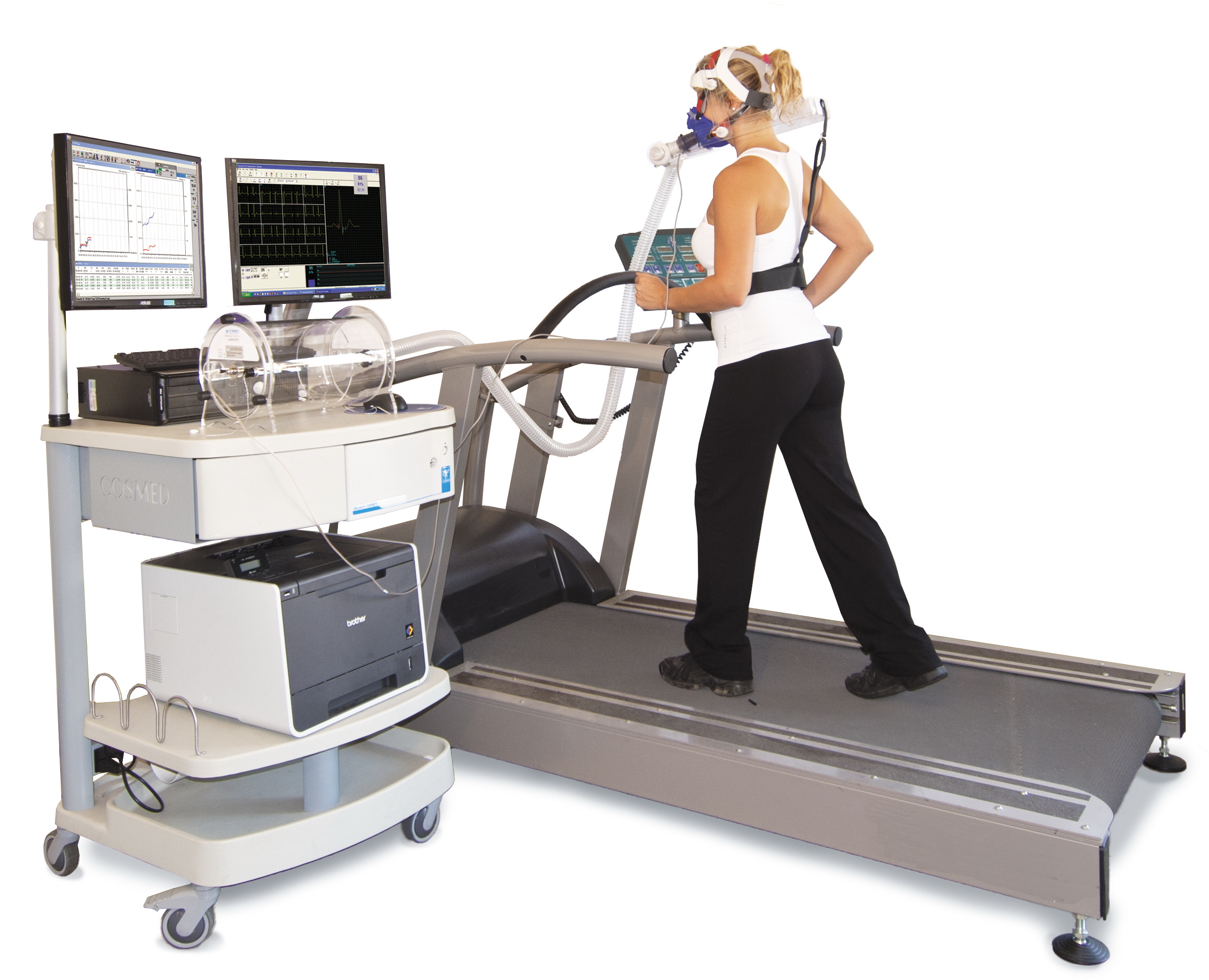
A cardiac stress test (also referred to as a cardiac diagnostic test, cardiopulmonary exercise test, or abbreviated CPX test) is a cardiological test that measures the heart's ability to respond to external stress in a controlled clinical environment. The stress response is induced by exercise or by intravenous pharmacological stimulation. Cardiac stress tests compare the coronary circulation while the patient is at rest with the same patient's circulation during maximum cardiac exertion, showing any abnormal blood flow to the myocardium (heart muscle tissue). The results can be interpreted as a reflection on the general physical condition of the test patient. This test can be used to diagnose coronary artery disease (also known as ischemic heart disease) and assess patient prognosis after a myocardial infarction (heart attack). Exercise-induced stressors are most commonly either exercise on a treadmill or pedalling a stationary exercise bicycle ergometer. The level of stress ... [...More Info...] [...Related Items...] OR: [Wikipedia] [Google] [Baidu] |
Myocardium
Cardiac muscle (also called heart muscle, myocardium, cardiomyocytes and cardiac myocytes) is one of three types of vertebrate muscle tissues, with the other two being skeletal muscle and smooth muscle. It is an involuntary, striated muscle that constitutes the main tissue of the wall of the heart. The cardiac muscle (myocardium) forms a thick middle layer between the outer layer of the heart wall (the pericardium) and the inner layer (the endocardium), with blood supplied via the coronary circulation. It is composed of individual cardiac muscle cells joined by intercalated discs, and encased by collagen fibers and other substances that form the extracellular matrix. Cardiac muscle contracts in a similar manner to skeletal muscle, although with some important differences. Electrical stimulation in the form of a cardiac action potential triggers the release of calcium from the cell's internal calcium store, the sarcoplasmic reticulum. The rise in calcium causes the c ... [...More Info...] [...Related Items...] OR: [Wikipedia] [Google] [Baidu] |
Ischemic Heart Disease
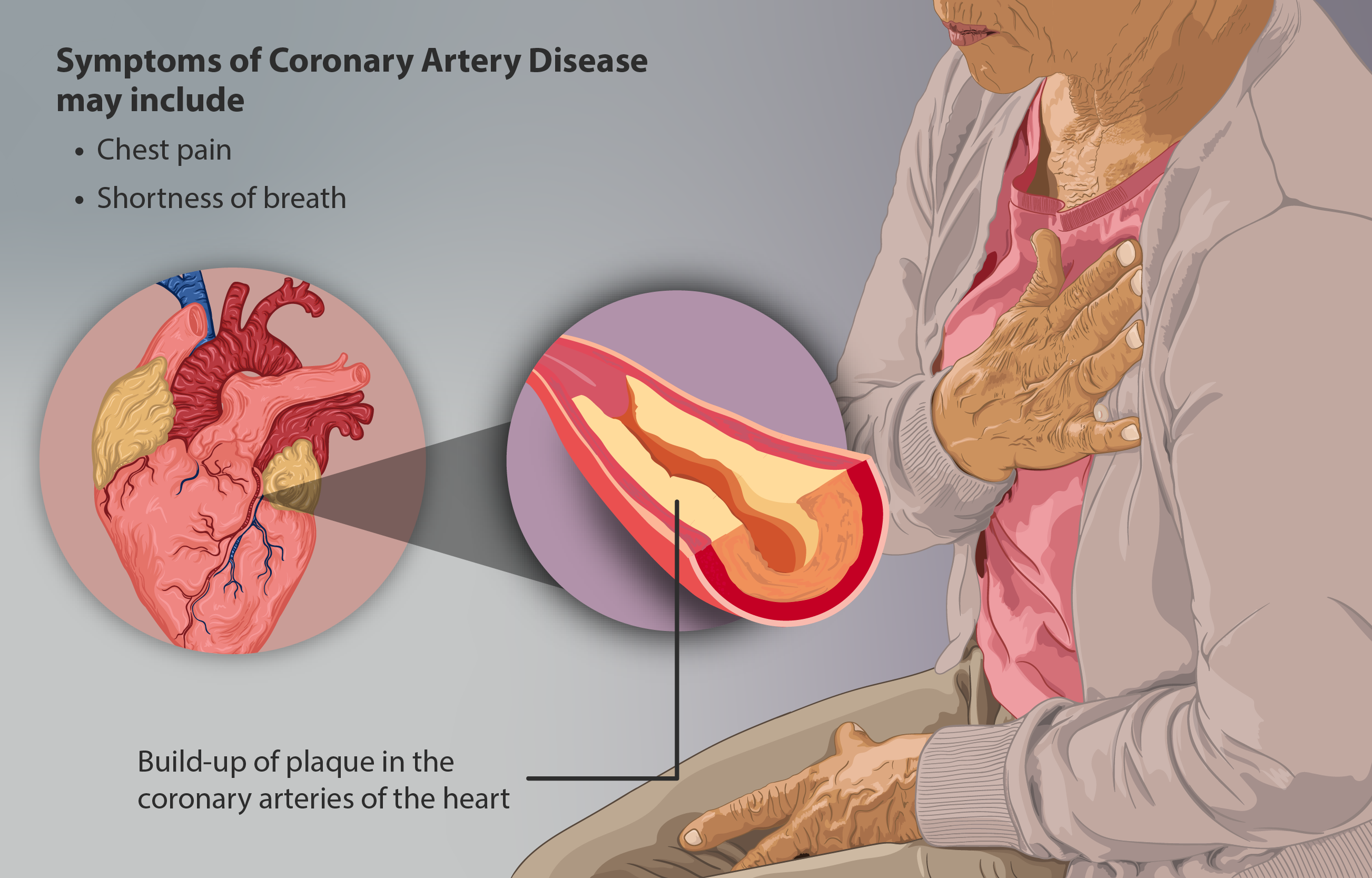
Coronary artery disease (CAD), also called coronary heart disease (CHD), ischemic heart disease (IHD), myocardial ischemia, or simply heart disease, involves the reduction of blood flow to the heart muscle due to build-up of atherosclerotic plaque in the arteries of the heart. It is the most common of the cardiovascular diseases. Types include stable angina, unstable angina, myocardial infarction, and sudden cardiac death. A common symptom is chest pain or discomfort which may travel into the shoulder, arm, back, neck, or jaw. Occasionally it may feel like heartburn. Usually symptoms occur with exercise or emotional stress, last less than a few minutes, and improve with rest. Shortness of breath may also occur and sometimes no symptoms are present. In many cases, the first sign is a heart attack. Other complications include heart failure or an abnormal heartbeat. Risk factors include high blood pressure, smoking, diabetes, lack of exercise, obesity, high blood cholesterol, ... [...More Info...] [...Related Items...] OR: [Wikipedia] [Google] [Baidu] |
Pulmonary Embolus
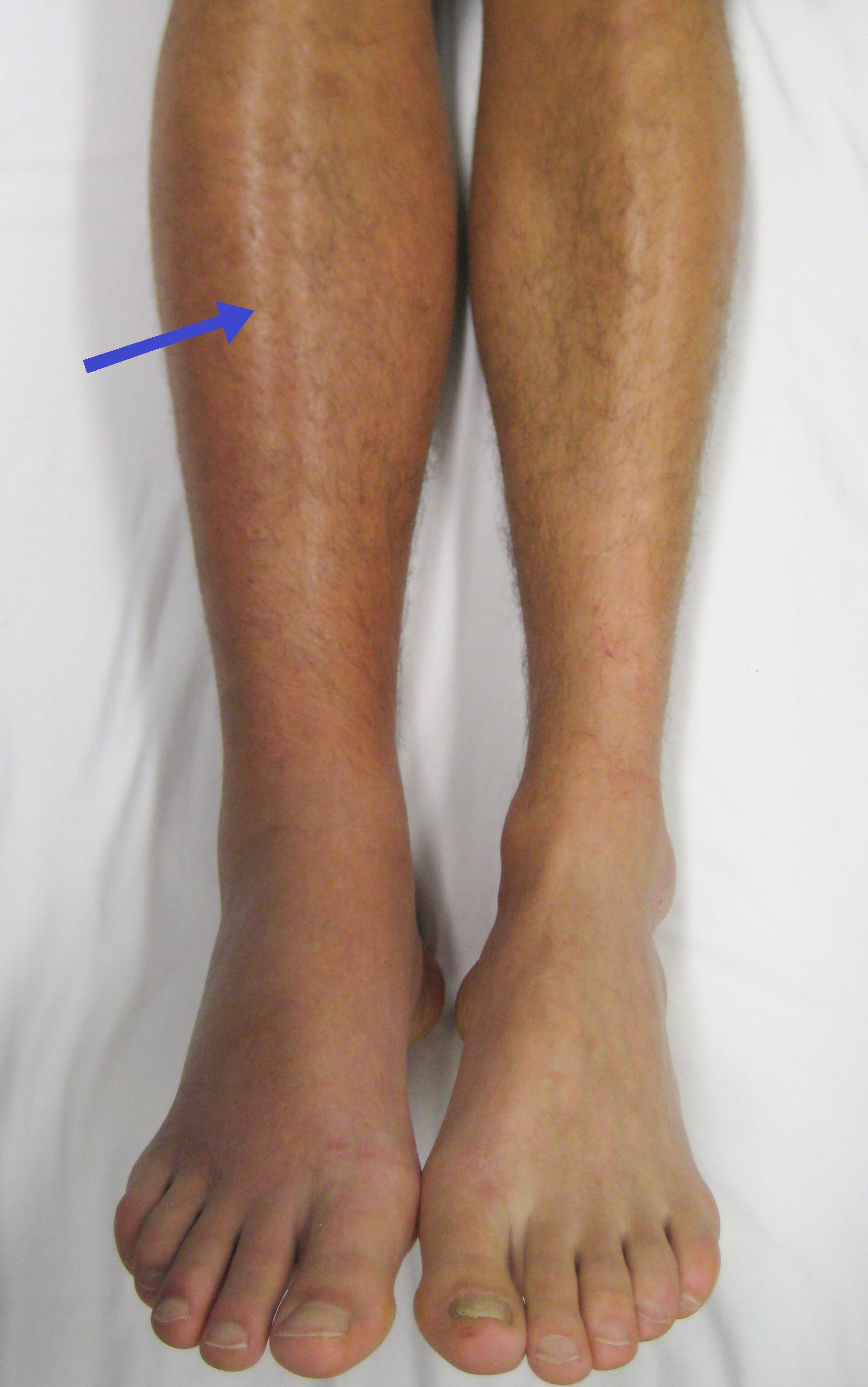
Pulmonary embolism (PE) is a blockage of an artery in the lungs by a substance that has moved from elsewhere in the body through the bloodstream (embolism). Symptoms of a PE may include shortness of breath, chest pain particularly upon breathing in, and coughing up blood. Symptoms of a blood clot in the leg may also be present, such as a red, warm, swollen, and painful leg. Signs of a PE include low blood oxygen levels, rapid breathing, rapid heart rate, and sometimes a mild fever. Severe cases can lead to passing out, abnormally low blood pressure, obstructive shock, and sudden death. PE usually results from a blood clot in the leg that travels to the lung. The risk of blood clots is increased by advanced age, cancer, prolonged bed rest and immobilization, smoking, stroke, long-haul travel over 4 hours, certain genetic conditions, estrogen-based medication, pregnancy, obesity, trauma or bone fracture, and after some types of surgery. A small proportion of cases are du ... [...More Info...] [...Related Items...] OR: [Wikipedia] [Google] [Baidu] |
Functional Neuroimaging
Functional neuroimaging is the use of neuroimaging technology to measure an aspect of brain function, often with a view to understanding the relationship between activity in certain brain areas and specific mental functions. It is primarily used as a research tool in cognitive neuroscience, cognitive psychology, neuropsychology, and social neuroscience. Overview Common methods of functional neuroimaging include * Positron emission tomography (PET) * Functional magnetic resonance imaging (fMRI) * Electroencephalography (EEG) * Magnetoencephalography (MEG) * Functional near-infrared spectroscopy (fNIRS) * Single-photon emission computed tomography (SPECT) * Functional ultrasound imaging (fUS) PET, fMRI, fNIRS and fUS can measure localized changes in cerebral blood flow related to neural activity. These changes are referred to as ''activations''. Regions of the brain which are activated when a subject performs a particular task may play a role in the neural computations which ... [...More Info...] [...Related Items...] OR: [Wikipedia] [Google] [Baidu] |
Myocardial Perfusion Imaging
Myocardial perfusion imaging or scanning (also referred to as MPI or MPS) is a nuclear medicine procedure that illustrates the function of the heart muscle (myocardium). It evaluates many heart conditions, such as coronary artery disease (CAD), hypertrophic cardiomyopathy and heart wall motion abnormalities. It can also detect regions of myocardial infarction by showing areas of decreased resting perfusion. The function of the myocardium is also evaluated by calculating the left ventricular ejection fraction (LVEF) of the heart. This scan is done in conjunction with a cardiac stress test. The diagnostic information is generated by provoking controlled regional ischemia in the heart with variable perfusion. Planar techniques, such as conventional scintigraphy, are rarely used. Rather, single-photon emission computed tomography (SPECT) is more common in the US. With multihead SPECT systems, imaging can often be completed in less than 10 minutes. With SPECT, inferior and posteri ... [...More Info...] [...Related Items...] OR: [Wikipedia] [Google] [Baidu] |


