|
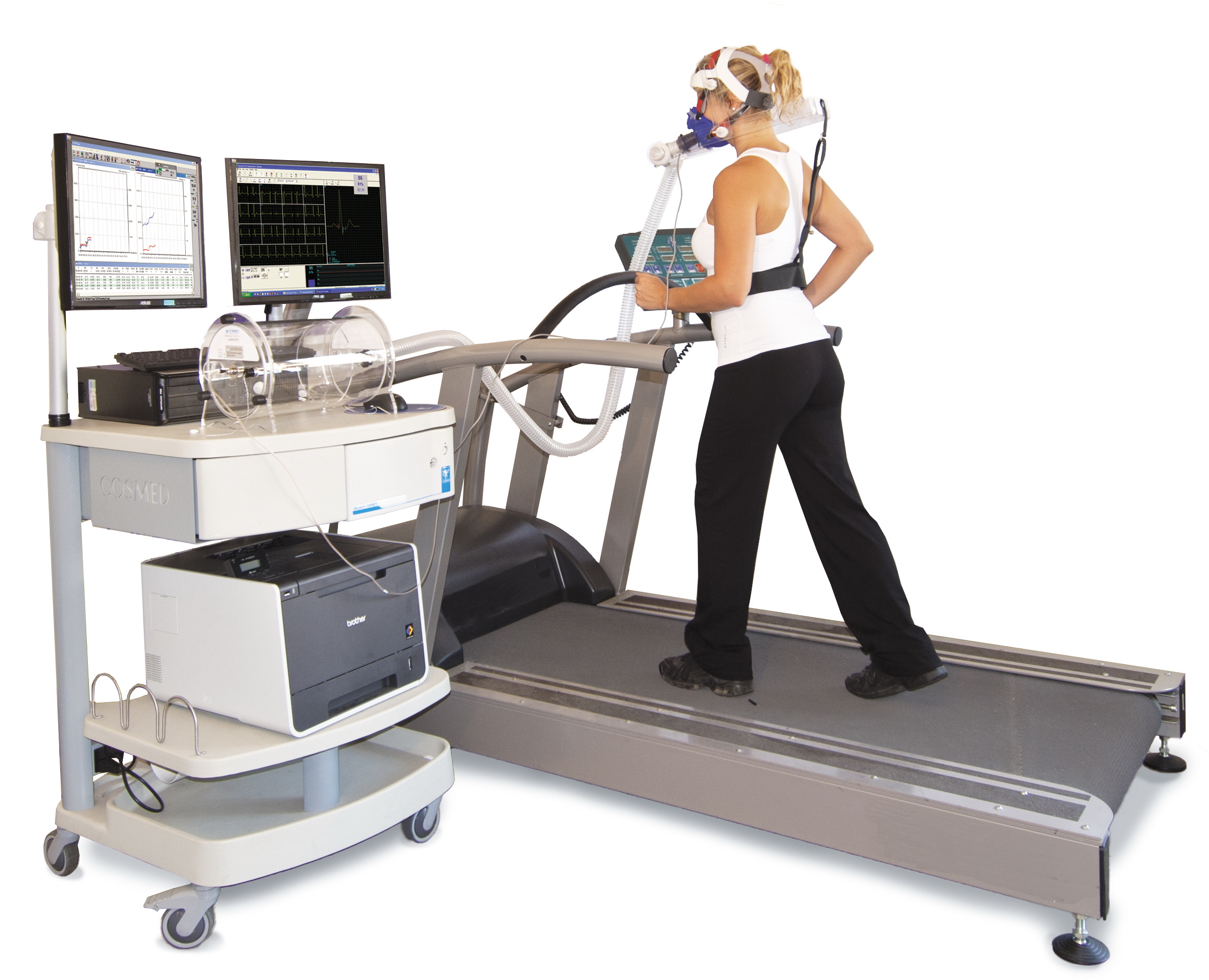
Cardiac Stress Test
A cardiac stress test (also referred to as a cardiac diagnostic test, cardiopulmonary exercise test, or abbreviated CPX test) is a cardiological test that measures the heart's ability to respond to external stress in a controlled clinical environment. The stress response is induced by exercise or by intravenous pharmacological stimulation. Cardiac stress tests compare the coronary circulation while the patient is at rest with the same patient's circulation during maximum cardiac exertion, showing any abnormal blood flow to the myocardium (heart muscle tissue). The results can be interpreted as a reflection on the general physical condition of the test patient. This test can be used to diagnose coronary artery disease (also known as ischemic heart disease) and assess patient prognosis after a myocardial infarction (heart attack). Exercise-induced stressors are most commonly either exercise on a treadmill or pedalling a stationary exercise bicycle ergometer. The level of stress ... [...More Info...] [...Related Items...] OR: [Wikipedia] [Google] [Baidu] |
Cardiology
Cardiology () is a branch of medicine that deals with disorders of the heart and the cardiovascular system. The field includes medical diagnosis and treatment of congenital heart defects, coronary artery disease, heart failure, valvular heart disease and electrophysiology. Physicians who specialize in this field of medicine are called cardiologists, a specialty of internal medicine. Pediatric cardiologists are pediatricians who specialize in cardiology. Physicians who specialize in cardiac surgery are called cardiothoracic surgeons or cardiac surgeons, a specialty of general surgery. Specializations All cardiologists study the disorders of the heart, but the study of adult and child heart disorders each require different training pathways. Therefore, an adult cardiologist (often simply called "cardiologist") is inadequately trained to take care of children, and pediatric cardiologists are not trained to treat adult heart disease. Surgical aspects are not included in ca ... [...More Info...] [...Related Items...] OR: [Wikipedia] [Google] [Baidu] |
Cardiac Catheterization
Cardiac catheterization (heart cath) is the insertion of a catheter into a chamber or vessel of the heart. This is done both for diagnostic and interventional purposes. A common example of cardiac catheterization is coronary catheterization that involves catheterization of the coronary arteries for coronary artery disease and myocardial infarctions ("heart attacks"). Catheterization is most often performed in special laboratories with fluoroscopy and highly maneuverable tables. These "cath labs" are often equipped with cabinets of catheters, stents, balloons, etc. of various sizes to increase efficiency. Monitors show the fluoroscopy imaging, electrocardiogram (ECG), pressure waves, and more. Uses Coronary angiography is a diagnostic procedure that allows visualization of the coronary vessels. Fluoroscopy is used to visualize the lumens of the arteries as a 2-D projection. Should these arteries show narrowing or blockage, then techniques exist to open these arteries. Per ... [...More Info...] [...Related Items...] OR: [Wikipedia] [Google] [Baidu] |
Perfusion
Perfusion is the passage of fluid through the circulatory system or lymphatic system to an organ or a tissue, usually referring to the delivery of blood to a capillary bed in tissue. Perfusion is measured as the rate at which blood is delivered to tissue, or volume of blood per unit time (blood flow) per unit tissue mass. The SI unit is m3/(s·kg), although for human organs perfusion is typically reported in ml/min/g. The word is derived from the French verb "perfuser" meaning to "pour over or through". All animal tissues require an adequate blood supply for health and life. Poor perfusion (malperfusion), that is, ischemia, causes health problems, as seen in cardiovascular disease, including coronary artery disease, cerebrovascular disease, peripheral artery disease, and many other conditions. Tests verifying that adequate perfusion exists are a part of a patient's assessment process that are performed by medical or emergency personnel. The most common methods include ev ... [...More Info...] [...Related Items...] OR: [Wikipedia] [Google] [Baidu] |
Sievert
The sievert (symbol: SvNot be confused with the sverdrup or the svedberg, two non-SI units that sometimes use the same symbol.) is a unit in the International System of Units (SI) intended to represent the stochastic health risk of ionizing radiation, which is defined as the probability of causing radiation-induced cancer and genetic damage. The sievert is important in dosimetry and radiation protection. It is named after Rolf Maximilian Sievert, a Swedish medical physicist renowned for work on radiation dose measurement and research into the biological effects of radiation. The sievert is used for radiation dose quantities such as equivalent dose and effective dose, which represent the risk of external radiation from sources outside the body, and committed dose, which represents the risk of internal irradiation due to inhaled or ingested radioactive substances. According to the International Commission on Radiological Protection (ICRP) one sievert results in a 5.5% probabilit ... [...More Info...] [...Related Items...] OR: [Wikipedia] [Google] [Baidu] |
Thallium(I) Chloride
Thallium(I) chloride, also known as thallous chloride, is a chemical compound with the formula TlCl. This colourless salt is an intermediate in the isolation of thallium from its ores. Typically, an acidic solution of thallium(I) sulfate is treated with hydrochloric acid to precipitate insoluble thallium(I) chloride. This solid crystallizes in the caesium chloride motif. The low solubility of TlCl is exploited in chemical synthesis: treatment of metal chloride complexes with TlPF6, gives the corresponding metal hexafluorophosphate derivative. The resulting TlCl precipitate is separated by filtration of the reaction mixture. The overall methodology is similar to the use of AgPF6, except that Tl+ is much less oxidizing. The crystalline structure is of cubic caesium chloride type at room temperature, but it lowers to the orthorhombic thallium iodide type upon cooling, the transition temperature being likely affected by the impurities. Nanometer-thin TlCl films grown on KBr subst ... [...More Info...] [...Related Items...] OR: [Wikipedia] [Google] [Baidu] |
Myoview
Technetium (99mTc) tetrofosmin is a drug used in nuclear medicine cardiac imaging. It is sold under the brand name Myoview (GE Healthcare). The radioisotope, technetium-99m, is chelated by two 1,2-''bis'' i-(2-ethoxyethyl)phosphinothane ligands which belong to the group of diphosphines and which are referred to as tetrofosmin. Tc-99m tetrofosmin is rapidly taken up by myocardial tissue and reaches its maximum level in approximately 5 minutes. About 66% of the total injected dose is excreted within 48 hours after injection (40% urine, 26% feces). Tc-99m tetrofosmin is indicated for use in scintigraphic imaging of the myocardium under stress and rest conditions. It is used to determine areas of reversible ischemia and infarcted tissue in the heart. It is also indicated to detect changes in perfusion induced by pharmacologic stress (adenosine, lexiscan, dobutamine Dobutamine is a medication used in the treatment of cardiogenic shock (as a result of inadequate tissue perfusion) ... [...More Info...] [...Related Items...] OR: [Wikipedia] [Google] [Baidu] |
Technetium (99mTc) Sestamibi
Technetium (99mTc) sestamibi (INN) (commonly sestamibi; USP: technetium Tc 99m sestamibi; trade name Cardiolite) is a pharmaceutical agent used in nuclear medicine imaging. The drug is a coordination complex consisting of the radioisotope technetium-99m bound to six (sesta=6) methoxyisobutylisonitrile (MIBI) ligands. The anion is not defined. The generic drug became available late September 2008. A scan of a patient using MIBI is commonly known as a "MIBI scan". Sestamibi is taken up by tissues with large numbers of mitochondria and negative plasma membrane potentials. Sestamibi is mainly used to image the myocardium (heart muscle). It is also used in the work-up of primary hyperparathyroidism to identify parathyroid adenomas, for radioguided surgery of the parathyroid and in the work-up of possible breast cancer. Cardiac imaging (MIBI scan) A ''MIBI scan'' or ''sestamibi scan'' is now a common method of cardiac imaging. Technetium (99mTc) sestamibi is a lipophilic cation ... [...More Info...] [...Related Items...] OR: [Wikipedia] [Google] [Baidu] |
Radioactive Tracer
A radioactive tracer, radiotracer, or radioactive label is a chemical compound in which one or more atoms have been replaced by a radionuclide so by virtue of its radioactive decay it can be used to explore the mechanism of chemical reactions by tracing the path that the radioisotope follows from reactants to products. Radiolabeling or radiotracing is thus the radioactive form of isotopic labeling. In biological contexts, use of radioisotope tracers are sometimes called radioisotope feeding experiments. Radioisotopes of hydrogen, carbon, phosphorus, sulfur, and iodine have been used extensively to trace the path of biochemical reactions. A radioactive tracer can also be used to track the distribution of a substance within a natural system such as a cell or tissue, or as a flow tracer to track fluid flow. Radioactive tracers are also used to determine the location of fractures created by hydraulic fracturing in natural gas production.Reis, John C. (1976). ''Environmental Con ... [...More Info...] [...Related Items...] OR: [Wikipedia] [Google] [Baidu] |
Myocardial Perfusion Imaging
Myocardial perfusion imaging or scanning (also referred to as MPI or MPS) is a nuclear medicine procedure that illustrates the function of the heart muscle (myocardium). It evaluates many heart conditions, such as coronary artery disease (CAD), hypertrophic cardiomyopathy and heart wall motion abnormalities. It can also detect regions of myocardial infarction by showing areas of decreased resting perfusion. The function of the myocardium is also evaluated by calculating the left ventricular ejection fraction (LVEF) of the heart. This scan is done in conjunction with a cardiac stress test. The diagnostic information is generated by provoking controlled regional ischemia in the heart with variable perfusion. Planar techniques, such as conventional scintigraphy, are rarely used. Rather, single-photon emission computed tomography (SPECT) is more common in the US. With multihead SPECT systems, imaging can often be completed in less than 10 minutes. With SPECT, inferior and posteri ... [...More Info...] [...Related Items...] OR: [Wikipedia] [Google] [Baidu] |


