|
Investing Layer Of Deep Cervical Fascia
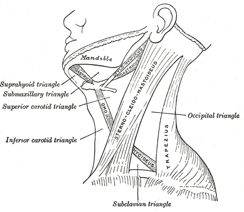
The investing layer of deep cervical fascia is the most superficial part of the deep cervical fascia, and encloses the whole neck. It is considered by some sources to be incomplete or nonexistent. Attachments It surrounds the neck like a collar, it splits around the sternocleidomastoid muscle and the trapezius muscle. It is attached as; * Posteriorly - Ligamentum nuchae * Anteriorly - Attached to the hyoid bone * Superiorly - (from backwards to forwards); ** External occipital protuberance and Superior nuchal line of occipital bone ** Mastoid process of Temporal bone ** External acoustic meatus ** Lower margin of the zygomatic arch ** Lower border of body of mandible from the angle of mandible to the symphysis menti * Inferiorly - (from backwards to forwards); ** Spine and acromial process of scapula ** Upper surface of the clavicle **Suprasternal notch of manubrium sterni Tracings * Horizontal extent - From ligamentum nuchae when traced forward, the fascia splits and enclo ... [...More Info...] [...Related Items...] OR: [Wikipedia] [Google] [Baidu] |
Cervical Vertebra
In tetrapods, cervical vertebrae (singular: vertebra) are the vertebrae of the neck, immediately below the skull. Truncal vertebrae (divided into thoracic and lumbar vertebrae in mammals) lie caudal (toward the tail) of cervical vertebrae. In sauropsid species, the cervical vertebrae bear cervical ribs. In lizards and saurischian dinosaurs, the cervical ribs are large; in birds, they are small and completely fused to the vertebrae. The vertebral transverse processes of mammals are homologous to the cervical ribs of other amniotes. Most mammals have seven cervical vertebrae, with the only three known exceptions being the manatee with six, the two-toed sloth with five or six, and the three-toed sloth with nine. In humans, cervical vertebrae are the smallest of the true vertebrae and can be readily distinguished from those of the thoracic or lumbar regions by the presence of a foramen (hole) in each transverse process, through which the vertebral artery, vertebral veins, and inferio ... [...More Info...] [...Related Items...] OR: [Wikipedia] [Google] [Baidu] |
Zygomatic Arch
In anatomy, the zygomatic arch, or cheek bone, is a part of the skull formed by the zygomatic process of the temporal bone (a bone extending forward from the side of the skull, over the opening of the ear) and the temporal process of the zygomatic bone (the side of the cheekbone), the two being united by an oblique suture (the zygomaticotemporal suture); the tendon of the temporal muscle passes medial to (i.e. through the middle of) the arch, to gain insertion into the coronoid process of the mandible (jawbone). The jugal point is the point at the anterior (towards face) end of the upper border of the zygomatic arch where the masseteric and maxillary edges meet at an angle, and where it meets the process of the zygomatic bone. The arch is typical of '' Synapsida'' (“fused arch”), a clade of amniotes that includes mammals and their extinct relatives, such as ''Moschops'' and '' Dimetrodon''. Structure The zygomatic process of the temporal arises by two roots: * an ''anter ... [...More Info...] [...Related Items...] OR: [Wikipedia] [Google] [Baidu] |
Temporal Styloid Process
The temporal styloid process is a slender bony process of the temporal bone extending downward and forward from the undersurface of the temporal bone just below the ear. The styloid process gives attachments to several muscles, and ligaments. Structure The styloid process is a slender and pointed bony process of the temporal bone projecting anteroinferiorly from the inferior surface of the temporal bone just below the ear. Its length normally ranges from just under 3 cm to just over 4 cm. It is usually nearly straight, but may be curved in some individuals. Its ''proximal'' (''tympanohyal'') ''part'' is ensheathed by the tympanic part of the temporal bone ''(vaginal process), whereas'' its ''distal (stylohyal)'' ''part'' gives attachment to several structures. Attachments The styloid process gives attachments to several muscles, and ligaments. It serves as an anchor point for several muscles associated with the tongue and larynx. * stylohyoid ligament * stylomandibu ... [...More Info...] [...Related Items...] OR: [Wikipedia] [Google] [Baidu] |
Parotid Gland
The parotid gland is a major salivary gland in many animals. In humans, the two parotid glands are present on either side of the mouth and in front of both ears. They are the largest of the salivary glands. Each parotid is wrapped around the mandibular ramus, and secretes serous saliva through the parotid duct into the mouth, to facilitate mastication and swallowing and to begin the digestion of starches. There are also two other types of salivary glands; they are submandibular and sublingual glands. Sometimes accessory parotid glands are found close to the main parotid glands. Etymology The word ''parotid'' literally means "beside the ear". From Greek παρωτίς (stem παρωτιδ-) : (gland) behind the ear < παρά - pará : in front, and οὖς - ous (stem ὠτ-, ōt-) : ear. Structure The parotid glands are a pair of mainly |
Submandibular Gland
The paired submandibular glands (historically known as submaxillary glands) are major salivary glands located beneath the floor of the mouth. They each weigh about 15 grams and contribute some 60–67% of unstimulated saliva secretion; on stimulation their contribution decreases in proportion as the parotid secretion rises to 50%. The average length of the normal human submandibular salivary gland is approximately 27mm, while the average width is approximately 14.3mm. Structure Lying superior to the digastric muscles, each submandibular gland is divided into superficial and deep lobes, which are separated by the mylohyoid muscle: * The superficial lobe comprises most of the gland, with the mylohyoid muscle runs under it * The deep lobe is the smaller part Secretions are delivered into the submandibular duct on the deep portion after which they hook around the posterior edge of the mylohyoid muscle and proceed on the superior surface laterally. The excretory ducts are then cross ... [...More Info...] [...Related Items...] OR: [Wikipedia] [Google] [Baidu] |
Sternocleidomastoid
The sternocleidomastoid muscle is one of the largest and most superficial cervical muscles. The primary actions of the muscle are rotation of the head to the opposite side and flexion of the neck. The sternocleidomastoid is innervated by the accessory nerve. Etymology and location It is given the name ''sternocleidomastoid'' because it originates at the manubrium of the sternum (''sterno-'') and the clavicle (''cleido-'') and has an insertion at the mastoid process of the temporal bone of the skull. Structure The sternocleidomastoid muscle originates from two locations: the manubrium of the sternum and the clavicle. It travels obliquely across the side of the neck and inserts at the mastoid process of the temporal bone of the skull by a thin aponeurosis. The sternocleidomastoid is thick and narrow at its centre, and broader and thinner at either end. The sternal head is a round fasciculus, tendinous in front, fleshy behind, arising from the upper part of the front of the manu ... [...More Info...] [...Related Items...] OR: [Wikipedia] [Google] [Baidu] |
Ligamentum Nuchae
The nuchal ligament is a ligament at the back of the neck that is continuous with the supraspinous ligament. Structure The nuchal ligament extends from the external occipital protuberance on the skull and median nuchal line to the spinous process of the seventh cervical vertebra in the lower part of the neck. From the anterior border of the nuchal ligament, a fibrous lamina is given off. This is attached to the posterior tubercle of the atlas, and to the spinous processes of the cervical vertebrae, and forms a septum between the muscles on either side of the neck. The trapezius and splenius capitis muscle attach to the nuchal ligament. Function It is a tendon-like structure that has developed independently in humans and other animals well adapted for running. In some four-legged animals, particularly ungulates, the nuchal ligament serves to sustain the weight of the head. Clinical significance In Chiari malformation treatment, decompression and duraplasty with a harvested n ... [...More Info...] [...Related Items...] OR: [Wikipedia] [Google] [Baidu] |
Manubrium Sterni
The sternum or breastbone is a long flat bone located in the central part of the chest. It connects to the ribs via cartilage and forms the front of the rib cage, thus helping to protect the heart, lungs, and major blood vessels from injury. Shaped roughly like a necktie, it is one of the largest and longest flat bones of the body. Its three regions are the manubrium, the body, and the xiphoid process. The word "sternum" originates from the Ancient Greek στέρνον (stérnon), meaning "chest". Structure The sternum is a narrow, flat bone, forming the middle portion of the front of the chest. The top of the sternum supports the clavicles (collarbones) and its edges join with the costal cartilages of the first two pairs of ribs. The inner surface of the sternum is also the attachment of the sternopericardial ligaments. Its top is also connected to the sternocleidomastoid muscle. The sternum consists of three main parts, listed from the top: * Manubrium * Body (gladiolus) * X ... [...More Info...] [...Related Items...] OR: [Wikipedia] [Google] [Baidu] |
Clavicle
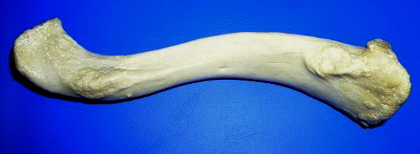
The clavicle, or collarbone, is a slender, S-shaped long bone approximately 6 inches (15 cm) long that serves as a strut between the shoulder blade and the sternum (breastbone). There are two clavicles, one on the left and one on the right. The clavicle is the only long bone in the body that lies horizontally. Together with the shoulder blade, it makes up the shoulder girdle. It is a palpable bone and, in people who have less fat in this region, the location of the bone is clearly visible. It receives its name from the Latin ''clavicula'' ("little key"), because the bone rotates along its axis like a key when the shoulder is abducted. The clavicle is the most commonly fractured bone. It can easily be fractured by impacts to the shoulder from the force of falling on outstretched arms or by a direct hit. Structure The collarbone is a thin doubly curved long bone that connects the arm to the trunk of the body. Located directly above the first rib, it acts as a strut to k ... [...More Info...] [...Related Items...] OR: [Wikipedia] [Google] [Baidu] |
Scapula
The scapula (plural scapulae or scapulas), also known as the shoulder blade, is the bone that connects the humerus (upper arm bone) with the clavicle (collar bone). Like their connected bones, the scapulae are paired, with each scapula on either side of the body being roughly a mirror image of the other. The name derives from the Classical Latin word for trowel or small shovel, which it was thought to resemble. In compound terms, the prefix omo- is used for the shoulder blade in medical terminology. This prefix is derived from ὦμος (ōmos), the Ancient Greek word for shoulder, and is cognate with the Latin , which in Latin signifies either the shoulder or the upper arm bone. The scapula forms the back of the shoulder girdle. In humans, it is a flat bone, roughly triangular in shape, placed on a posterolateral aspect of the thoracic cage. Structure The scapula is a thick, flat bone lying on the thoracic wall that provides an attachment for three groups of muscles: intrin ... [...More Info...] [...Related Items...] OR: [Wikipedia] [Google] [Baidu] |
Inferiorly
Standard anatomical terms of location are used to unambiguously describe the anatomy of animals, including humans. The terms, typically derived from Latin or Greek language, Greek roots, describe something in its standard anatomical position. This position provides a definition of what is at the front ("anterior"), behind ("posterior") and so on. As part of defining and describing terms, the body is described through the use of anatomical planes and anatomical axis, anatomical axes. The meaning of terms that are used can change depending on whether an organism is bipedal or quadrupedal. Additionally, for some animals such as invertebrates, some terms may not have any meaning at all; for example, an animal that is radially symmetrical will have no anterior surface, but can still have a description that a part is close to the middle ("proximal") or further from the middle ("distal"). International organisations have determined vocabularies that are often used as standard vocabular ... [...More Info...] [...Related Items...] OR: [Wikipedia] [Google] [Baidu] |
Symphysis Menti
In human anatomy, the facial skeleton of the skull the external surface of the mandible is marked in the median line by a faint ridge, indicating the mandibular symphysis (Latin: ''symphysis menti'') or line of junction where the two lateral halves of the mandible typically fuse at an early period of life (1-2 years). It is not a true symphysis as there is no cartilage between the two sides of the mandible. This ridge divides below and encloses a triangular eminence, the mental protuberance, the base of which is depressed in the center but raised on either side to form the mental tubercle. The lowest (most inferior) end of the mandibular symphysis — the point of the chin — is called the "menton". It serves as the origin for the geniohyoid and the genioglossus muscles. Other animals Solitary mammalian carnivores that rely on a powerful canine bite to subdue their prey have a strong mandibular symphysis, while pack hunters delivering shallow bites have a weaker one. When filter ... [...More Info...] [...Related Items...] OR: [Wikipedia] [Google] [Baidu] |


