|
Parotid Gland
The parotid gland is a major salivary gland in many animals. In humans, the two parotid glands are present on either side of the mouth and in front of both ears. They are the largest of the salivary glands. Each parotid is wrapped around the mandibular ramus, and secretes serous saliva through the parotid duct into the mouth, to facilitate mastication and swallowing and to begin the digestion of starches. There are also two other types of salivary glands; they are submandibular and sublingual glands. Sometimes accessory parotid glands are found close to the main parotid glands. Etymology The word ''parotid'' literally means "beside the ear". From Greek παρωτίς (stem παρωτιδ-) : (gland) behind the ear < παρά - pará : in front, and οὖς - ous (stem ὠτ-, ōt-) : ear. Structure The parotid glands are a pair of mainly serous salivary gland ...[...More Info...] [...Related Items...] OR: [Wikipedia] [Google] [Baidu] |
Digestive System
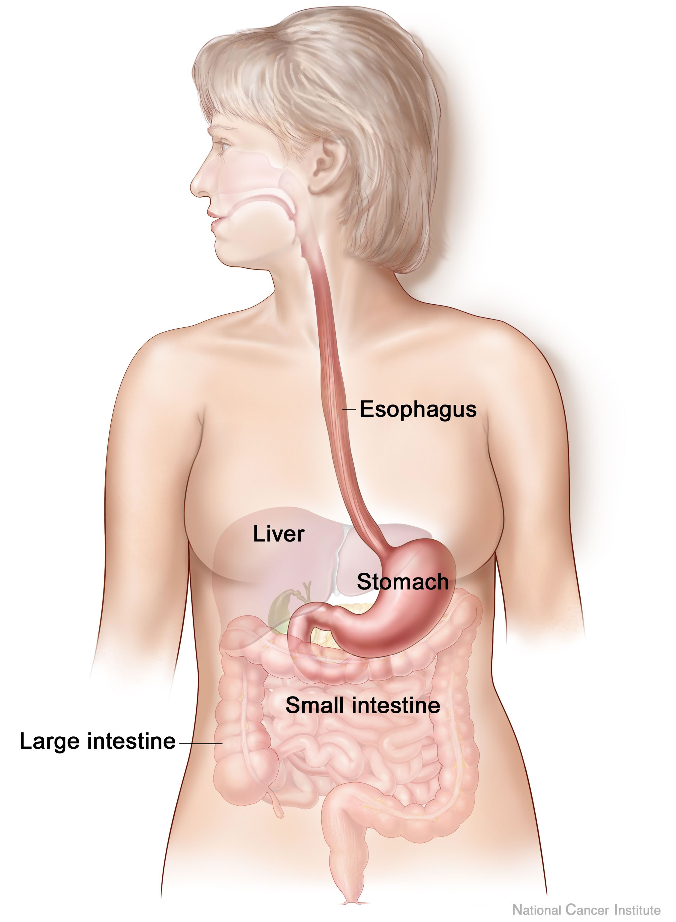
The human digestive system consists of the gastrointestinal tract plus the accessory organs of digestion (the tongue, salivary glands, pancreas, liver, and gallbladder). Digestion involves the breakdown of food into smaller and smaller components, until they can be absorbed and assimilated into the body. The process of digestion has three stages: the cephalic phase, the gastric phase, and the intestinal phase. The first stage, the cephalic phase of digestion, begins with secretions from gastric glands in response to the sight and smell of food. This stage includes the mechanical breakdown of food by chewing, and the chemical breakdown by digestive enzymes, that takes place in the mouth. Saliva contains the digestive enzymes amylase, and lingual lipase, secreted by the salivary and serous glands on the tongue. Chewing, in which the food is mixed with saliva, begins the mechanical process of digestion. This produces a bolus which is swallowed down the esophagus to ... [...More Info...] [...Related Items...] OR: [Wikipedia] [Google] [Baidu] |
Palpated
Palpation is the process of using one's hands to check the body, especially while perceiving/diagnosing a disease or illness. Usually performed by a health care practitioner, it is the process of feeling an object in or on the body to determine its size, shape, firmness, or location (for example, a veterinarian can feel the stomach of a pregnant animal to ensure good health and successful delivery). Palpation is an important part of the physical examination; the sense of touch is just as important in this examination as the sense of sight is. Physicians develop great skill in palpating problems below the surface of the body, becoming able to detect things that untrained persons would not. Mastery of anatomy and much practice are required to achieve a high level of skill. The concept of being able to detect or notice subtle tactile signs and to recognize their significance or implications is called appreciating them (just as in general vocabulary one can speak of appreciating th ... [...More Info...] [...Related Items...] OR: [Wikipedia] [Google] [Baidu] |
Ectopia (medicine)
An ectopia () is a displacement or malposition of an organ or other body part, which is then referred to as ectopic ({{IPAc-en, ɛ, k, ˈ, t, ɒ, p, ɪ, k). Most ectopias are congenital, but some may happen later in life. Examples * Ectopic ACTH syndrome, also known as small-cell carcinoma. * Ectopic calcification, a pathologic deposition of calcium salts in tissues or bone growth in soft tissues * Cerebellar tonsillar ectopia, aka Chiari malformation, a herniation of the brain through the foramen magnum, which may be congenital or caused by trauma. *Ectopic cilia, a hair growing where it isn't supposed to be, commonly an eyelash on an abnormal spot on the eyelid, distichia * Ectopia cordis, the displacement of the heart outside the body during fetal development *Ectopic enamel, a tooth abnormality, where enamel is found in an unusual location, such as at the root of a tooth * Ectopic expression, the expression of a gene in an abnormal place in an organism * Ectopic hormone, a ho ... [...More Info...] [...Related Items...] OR: [Wikipedia] [Google] [Baidu] |
List Of Anatomical Variations
This article lists anatomical variations that are not deemed inherently pathological. {{incomplete list, date=December 2013 Accessory features Bones * Cervical rib * Fabella * Foramen tympanicum * Supracondylar process of the humerus * Sternal foramen * Stafne bone cavity * Episternal ossicles * Fossa navicularis magna * Transverse basilar fissure - or ''Saucer's fissure'' * Canalis basilaris medianus * Craniopharyngeal canal * Intermediate condylar canal * Foramen arcuale * Os odontoideum * Os acromiale * Ossiculum terminale (of dens) * Scapular foramina and tunnels Muscles * Accessory soleus muscle * Axillary arch * Epitrochleoanconeus muscle - or ''anconeous epitrochlearis'' * Extensor medii proprius muscle * Extensor digitorum brevis manus muscle * Extensor indicis et medii communis muscle * Extensor pollicis et indicis communis muscle * Extensor carpi radialis tertius muscle - or ''extensor carpi radialis accessorius'' * Linburg-Comstock variation - o ... [...More Info...] [...Related Items...] OR: [Wikipedia] [Google] [Baidu] |
Maxillary Artery
The maxillary artery supplies deep structures of the face. It branches from the external carotid artery just deep to the neck of the mandible. Structure The maxillary artery, the larger of the two terminal branches of the external carotid artery, arises behind the neck of the mandible, and is at first imbedded in the substance of the parotid gland; it passes forward between the ramus of the mandible and the sphenomandibular ligament, and then runs, either superficial or deep to the lateral pterygoid muscle, to the pterygopalatine fossa. It supplies the deep structures of the face, and may be divided into mandibular, pterygoid, and pterygopalatine portions. First portion The ''first'' or ''mandibular '' or ''bony'' portion passes horizontally forward, between the neck of the mandible and the sphenomandibular ligament, where it lies parallel to and a little below the auriculotemporal nerve; it crosses the inferior alveolar nerve, and runs along the lower border of the lateral ... [...More Info...] [...Related Items...] OR: [Wikipedia] [Google] [Baidu] |
Great Auricular Nerve
The great auricular nerve is a cutaneous nerve of the head. It originates from the cervical plexus, with branches of spinal nerves C2 and C3. It provides sensory nerve supply to the skin over the parotid gland and the mastoid process of the temporal bone, and surfaces of the outer ear. Pain resulting from parotitis is caused by an impingement on the great auricular nerve. Structure The great auricular nerve is the largest of the ascending branches of the cervical plexus. It arises from the second and third cervical nerves. It winds around the posterior border of the sternocleidomastoid muscle, and, after perforating the deep fascia, ascends upon that muscle beneath the platysma muscle to the parotid gland. Here, it divides into an anterior and a posterior branch. Branches * The anterior branch (ramus anterior; facial branch) is distributed to the skin of the face over the parotid gland. It communicates with the facial nerve inside the parotid gland. * The posterior ... [...More Info...] [...Related Items...] OR: [Wikipedia] [Google] [Baidu] |
Superficial Temporal Artery
In human anatomy, the superficial temporal artery is a major artery of the head. It arises from the external carotid artery when it splits into the superficial temporal artery and maxillary artery. Its pulse can be felt above the zygomatic arch, above and in front of the tragus of the ear. Structure The superficial temporal artery is the smaller of two end branches that split superiorly from the external carotid. Based on its direction, the superficial temporal artery appears to be a continuation of the external carotid. It begins within the parotid gland, behind the neck of the mandible, and passes superficially over the posterior root of the zygomatic process of the temporal bone; about 5 cm above this process it divides into two branches: ''a. frontal'', and ''a. parietal''. Branches The parietal branch of the superficial temporal artery (posterior temporal) is a small artery in the head. It is larger than the frontal branch and curves upward and backward on the s ... [...More Info...] [...Related Items...] OR: [Wikipedia] [Google] [Baidu] |
External Carotid Artery
The external carotid artery is a major artery of the head and neck. It arises from the common carotid artery when it splits into the external and internal carotid artery. External carotid artery supplies blood to the face and neck. Structure The external carotid artery begins at the upper border of thyroid cartilage, and curves, passing forward and upward, and then inclining backward to the space behind the neck of the mandible, where it divides into the superficial temporal and maxillary artery within the parotid gland. It rapidly diminishes in size as it travels up the neck, owing to the number and large size of its branches. At its origin, this artery is closer to the skin and more medial than the internal carotid, and is situated within the carotid triangle. Development In children, the external carotid artery is somewhat smaller than the internal carotid; but in the adult, the two vessels are of nearly equal size. Relations At the origin, external carotid artery ... [...More Info...] [...Related Items...] OR: [Wikipedia] [Google] [Baidu] |
Retromandibular Vein
The retromandibular vein (temporomaxillary vein, posterior facial vein) is a major vein of the face. Anatomy Origin The retromandibular vein is formed by the union of the superficial temporal and maxillary veins. Course It descends in the substance of the parotid gland, superficial to the external carotid artery (but beneath the facial nerve), between the ramus of the mandible and the sternocleidomastoideus muscle. It terminates by dividing into two branches: * an ''anterior'', which passes forward and joins anterior facial vein, to form the common facial vein, which then drains into the internal jugular vein. * a ''posterior'', which is joined by the posterior auricular vein and becomes the external jugular vein. Function The retromandibular vein provides venous drainage to the superior cranium The skull is a bone protective cavity for the brain. The skull is composed of four types of bone i.e., cranial bones, facial bones, ear ossicles and hyoid bone. Ho ... [...More Info...] [...Related Items...] OR: [Wikipedia] [Google] [Baidu] |
Facial Nerve
The facial nerve, also known as the seventh cranial nerve, cranial nerve VII, or simply CN VII, is a cranial nerve that emerges from the pons of the brainstem, controls the muscles of facial expression, and functions in the conveyance of taste sensations from the anterior two-thirds of the tongue. The nerve typically travels from the pons through the facial canal in the temporal bone and exits the skull at the stylomastoid foramen. It arises from the brainstem from an area posterior to the cranial nerve VI (abducens nerve) and anterior to cranial nerve VIII (vestibulocochlear nerve). The facial nerve also supplies preganglionic parasympathetic fibers to several head and neck ganglia. The facial and intermediate nerves can be collectively referred to as the nervus intermediofacialis. The path of the facial nerve can be divided into six segments: # intracranial (cisternal) segment # meatal (canalicular) segment (within the internal auditory canal) # labyrinthine segmen ... [...More Info...] [...Related Items...] OR: [Wikipedia] [Google] [Baidu] |
Anatomical Terms Of Location
Standard anatomical terms of location are used to unambiguously describe the anatomy of animals, including humans. The terms, typically derived from Latin or Greek roots, describe something in its standard anatomical position. This position provides a definition of what is at the front ("anterior"), behind ("posterior") and so on. As part of defining and describing terms, the body is described through the use of anatomical planes and anatomical axes. The meaning of terms that are used can change depending on whether an organism is bipedal or quadrupedal. Additionally, for some animals such as invertebrates, some terms may not have any meaning at all; for example, an animal that is radially symmetrical will have no anterior surface, but can still have a description that a part is close to the middle ("proximal") or further from the middle ("distal"). International organisations have determined vocabularies that are often used as standard vocabularies for subdisciplines o ... [...More Info...] [...Related Items...] OR: [Wikipedia] [Google] [Baidu] |
Maxillary Second Molar
The maxillary second molar is the tooth located distally (away from the midline of the face) from both the maxillary first molars of the mouth but mesial (toward the midline of the face) from both maxillary third molars. This is true only in permanent teeth. In deciduous (baby) teeth, the maxillary second molar is the last tooth in the mouth and does not have a third molar behind it. The function of this molar is similar to that of all molars in regard to grinding being the principal action during mastication, commonly known as chewing. There are usually four cusps on maxillary molars, two on the buccal (side nearest the cheek) and two palatal (side nearest the palate). There are great differences between the deciduous (baby) maxillary molars and those of the permanent maxillary molars, even though their function are similar. The permanent maxillary molars are not considered to have any teeth that precede it. Despite being named molars, the deciduous molars are followed b ... [...More Info...] [...Related Items...] OR: [Wikipedia] [Google] [Baidu] |


