|
Iniencephaly
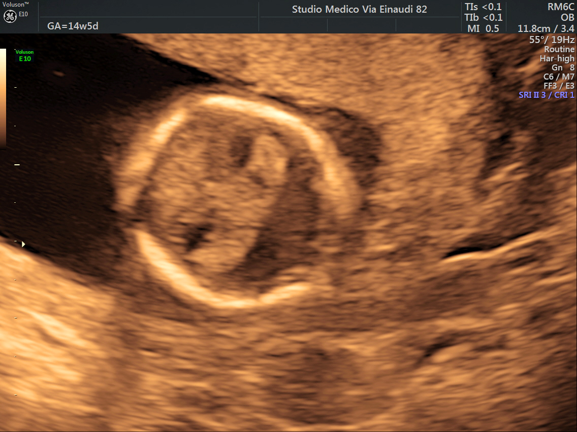
Iniencephaly is a rare type of cephalic disorder characterised by three common characteristics: a defect to the occipital bone, spina bifida of the cervical vertebrae and retroflexion (backward bending) of the head on the cervical spine. Stillbirth is the most common outcome, with a few rare examples of live birth, after which death invariably occurs within a short time. The disorder was first described by Étienne Geoffroy Saint-Hilaire in 1836. The name is derived from the Ancient Greek word ἰνίον ''inion'', for the occipital bone/nape of the neck. Classifications There are two types of iniencephaly. The more severe group is iniencephaly apertus (open iniencephaly), involving the development of an encephalocele. In the other group, iniencephaly clausus (closed iniencephaly), the encephalocele is absent. Signs and symptoms The affected infant tends to be short, with a disproportionately large head. The fetal head of infants born with iniencephaly are hyperextended whil ... [...More Info...] [...Related Items...] OR: [Wikipedia] [Google] [Baidu] |
Klippel–Feil Syndrome
Klippel–Feil syndrome (KFS), also known as cervical vertebral fusion syndrome, is a rare congenital condition characterized by the abnormal fusion of any two of the seven bones in the neck (cervical vertebrae). It results in a limited ability to move the neck and shortness of the neck, resulting in the appearance of a low hairline. The syndrome is difficult to diagnose, as it occurs in a group of patients affected with many different abnormalities who can only be unified by the presence of fused or segmental cervical vertebrae. KFS is not always genetic and not always known about on the date of birth. The disease was initially reported in 1884 by Maurice Klippel and André Feil from France. In 1919, in his Doctor of Philosophy thesis, André Feil suggested another classification of the syndrome, encompassing not only deformation of the cervical spine, but also deformation of the lumbar and thoracic spine. Signs and symptoms KFS is associated with many other abnormalities o ... [...More Info...] [...Related Items...] OR: [Wikipedia] [Google] [Baidu] |
Cortex (anatomy)
In anatomy and zoology, the cortex (plural cortices) is the outermost (or superficial) layer of an organ. Organs with well-defined cortical layers include kidneys, adrenal glands, ovaries, the thymus, and portions of the brain, including the cerebral cortex, the best-known of all cortices. Etymology The word is of Latin origin and means bark, rind, shell or husk. Notable examples * The renal cortex, between the renal capsule and the renal medulla; assists in ultrafiltration * The adrenal cortex, situated along the perimeter of the adrenal gland; mediates the stress response through the production of various hormones * The thymic cortex, mainly composed of lymphocytes; functions as a site for somatic recombination of T cell receptors, and positive selection * The cerebral cortex, the outer layer of the cerebrum, plays a key role in memory, attention, perceptual awareness, thought, language, and consciousness. * Cortical bone is the hard outer layer of bone; distinct from the ... [...More Info...] [...Related Items...] OR: [Wikipedia] [Google] [Baidu] |
Hernia
A hernia is the abnormal exit of tissue or an organ (anatomy), organ, such as the bowel, through the wall of the cavity in which it normally resides. Various types of hernias can occur, most commonly involving the abdomen, and specifically the groin. Groin hernias are most commonly of the inguinal hernia, inguinal type but may also be femoral hernia, femoral. Other types of hernias include Hiatal hernia, hiatus, incisional hernia, incisional, and umbilical hernias. Symptoms are present in about 66% of people with groin hernias. This may include pain or discomfort in the lower abdomen, especially with coughing, exercise, or Urination, urinating or Defecation, defecating. Often, it gets worse throughout the day and improves when lying down. A bulge may appear at the site of hernia, that becomes larger when bending down. Groin hernias occur more often on the right than left side. The main concern is Strangulation (bowel), bowel strangulation, where the blood supply to part of the bowe ... [...More Info...] [...Related Items...] OR: [Wikipedia] [Google] [Baidu] |
Cardiovascular Disorders
Cardiovascular disease (CVD) is a class of diseases that involve the heart or blood vessels. CVD includes coronary artery diseases (CAD) such as angina and myocardial infarction (commonly known as a heart attack). Other CVDs include stroke, heart failure, hypertensive heart disease, rheumatic heart disease, cardiomyopathy, abnormal heart rhythms, congenital heart disease, valvular heart disease, carditis, aortic aneurysms, peripheral artery disease, thromboembolic disease, and venous thrombosis. The underlying mechanisms vary depending on the disease. It is estimated that dietary risk factors are associated with 53% of CVD deaths. Coronary artery disease, stroke, and peripheral artery disease involve atherosclerosis. This may be caused by high blood pressure, smoking, diabetes mellitus, lack of exercise, obesity, high blood cholesterol, poor diet, excessive alcohol consumption, and poor sleep, among other things. High blood pressure is estimated to account for approximately ... [...More Info...] [...Related Items...] OR: [Wikipedia] [Google] [Baidu] |
Gastroschisis
Gastroschisis is a birth defect in which the baby's intestines extend outside of the abdomen through a hole next to the belly button. The size of the hole is variable, and other organs including the stomach and liver may also occur outside the baby's body. Complications may include feeding problems, prematurity, intestinal atresia, and intrauterine growth restriction. The cause is typically unknown. Rates are higher in babies born to mothers who smoke, drink alcohol, or are younger than 20 years old. Ultrasounds during pregnancy may make the diagnosis. Otherwise diagnosis occurs at birth. It differs from omphalocele in that there is no covering membrane over the intestines. Treatment involves surgery. This typically occurs shortly after birth. In those with large defects the exposed organs may be covered with a special material and slowly moved back into the abdomen. The condition affects about 4 per 10,000 newborns. Rates of the condition appear to be increasing. Signs and ... [...More Info...] [...Related Items...] OR: [Wikipedia] [Google] [Baidu] |
Omphalocele
Omphalocele or omphalocoele also called exomphalos, is a rare abdominal wall defect. Beginning at the 6th week of development, rapid elongation of the gut and increased liver size reduces intra abdominal space, which pushes intestinal loops out of the abdominal cavity. Around 10th week, the intestine returns to the abdominal cavity and the process is completed by the 12th week. Persistence of intestine or the presence of other abdominal viscera (e.g. stomach, liver) in the umbilical cord results in an omphalocele. Omphalocele occurs in 1 in 4,000 births and is associated with a high rate of mortality (25%) and severe malformations, such as cardiac anomalies (50%), neural tube defect (40%), exstrophy of the bladder and Beckwith–Wiedemann syndrome. Approximately 15% of live-born infants with omphalocele have chromosomal abnormalities. About 30% of infants with an omphalocele have other congenital abnormalities. Signs and symptoms The sac, which is formed from an outpouching of t ... [...More Info...] [...Related Items...] OR: [Wikipedia] [Google] [Baidu] |
Pulmonary Hypoplasia
Pulmonary hypoplasia is incomplete development of the lungs, resulting in an abnormally low number or size of bronchopulmonary segments or alveoli. A congenital malformation, it most often occurs secondary to other fetal abnormalities that interfere with normal development of the lungs. Primary (idiopathic) pulmonary hypoplasia is rare and usually not associated with other maternal or fetal abnormalities. Incidence of pulmonary hypoplasia ranges from 9–11 per 10,000 live births and 14 per 10,000 births. Pulmonary hypoplasia is a relatively common cause of neonatal death. It also is a common finding in stillbirths, although not regarded as a cause of these. Causes Causes of pulmonary hypoplasia include a wide variety of congenital malformations and other conditions in which pulmonary hypoplasia is a complication. These include congenital diaphragmatic hernia, congenital cystic adenomatoid malformation, fetal hydronephrosis, caudal regression syndrome, mediastinal tumor, a ... [...More Info...] [...Related Items...] OR: [Wikipedia] [Google] [Baidu] |
Low-set Ears
Low-set ears are a clinical feature in which the ears are positioned lower on the head than usual. They are present in many congenital conditions. Low-set ears are defined as outer ears positioned two or more standard deviations lower than the population average. Clinically, if the point at which the helix of the outer ear meets the cranium is at or below the line connecting the inner canthi of eyes(bicanthal plane), the ears are considered low set. Low-set ears can be associated with conditions such as: *Down syndrome *Turner syndrome *Noonan syndrome *Patau syndrome *DiGeorge syndrome *Cri du chat syndrome *Edwards syndrome *Fragile X syndrome *Okamoto syndrome It is usually bilateral, but it can be unilateral in Goldenhar syndrome. See also *LEOPARD syndrome Noonan syndrome with multiple lentigines (NSML) which is part of a group called Ras/MAPK pathway syndromes, is a rare autosomal dominant, multisystem disease caused by a mutation in the protein tyrosine phosphatase, non-r ... [...More Info...] [...Related Items...] OR: [Wikipedia] [Google] [Baidu] |
Holoprosencephaly
Holoprosencephaly (HPE) is a cephalic disorder in which the prosencephalon (the forebrain of the embryo) fails to develop into two hemispheres, typically occurring between the 18th and 28th day of gestation. Normally, the forebrain is formed and the face begins to develop in the fifth and sixth weeks of human pregnancy. The condition also occurs in other species. Holoprosencephaly is estimated to occur in approximately 1 in every 250 conceptions and most cases are not compatible with life and result in fetal death in utero due to deformities to the skull and brain. However, holoprosencephaly is still estimated to occur in approximately 1 in every 8,000 live births. When the embryo's forebrain does not divide to form bilateral cerebral hemispheres (the left and right halves of the brain), it causes defects in the development of the face and in brain structure and function. The severity of holoprosencephaly is highly variable. In less severe cases, babies are born with normal or ... [...More Info...] [...Related Items...] OR: [Wikipedia] [Google] [Baidu] |
Club Foot
Clubfoot is a birth defect where one or both feet are rotated inward and downward. Congenital clubfoot is the most common congenital malformation of the foot with an incidence of 1 per 1000 births. In approximately 50% of cases, clubfoot affects both feet, but it can present unilaterally causing one leg or foot to be shorter than the other. Most of the time, it is not associated with other problems. Without appropriate treatment, the foot deformity will persist and lead to pain and impaired ability to walk, which can have a dramatic impact on the quality of life. The exact cause is usually not identified. Both genetic and environmental factors are believed to be involved. There are two main types of congenital clubfoot: idiopathic (80% of cases) and secondary clubfoot (20% of cases). The idiopathic congenital clubfoot is a multifactorial condition that includes environmental, vascular, positional, and genetic factors. There appears to be hereditary component for this birth d ... [...More Info...] [...Related Items...] OR: [Wikipedia] [Google] [Baidu] |
Arthrogryposis
Arthrogryposis (AMC) describes congenital joint contracture in two or more areas of the body. It derives its name from Greek, literally meaning "curving of joints" (', "joint"; ', late Latin form of late Greek ', "hooking"). Children born with one or more joint contractures have abnormal fibrosis of the muscle tissue causing muscle shortening, and therefore are unable to perform active extension and flexion in the affected joint or joints. AMC has been divided into three groups: amyoplasia, distal arthrogryposis, and syndromic. Amyoplasia is characterized by severe joint contractures and muscle weakness. Distal arthrogryposis mainly involves the hands and feet. Types of arthrogryposis with a primary neurological or muscle disease belong to the syndromic group. Signs and symptoms Often, every joint in a patient with arthrogryposis is affected; in 84% all limbs are involved, in 11% only the legs, and in 4% only the arms are involved. Every joint in the body, when affected, displays ... [...More Info...] [...Related Items...] OR: [Wikipedia] [Google] [Baidu] |
Cleft Palate
A cleft lip contains an opening in the upper lip that may extend into the nose. The opening may be on one side, both sides, or in the middle. A cleft palate occurs when the palate (the roof of the mouth) contains an opening into the nose. The term orofacial cleft refers to either condition or to both occurring together. These disorders can result in feeding problems, speech problems, hearing problems, and frequent ear infections. Less than half the time the condition is associated with other disorders. Cleft lip and palate are the result of tissues of the face not joining properly during development. As such, they are a type of birth defect. The cause is unknown in most cases. Risk factors include smoking during pregnancy, diabetes, obesity, an older mother, and certain medications (such as some used to treat seizures). Cleft lip and cleft palate can often be diagnosed during pregnancy with an ultrasound exam. A cleft lip or palate can be successfully treated with surgery. ... [...More Info...] [...Related Items...] OR: [Wikipedia] [Google] [Baidu] |


