|
Holoprosencephaly
Holoprosencephaly (HPE) is a cephalic disorder in which the prosencephalon (the forebrain of the embryo) fails to develop into two hemispheres, typically occurring between the 18th and 28th day of gestation. Normally, the forebrain is formed and the face begins to develop in the fifth and sixth weeks of human pregnancy. The condition also occurs in other species. Holoprosencephaly is estimated to occur in approximately 1 in every 250 conceptions and most cases are not compatible with life and result in fetal death in utero due to deformities to the skull and brain. However, holoprosencephaly is still estimated to occur in approximately 1 in every 8,000 live births. When the embryo's forebrain does not divide to form bilateral cerebral hemispheres (the left and right halves of the brain), it causes defects in the development of the face and in brain structure and function. The severity of holoprosencephaly is highly variable. In less severe cases, babies are born with normal or ... [...More Info...] [...Related Items...] OR: [Wikipedia] [Google] [Baidu] |
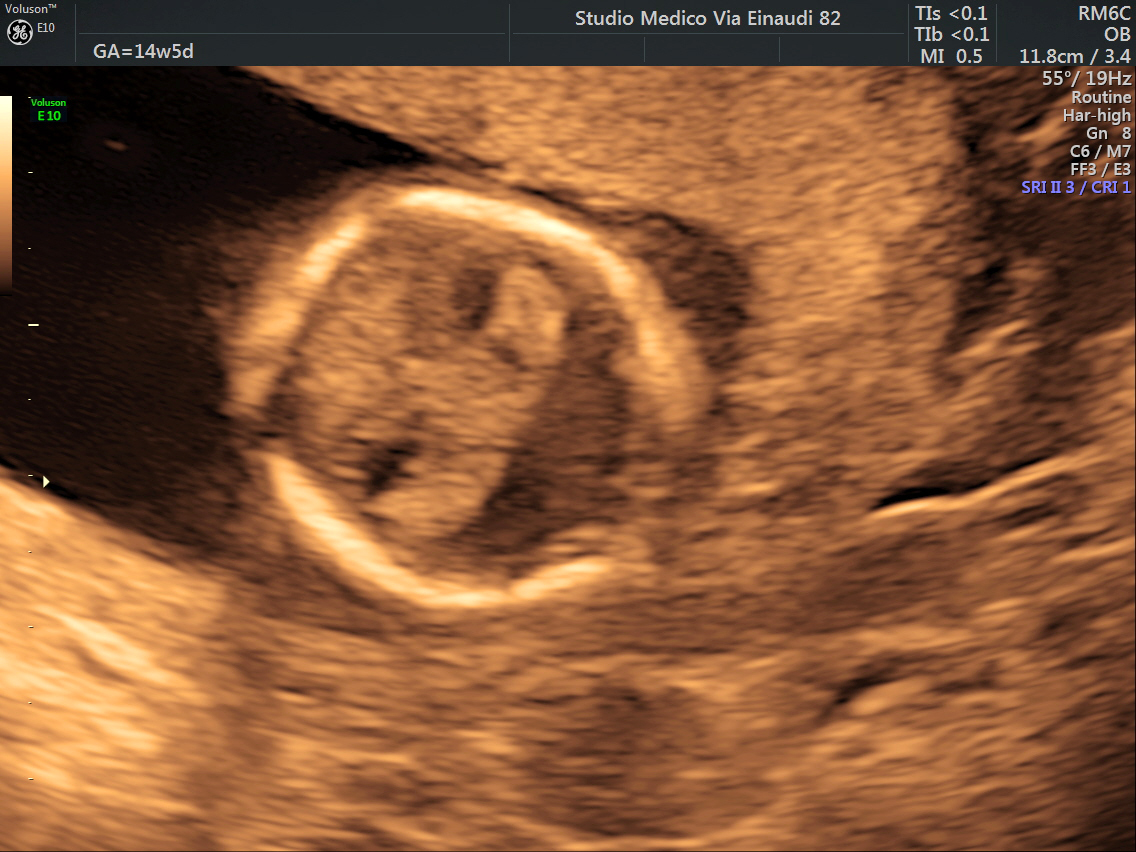
Holoprosencephaly Fetus 14 Weks US By Dr
Holoprosencephaly (HPE) is a cephalic disorder in which the prosencephalon (the forebrain of the embryo) fails to develop into two hemispheres, typically occurring between the 18th and 28th day of gestation. Normally, the forebrain is formed and the face begins to develop in the fifth and sixth weeks of human pregnancy. The condition also occurs in other species. Holoprosencephaly is estimated to occur in approximately 1 in every 250 conceptions and most cases are not compatible with life and result in fetal death in utero due to deformities to the skull and brain. However, holoprosencephaly is still estimated to occur in approximately 1 in every 8,000 live births. When the embryo's forebrain does not divide to form bilateral cerebral hemispheres (the left and right halves of the brain), it causes defects in the development of the face and in brain structure and function. The severity of holoprosencephaly is highly variable. In less severe cases, babies are born with normal or n ... [...More Info...] [...Related Items...] OR: [Wikipedia] [Google] [Baidu] |
Sonic Hedgehog
Sonic hedgehog protein (SHH) is encoded for by the ''SHH'' gene. The protein is named after the character ''Sonic the Hedgehog''. This signaling molecule is key in regulating embryonic morphogenesis in all animals. SHH controls organogenesis and the organization of the central nervous system, limbs, digits and many other parts of the body. Sonic hedgehog is a morphogen that patterns the developing embryo using a concentration gradient characterized by the French flag model. This model has a non-uniform distribution of SHH molecules which governs different cell fates according to concentration. Mutations in this gene can cause holoprosencephaly, a failure of splitting in the cerebral hemispheres, as demonstrated in an experiment using SHH knock-out mice in which the forebrain midline failed to develop and instead only a single fused telencephalic vesicle resulted. Sonic hedgehog still plays a role in differentiation, proliferation, and maintenance of adult tissues. Abnormal activ ... [...More Info...] [...Related Items...] OR: [Wikipedia] [Google] [Baidu] |
Cyclopia
Cyclopia (named after the Greek mythology character cyclopes) is the most extreme form of holoprosencephaly and is a congenital disorder (birth defect) characterized by the failure of the embryonic prosencephalon to properly divide the orbits of the eye into two cavities. Its incidence is 1 in 16,000 in born animals and 1 in 200 in miscarried fetuses. Signs and symptoms Typically, the nose is either missing or not functional. This deformity (called proboscis) forms above the center eye and is characteristic of a form of cyclopia called rhinencephaly or rhinocephaly. Most such embryos are either naturally aborted or are stillborn upon delivery. Although cyclopia is rare, several cyclopic human babies are preserved in medical museums (e.g. The Vrolik Museum, Amsterdam, Trivandrum Medical College). Some extreme cases of cyclopia have been documented in farm animals (horses, sheep, pigs, and sometimes chickens). In such cases, the nose and mouth fail to form, or the nose grows ... [...More Info...] [...Related Items...] OR: [Wikipedia] [Google] [Baidu] |
Brain Development
The development of the nervous system, or neural development (neurodevelopment), refers to the processes that generate, shape, and reshape the nervous system of animals, from the earliest stages of embryonic development to adulthood. The field of neural development draws on both neuroscience and developmental biology to describe and provide insight into the cellular and molecular mechanisms by which complex nervous systems develop, from nematodes and Drosophila melanogaster, fruit flies to mammals. Defects in neural development can lead to malformations such as holoprosencephaly, and a wide variety of neurological disorders including paresis, limb paresis and paralysis, balance and vision disorders, and seizures, and in humans other disorders such as Rett syndrome, Down syndrome and intellectual disability. Overview of vertebrate brain development The vertebrate central nervous system (CNS) is derived from the ectoderm—the outermost germ layer of the embryo. A part of the ... [...More Info...] [...Related Items...] OR: [Wikipedia] [Google] [Baidu] |
Congenital Abnormality
A birth defect, also known as a congenital disorder, is an abnormal condition that is present at birth regardless of its cause. Birth defects may result in disabilities that may be physical, intellectual, or developmental. The disabilities can range from mild to severe. Birth defects are divided into two main types: structural disorders in which problems are seen with the shape of a body part and functional disorders in which problems exist with how a body part works. Functional disorders include metabolic and degenerative disorders. Some birth defects include both structural and functional disorders. Birth defects may result from genetic or chromosomal disorders, exposure to certain medications or chemicals, or certain infections during pregnancy. Risk factors include folate deficiency, drinking alcohol or smoking during pregnancy, poorly controlled diabetes, and a mother over the age of 35 years old. Many are believed to involve multiple factors. Birth defects may be visib ... [...More Info...] [...Related Items...] OR: [Wikipedia] [Google] [Baidu] |
Prosencephalon
In the anatomy of the brain of vertebrates, the forebrain or prosencephalon is the Anatomical terms of location#Directional terms, rostral (forward-most) portion of the brain. The forebrain (prosencephalon), the midbrain (mesencephalon), and hindbrain (rhombencephalon) are the three Brain vesicle, primary brain vesicles during the early development of the nervous system. The forebrain controls body temperature, reproductive functions, eating, sleeping, and the display of emotions. At the five-vesicle stage, the forebrain separates into the diencephalon (thalamus, hypothalamus, subthalamus, and epithalamus) and the telencephalon which develops into the cerebrum. The cerebrum consists of the cerebral cortex, underlying white matter, and the basal ganglia. In humans, by 5 weeks in utero it is visible as a single portion toward the front of the fetus. At 8 weeks in utero, the forebrain splits into the left and right cerebral hemispheres. When the embryonic forebrain fails to divide ... [...More Info...] [...Related Items...] OR: [Wikipedia] [Google] [Baidu] |
Forebrain
In the anatomy of the brain of vertebrates, the forebrain or prosencephalon is the Anatomical terms of location#Directional terms, rostral (forward-most) portion of the brain. The forebrain (prosencephalon), the midbrain (mesencephalon), and hindbrain (rhombencephalon) are the three Brain vesicle, primary brain vesicles during the early development of the nervous system. The forebrain controls body temperature, reproductive functions, eating, sleeping, and the display of emotions. At the five-vesicle stage, the forebrain separates into the diencephalon (thalamus, hypothalamus, subthalamus, and epithalamus) and the telencephalon which develops into the cerebrum. The cerebrum consists of the cerebral cortex, underlying white matter, and the basal ganglia. In humans, by 5 weeks in utero it is visible as a single portion toward the front of the fetus. At 8 weeks in utero, the forebrain splits into the left and right cerebral hemispheres. When the embryonic forebrain fails to divide ... [...More Info...] [...Related Items...] OR: [Wikipedia] [Google] [Baidu] |
Cytogenetics
Cytogenetics is essentially a branch of genetics, but is also a part of cell biology/cytology (a subdivision of human anatomy), that is concerned with how the chromosomes relate to cell behaviour, particularly to their behaviour during mitosis and meiosis. Techniques used include karyotyping, analysis of G-banded chromosomes, other cytogenetic banding techniques, as well as molecular cytogenetics such as fluorescent ''in situ'' hybridization (FISH) and comparative genomic hybridization (CGH). History Beginnings Chromosomes were first observed in plant cells by Carl Nägeli in 1842. Their behavior in animal (salamander) cells was described by Walther Flemming, the discoverer of mitosis, in 1882. The name was coined by another German anatomist, von Waldeyer in 1888. The next stage took place after the development of genetics in the early 20th century, when it was appreciated that the set of chromosomes (the karyotype) was the carrier of the genes. Levitsky seems to have been t ... [...More Info...] [...Related Items...] OR: [Wikipedia] [Google] [Baidu] |
Genetic Counseling
Genetic counseling is the process of investigating individuals and families affected by or at risk of genetic disorders to help them understand and adapt to the medical, psychological and familial implications of genetic contributions to disease; this field is considered necessary for the implementation of genomic medicine. The process integrates: * Interpretation of family and medical histories to assess the chance of disease occurrence or recurrence * Education about inheritance, testing, management, prevention, resources * Counseling to promote informed choices, adaptation to the risk or condition and support in reaching out to relatives that are also at risk History The practice of advising people about inherited traits began around the turn of the 20th century, shortly after William Bateson suggested that the new medical and biological study of heredity be called "genetics". Heredity became intertwined with social reforms when the field of modern eugenics took form. Althoug ... [...More Info...] [...Related Items...] OR: [Wikipedia] [Google] [Baidu] |
Olfactory Bulb
The olfactory bulb (Latin: ''bulbus olfactorius'') is a grey matter, neural structure of the vertebrate forebrain involved in olfaction, the sense of odor, smell. It sends olfactory information to be further processed in the amygdala, the orbitofrontal cortex (OFC) and the hippocampus where it plays a role in emotion, memory and learning. The bulb is divided into two distinct structures: the main olfactory bulb and the accessory olfactory bulb. The main olfactory bulb connects to the amygdala via the piriform cortex of the primary olfactory cortex and directly projects from the main olfactory bulb to specific amygdala areas. The accessory olfactory bulb resides on the dorsal-posterior region of the main olfactory bulb and forms a parallel pathway. Destruction of the olfactory bulb results in ipsilateral anosmia, while irritative lesions of the uncus can result in olfactory and gustatory hallucinations. Structure In most vertebrates, the olfactory bulb is the most Anatomical term ... [...More Info...] [...Related Items...] OR: [Wikipedia] [Google] [Baidu] |
Lobes Of The Brain
The lobes of the brain are the major identifiable zones of the cerebral cortex, and they comprise the surface of each hemisphere of the cerebrum. The two hemispheres are roughly symmetrical in structure, and are connected by the corpus callosum. They traditionally have been divided into four lobes, but are today considered as having six lobes each. The lobes are large areas that are anatomically distinguishable, and are also functionally distinct to some degree. Each lobe of the brain has numerous ridges, or gyri, and furrows, the sulci that constitute further subzones of the cortex. The expression "lobes of the brain" usually refers only to those of the cerebrum, not to the distinct areas of the cerebellum. Frontal lobe The frontal lobe is located at the front of each cerebral hemisphere and positioned in front of the parietal lobe and above and in front of the temporal lobe. It is separated from the parietal lobe by a space between tissues called the central sulcus, and from t ... [...More Info...] [...Related Items...] OR: [Wikipedia] [Google] [Baidu] |
Corpus Callosum
The corpus callosum (Latin for "tough body"), also callosal commissure, is a wide, thick nerve tract, consisting of a flat bundle of commissural fibers, beneath the cerebral cortex in the brain. The corpus callosum is only found in placental mammals. It spans part of the longitudinal fissure, connecting the left and right cerebral hemispheres, enabling communication between them. It is the largest white matter structure in the human brain, about in length and consisting of 200–300 million axonal projections. A number of separate nerve tracts, classed as subregions of the corpus callosum, connect different parts of the hemispheres. The main ones are known as the genu, the rostrum, the trunk or body, and the splenium. Structure The corpus callosum forms the floor of the longitudinal fissure that separates the two cerebral hemispheres. Part of the corpus callosum forms the roof of the lateral ventricles. The corpus callosum has four main parts – individual nerve tracts ... [...More Info...] [...Related Items...] OR: [Wikipedia] [Google] [Baidu] |


