|
Hypercoagulable State
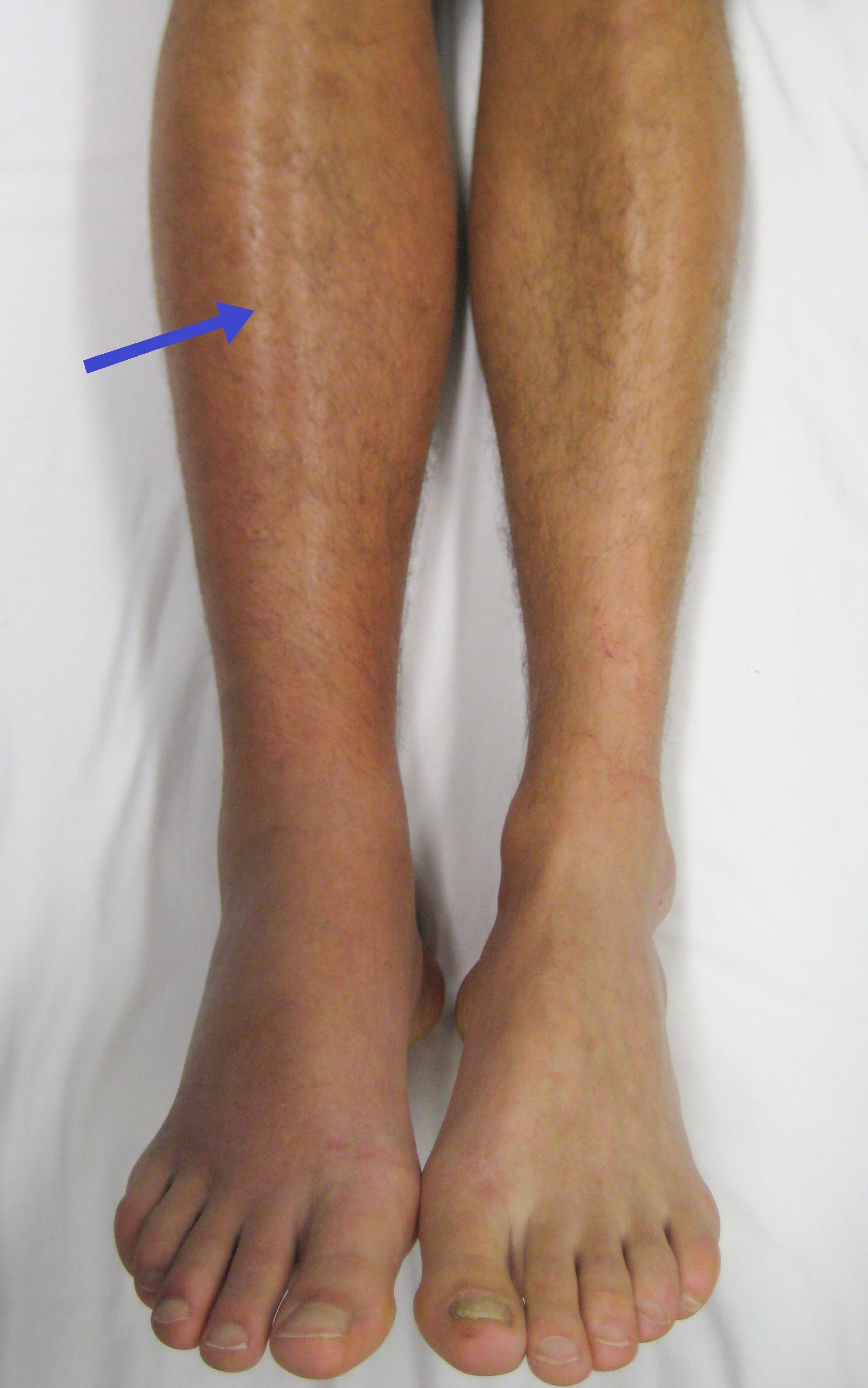
Thrombophilia (sometimes called hypercoagulability or a prothrombotic state) is an abnormality of blood coagulation that increases the risk of thrombosis (blood clots in blood vessels). Such abnormalities can be identified in 50% of people who have an episode of thrombosis (such as deep vein thrombosis in the leg) that was not provoked by other causes. A significant proportion of the population has a detectable thrombophilic abnormality, but most of these develop thrombosis only in the presence of an additional risk factor. There is no specific treatment for most thrombophilias, but recurrent episodes of thrombosis may be an indication for long-term preventive anticoagulation. The first major form of thrombophilia to be identified by medical science, antithrombin deficiency, was identified in 1965, while the most common abnormalities (including factor V Leiden) were described in the 1990s. Signs and symptoms The most common conditions associated with thrombophilia are deep v ... [...More Info...] [...Related Items...] OR: [Wikipedia] [Google] [Baidu] |
Blood Coagulation
Coagulation, also known as clotting, is the process by which blood changes from a liquid to a gel, forming a blood clot. It potentially results in hemostasis, the cessation of blood loss from a damaged vessel, followed by repair. The mechanism of coagulation involves activation, adhesion and aggregation of platelets, as well as deposition and maturation of fibrin. Coagulation begins almost instantly after an injury to the endothelium lining a blood vessel. Exposure of blood to the subendothelial space initiates two processes: changes in platelets, and the exposure of subendothelial tissue factor to plasma factor VII, which ultimately leads to cross-linked fibrin formation. Platelets immediately form a plug at the site of injury; this is called ''primary hemostasis. Secondary hemostasis'' occurs simultaneously: additional coagulation (clotting) factors beyond factor VII ( listed below) respond in a cascade to form fibrin strands, which strengthen the platelet plug. Disorders of ... [...More Info...] [...Related Items...] OR: [Wikipedia] [Google] [Baidu] |
Cerebral Venous Sinus Thrombosis
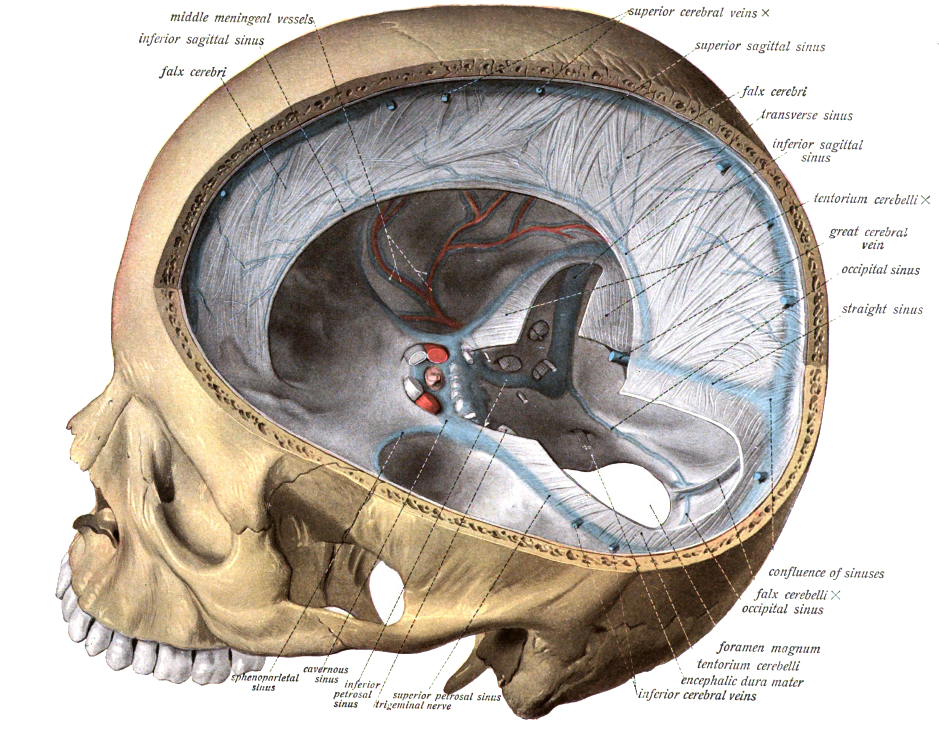
Cerebral venous sinus thrombosis (CVST), cerebral venous and sinus thrombosis or cerebral venous thrombosis (CVT), is the presence of a blood clot in the dural venous sinuses (which drain blood from the brain), the cerebral veins, or both. Symptoms may include severe headache, visual symptoms, any of the symptoms of stroke such as weakness of the face and limbs on one side of the body, and seizures, which occur in around 40% of patients. The diagnosis is usually by computed tomography (CT scan) or magnetic resonance imaging (MRI) to demonstrate obstruction of the venous sinuses. After confirmation of the diagnosis, investigations may be performed to determine the underlying cause, especially if one is not readily apparent. Treatment is typically with anticoagulants (medications that suppress blood clotting) such as low molecular weight heparin. Rarely, thrombolysis (enzymatic destruction of the blood clot) or mechanical thrombectomy is used, although evidence for this therap ... [...More Info...] [...Related Items...] OR: [Wikipedia] [Google] [Baidu] |
Abruptio Placentae
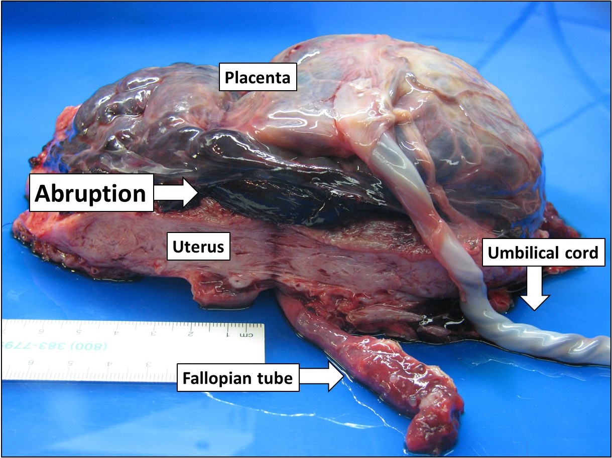
Placental abruption is when the placenta separates early from the uterus, in other words separates before childbirth. It occurs most commonly around 25 weeks of pregnancy. Symptoms may include vaginal bleeding, lower abdominal pain, and dangerously low blood pressure. Complications for the mother can include disseminated intravascular coagulopathy and kidney failure. Complications for the baby can include fetal distress, low birthweight, preterm delivery, and stillbirth. The cause of placental abruption is not entirely clear. Risk factors include smoking, pre-eclampsia, prior abruption (most important and predictive risk factor), trauma during pregnancy, cocaine use, and previous cesarean section. Diagnosis is based on symptoms and supported by ultrasound. It is classified as a complication of pregnancy. For small abruption, bed rest may be recommended, while for more significant abruptions or those that occur near term, delivery may be recommended. If everything is stable, ... [...More Info...] [...Related Items...] OR: [Wikipedia] [Google] [Baidu] |
Pre-eclampsia
Pre-eclampsia is a disorder of pregnancy characterized by the onset of high blood pressure and often a significant amount of protein in the urine. When it arises, the condition begins after 20 weeks of pregnancy. In severe cases of the disease there may be red blood cell breakdown, a low blood platelet count, impaired liver function, kidney dysfunction, swelling, shortness of breath due to fluid in the lungs, or visual disturbances. Pre-eclampsia increases the risk of undesirable outcomes for both the mother and the fetus. If left untreated, it may result in seizures at which point it is known as eclampsia. Risk factors for pre-eclampsia include obesity, prior hypertension, older age, and diabetes mellitus. It is also more frequent in a woman's first pregnancy and if she is carrying twins. The underlying mechanism involves abnormal formation of blood vessels in the placenta amongst other factors. Most cases are diagnosed before delivery. Commonly, pre-eclampsia continues i ... [...More Info...] [...Related Items...] OR: [Wikipedia] [Google] [Baidu] |
Stillbirth
Stillbirth is typically defined as fetal death at or after 20 or 28 weeks of pregnancy, depending on the source. It results in a baby born without signs of life. A stillbirth can result in the feeling of guilt or grief in the mother. The term is in contrast to miscarriage, which is an early pregnancy loss, and Sudden Infant Death Syndrome, where the baby dies a short time after being born alive. Often the cause is unknown. Causes may include pregnancy complications such as pre-eclampsia and birth complications, problems with the placenta or umbilical cord, birth defects, infections such as malaria and syphilis, and poor health in the mother. Risk factors include a mother's age over 35, smoking, drug use, use of assisted reproductive technology, and first pregnancy. Stillbirth may be suspected when no fetal movement is felt. Confirmation is by ultrasound. Worldwide prevention of most stillbirths is possible with improved health systems. Around half of stillbirths occur durin ... [...More Info...] [...Related Items...] OR: [Wikipedia] [Google] [Baidu] |
Intrauterine Growth Restriction
Intrauterine growth restriction (IUGR), or fetal growth restriction, refers to poor growth of a fetus while in the womb during pregnancy. IUGR is defined by clinical features of malnutrition and evidence of reduced growth regardless of an infant's birth weight percentile. The causes of IUGR are broad and may involve maternal, fetal, or placental complications. At least 60% of the 4 million neonatal deaths that occur worldwide every year are associated with low birth weight (LBW), caused by intrauterine growth restriction (IUGR), preterm delivery, and genetic abnormalities, demonstrating that under-nutrition is already a leading health problem at birth. Intrauterine growth restriction can result in a baby being small for gestational age (SGA), which is most commonly defined as a weight below the 10th percentile for the gestational age. At the end of pregnancy, it can result in a low birth weight. Types There are two major categories of IUGR: pseudo IUGR and true IUGR With pseudo ... [...More Info...] [...Related Items...] OR: [Wikipedia] [Google] [Baidu] |
Recurrent Miscarriage
Recurrent miscarriage is three or more consecutive pregnancy losses. In contrast, infertility is the inability to conceive. In many cases the cause of RPL is unknown. After three or more losses, a thorough evaluation is recommended by American Society of Reproductive Medicine. About 1% of couples trying to have children are affected by recurrent miscarriage. Causes There are various causes for recurrent miscarriage, and some can be treated. Some couples never have a cause identified, often after extensive investigations. About 50–75% of cases of recurrent miscarriage are unexplained. Chromosomal disorders A balanced translocation or Robertsonian translocation in one of the partners leads to unviable fetuses that are miscarried. This explains why a karyogram is often performed in both partners if a woman has experienced repeated miscarriages. Aneuploidy may be a cause of a random spontaneous as well as recurrent pregnancy loss. Aneuploidy is more common with advanced re ... [...More Info...] [...Related Items...] OR: [Wikipedia] [Google] [Baidu] |
Stroke
A stroke is a medical condition in which poor blood flow to the brain causes cell death. There are two main types of stroke: ischemic, due to lack of blood flow, and hemorrhagic, due to bleeding. Both cause parts of the brain to stop functioning properly. Signs and symptoms of a stroke may include an inability to move or feel on one side of the body, problems understanding or speaking, dizziness, or loss of vision to one side. Signs and symptoms often appear soon after the stroke has occurred. If symptoms last less than one or two hours, the stroke is a transient ischemic attack (TIA), also called a mini-stroke. A hemorrhagic stroke may also be associated with a severe headache. The symptoms of a stroke can be permanent. Long-term complications may include pneumonia and loss of bladder control. The main risk factor for stroke is high blood pressure. Other risk factors include high blood cholesterol, tobacco smoking, obesity, diabetes mellitus, a previous TIA, end-st ... [...More Info...] [...Related Items...] OR: [Wikipedia] [Google] [Baidu] |
Myocardial Infarction
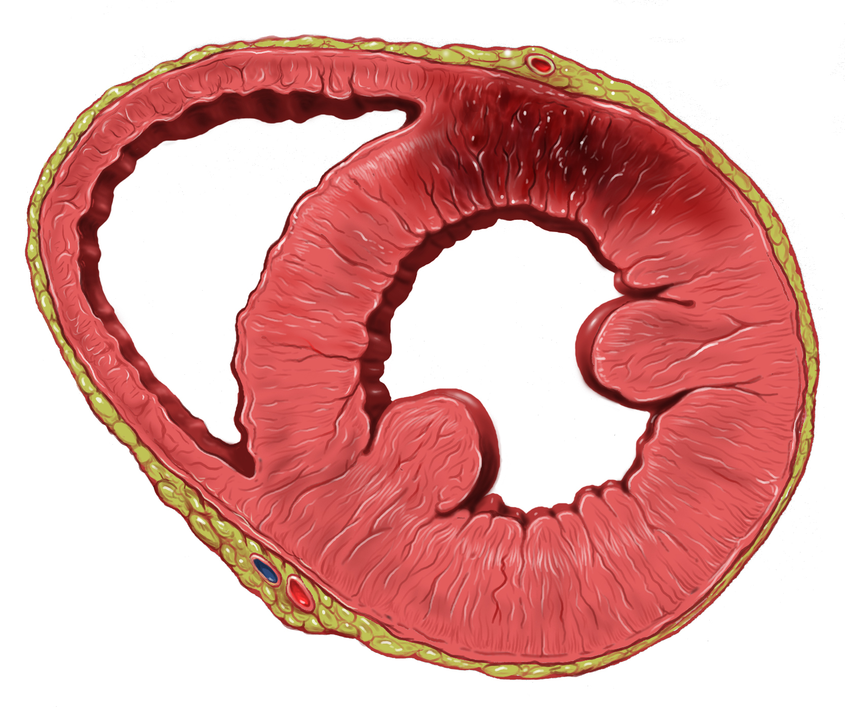
A myocardial infarction (MI), commonly known as a heart attack, occurs when blood flow decreases or stops to the coronary artery of the heart, causing damage to the heart muscle. The most common symptom is chest pain or discomfort which may travel into the shoulder, arm, back, neck or jaw. Often it occurs in the center or left side of the chest and lasts for more than a few minutes. The discomfort may occasionally feel like heartburn. Other symptoms may include shortness of breath, nausea, feeling faint, a cold sweat or feeling tired. About 30% of people have atypical symptoms. Women more often present without chest pain and instead have neck pain, arm pain or feel tired. Among those over 75 years old, about 5% have had an MI with little or no history of symptoms. An MI may cause heart failure, an irregular heartbeat, cardiogenic shock or cardiac arrest. Most MIs occur due to coronary artery disease. Risk factors include high blood pressure, smoking, diabetes, ... [...More Info...] [...Related Items...] OR: [Wikipedia] [Google] [Baidu] |
Arterial Thrombosis
Thrombosis (from Ancient Greek "clotting") is the formation of a blood clot inside a blood vessel, obstructing the flow of blood through the circulatory system. When a blood vessel (a vein or an artery) is injured, the body uses platelets (thrombocytes) and fibrin to form a blood clot to prevent blood loss. Even when a blood vessel is not injured, blood clots may form in the body under certain conditions. A clot, or a piece of the clot, that breaks free and begins to travel around the body is known as an embolus. Thrombosis may occur in veins (venous thrombosis) or in arteries ( arterial thrombosis). Venous thrombosis (sometimes called DVT, deep vein thrombosis) leads to a blood clot in the affected part of the body, while arterial thrombosis (and, rarely, severe venous thrombosis) affects the blood supply and leads to damage of the tissue supplied by that artery ( ischemia and necrosis). A piece of either an arterial or a venous thrombus can break off as an embolus, which co ... [...More Info...] [...Related Items...] OR: [Wikipedia] [Google] [Baidu] |
Renal Vein Thrombosis
Renal vein thrombosis (RVT) is the formation of a clot in the vein that drains blood from the kidneys, ultimately leading to a reduction in the drainage of one or both kidneys and the possible migration of the clot to other parts of the body. First described by German pathologist Friedrich Daniel von Recklinghausen in 1861, RVT most commonly affects two subpopulations: newly born infants with blood clotting abnormalities or dehydration and adults with nephrotic syndrome. Nephrotic syndrome, a kidney disorder, causes excessive loss of protein in the urine, low levels of albumin in the blood, a high level of cholesterol in the blood and swelling, triggering a hypercoagulable state and increasing chances of clot formation. Other less common causes include hypercoagulable state, cancer, kidney transplantation, Behcet syndrome, antiphospholipid antibody syndrome or blunt trauma to the back or abdomen. Treatment of RVT mainly focuses on preventing further blood clots in the kidneys ... [...More Info...] [...Related Items...] OR: [Wikipedia] [Google] [Baidu] |


