|
Renal Vein Thrombosis
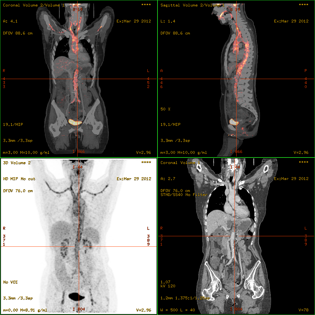
Renal vein thrombosis (RVT) is the formation of a clot in the vein that drains blood from the kidneys, ultimately leading to a reduction in the drainage of one or both kidneys and the possible migration of the clot to other parts of the body. First described by German pathologist Friedrich Daniel von Recklinghausen in 1861, RVT most commonly affects two subpopulations: newly born infants with blood clotting abnormalities or dehydration and adults with nephrotic syndrome. Nephrotic syndrome, a kidney disorder, causes excessive loss of protein in the urine, low levels of albumin in the blood, a high level of cholesterol in the blood and swelling, triggering a hypercoagulable state and increasing chances of clot formation. Other less common causes include hypercoagulable state, cancer, kidney transplantation, Behcet syndrome, antiphospholipid antibody syndrome or blunt trauma to the back or abdomen. Treatment of RVT mainly focuses on preventing further blood clots in the kidney ... [...More Info...] [...Related Items...] OR: [Wikipedia] [Google] [Baidu] |
Friedrich Daniel Von Recklinghausen
Friedrich Daniel von Recklinghausen (; December 2, 1833 – August 26, 1910) was a German pathologist born in Gütersloh, Westphalia. He was the father of physiologist Heinrich von Recklinghausen (1867–1942). Early life Recklinghausen was born in Gütersloh, Germany, in 1833. He was the son of Friedrich Christoph von Recklinghausen and Friederike Charlotte Zumwinkel. His father was an elementary school teacher and a sexton. His mother died shortly after his birth in 1833. The Recklinghausens were a patrician family who put multiple councilors and mayors in their positions. He went to the elementary school where his father taught in Gütersloh. He then attended high school at Ratsgymnasium, Bielefeld. Academic background Starting in 1852, Recklinghausen studied medicine at the Universities of Bonn, Würzburg, and Berlin, earning his doctorate at the latter institution in 1855. Afterwards he studied pathological anatomy under Rudolf Virchow, the father of modern pathology, and ... [...More Info...] [...Related Items...] OR: [Wikipedia] [Google] [Baidu] |
Vasculitis
Vasculitis is a group of disorders that destroy blood vessels by inflammation. Both arteries and veins are affected. Lymphangitis (inflammation of lymphatic vessels) is sometimes considered a type of vasculitis. Vasculitis is primarily caused by leukocyte migration and resultant damage. Although both occur in vasculitis, inflammation of veins ( phlebitis) or arteries ( arteritis) on their own are separate entities. Signs and symptoms Possible signs and symptoms include: * General symptoms: Fever, unintentional weight loss * Skin: Palpable purpura, livedo reticularis * Muscles and joints: Muscle pain or inflammation, joint pain or joint swelling * Nervous system: Mononeuritis multiplex, headache, stroke, tinnitus, reduced visual acuity, acute visual loss * Heart and arteries: Heart attack, high blood pressure, gangrene * Respiratory tract: Nosebleeds, bloody cough, lung infiltrates * GI tract: Abdominal pain, bloody stool, perforations (hole in the GI tract) * Kidneys: ... [...More Info...] [...Related Items...] OR: [Wikipedia] [Google] [Baidu] |
Heparin
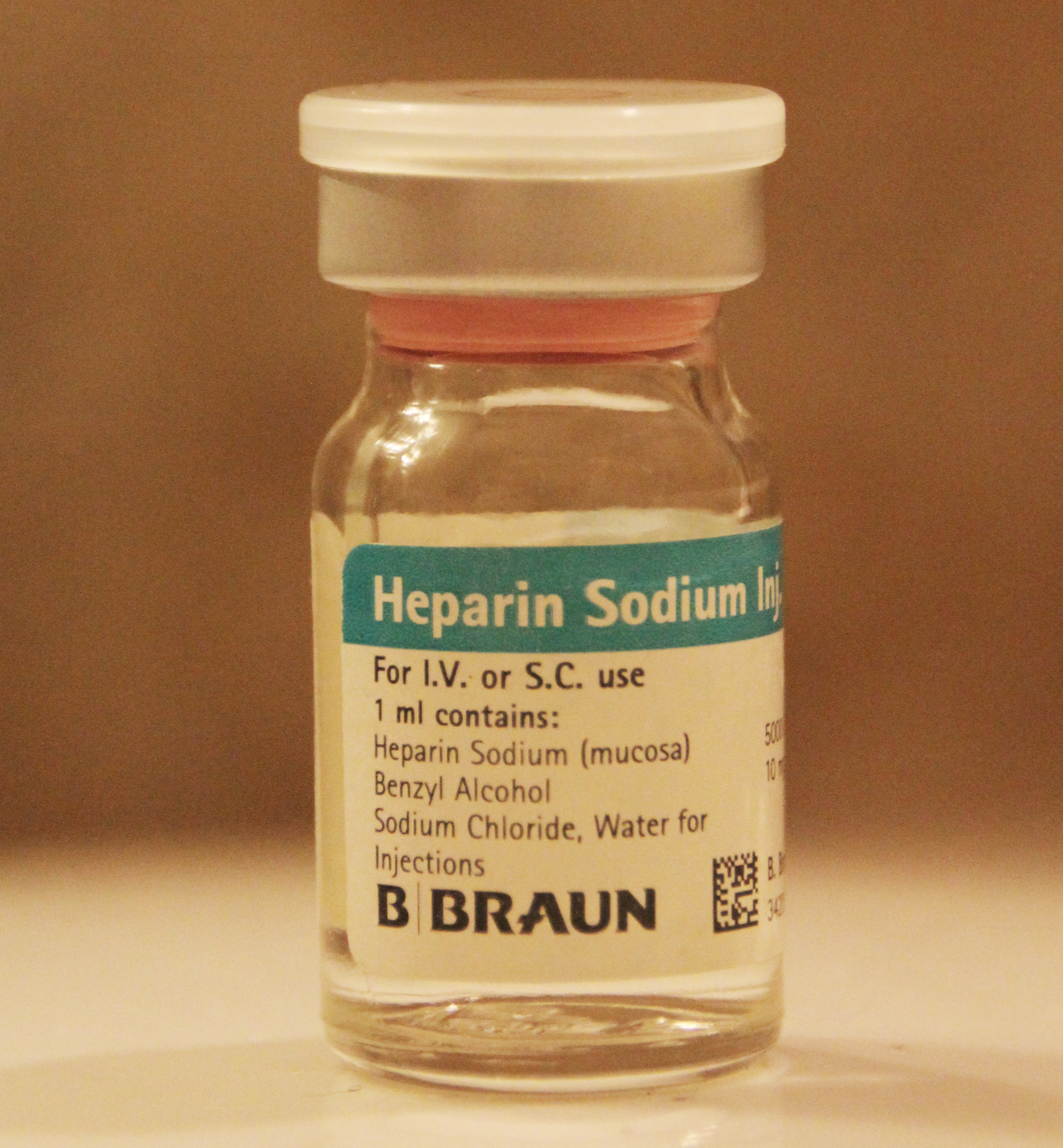
Heparin, also known as unfractionated heparin (UFH), is a medication and naturally occurring glycosaminoglycan. Since heparins depend on the activity of antithrombin, they are considered anticoagulants. Specifically it is also used in the treatment of heart attacks and unstable angina. It is given intravenously or by injection under the skin. Other uses for its anticoagulant properties include inside blood specimen test tubes and kidney dialysis machines. Common side effects include bleeding, pain at the injection site, and low blood platelets. Serious side effects include heparin-induced thrombocytopenia. Greater care is needed in those with poor kidney function. Heparin is contraindicated for suspected cases of vaccine-induced pro-thrombotic immune thrombocytopenia (VIPIT) secondary to SARS-CoV-2 vaccination, as heparin may further increase the risk of bleeding in an anti-PF4/heparin complex autoimmune manner, in favor of alternative anticoagulant medications (such ... [...More Info...] [...Related Items...] OR: [Wikipedia] [Google] [Baidu] |
Magnetic Resonance Angiography
Magnetic resonance angiography (MRA) is a group of techniques based on magnetic resonance imaging (MRI) to image blood vessels. Magnetic resonance angiography is used to generate images of arteries (and less commonly veins) in order to evaluate them for stenosis (abnormal narrowing), occlusions, aneurysms (vessel wall dilatations, at risk of rupture) or other abnormalities. MRA is often used to evaluate the arteries of the neck and brain, the thoracic and abdominal aorta, the renal arteries, and the legs (the latter exam is often referred to as a "run-off"). Acquisition A variety of techniques can be used to generate the pictures of blood vessels, both arteries and veins, based on flow effects or on contrast (inherent or pharmacologically generated). The most frequently applied MRA methods involve the use intravenous contrast agents, particularly those containing gadolinium to shorten the ''T''1 of blood to about 250 ms, shorter than the ''T''1 of all other tissues (except fat ... [...More Info...] [...Related Items...] OR: [Wikipedia] [Google] [Baidu] |
CT Angiography
Computed tomography angiography (also called CT angiography or CTA) is a computed tomography technique used for angiography—the visualization of arteries and veins—throughout the human body. Using contrast injected into the blood vessels, images are created to look for blockages, aneurysms (dilations of walls), dissections (tearing of walls), and stenosis (narrowing of vessel). CTA can be used to visualize the vessels of the heart, the aorta and other large blood vessels, the lungs, the kidneys, the head and neck, and the arms and legs. CTA can also be used to localise arterial or venous bleed of the gastrointestinal system. Medical uses CTA can be used to examine blood vessels in many key areas of the body including the brain, kidneys, pelvis, and the lungs. Coronary CT angiography Coronary CT angiography (CCTA) is the use of CT angiography to assess the arteries of the heart. The patient receives an intravenous injection of contrast and then the heart is scanned using a ... [...More Info...] [...Related Items...] OR: [Wikipedia] [Google] [Baidu] |
Ultrasound Imaging
Medical ultrasound includes diagnostic techniques (mainly imaging techniques) using ultrasound, as well as therapeutic applications of ultrasound. In diagnosis, it is used to create an image of internal body structures such as tendons, muscles, joints, blood vessels, and internal organs, to measure some characteristics (e.g. distances and velocities) or to generate an informative audible sound. Its aim is usually to find a source of disease or to exclude pathology. The usage of ultrasound to produce visual images for medicine is called medical ultrasonography or simply sonography. The practice of examining pregnant women using ultrasound is called obstetric ultrasonography, and was an early development of clinical ultrasonography. Ultrasound is composed of sound waves with frequencies which are significantly higher than the range of human hearing (>20,000 Hz). Ultrasonic images, also known as sonograms, are created by sending pulses of ultrasound into tissue using ... [...More Info...] [...Related Items...] OR: [Wikipedia] [Google] [Baidu] |
NutCracker2
A nutcracker is a tool designed to open nuts by cracking their shells. There are many designs, including levers, screws, and ratchets. The lever version is also used for cracking lobster and crab shells. A decorative version portrays a person whose mouth forms the jaws of the nutcracker. Functions Nuts were historically opened using a hammer and anvil, often made of stone. Some nuts such as walnuts can also be opened by hand, by holding the nut in the palm of the hand and applying pressure with the other palm or thumb, or using another nut. Manufacturers produce modern functional nutcrackers usually somewhat resembling pliers, but with the pivot point at the end beyond the nut, rather than in the middle. These are also used for cracking the shells of crab and lobster to make the meat inside available for eating. Hinged lever nutcrackers, often called a "pair of nutcrackers", may date back to Ancient Greece. By the 14th century in Europe, nutcrackers were documented in Eng ... [...More Info...] [...Related Items...] OR: [Wikipedia] [Google] [Baidu] |
Focal Segmental Glomerulosclerosis
Focal segmental glomerulosclerosis (FSGS) is a histopathologic finding of scarring (sclerosis) of glomeruli and damage to renal podocytes.Rosenberg, Avi Z.; Kopp, Jeffrey B. (2017-03-07). "Focal Segmental Glomerulosclerosis". ''Clinical Journal of the American Society of Nephrology''. 12 (3): 502–517. doi:10.2215/CJN.05960616. ISSN 1555-9041. PMC 5338705. PMID 28242845.D'Agati V. The many masks of focal segmental glomerulosclerosis. Kidney Int. 1994 Oct;46(4):1223-41. doi: 10.1038/ki.1994.388. . This process damages the filtration function of the kidney, resulting in protein loss in the urine. FSGS is a leading cause of excess protein loss— nephrotic syndrome—in children and adults.Kitiyakara C, Eggers P, Kopp JB. Twenty-one-year trend in ESRD due to focal segmental glomerulosclerosis in the United States. Am J Kidney Dis. 2004 Nov;44(5):815-25. . Signs and symptoms include proteinuria, water retention, and edema.Rydel JJ, Korbet SM, Borok RZ, Schwartz MM. Focal segment ... [...More Info...] [...Related Items...] OR: [Wikipedia] [Google] [Baidu] |
Minimal Change Disease
Minimal change disease (also known as MCD, minimal change glomerulopathy, and nil disease, among others) is a disease affecting the kidneys which causes a nephrotic syndrome. Nephrotic syndrome leads to the loss of significant amounts of protein in the urine, which causes the widespread edema (soft tissue swelling) and impaired kidney function commonly experienced by those affected by the disease. It is most common in children and has a peak incidence at 2 to 6 years of age. MCD is responsible for 10–25% of nephrotic syndrome cases in adults. It is also the most common cause of nephrotic syndrome of unclear cause (idiopathic) in children. Signs and symptoms The clinical signs of minimal change disease are proteinuria (abnormal excretion of proteins, mainly albumin, into the urine), edema (swelling of soft tissues as a consequence of water retention), weight gain, and hypoalbuminaemia (low serum albumin). These signs are referred to collectively as nephrotic syndrome. The ... [...More Info...] [...Related Items...] OR: [Wikipedia] [Google] [Baidu] |
Membranous Glomerulonephritis
Membranous glomerulonephritis (MGN) is a slowly progressive disease of the kidney affecting mostly people between ages of 30 and 50 years, usually white people (i.e., those of European, Middle Eastern, or North African ancestry.). It is the second most common cause of nephrotic syndrome in adults, with focal segmental glomerulosclerosis (FSGS) recently becoming the most common. Signs and symptoms Most people will present as nephrotic syndrome, with the triad of albuminuria, edema and low serum albumin (with or without kidney failure). High blood pressure and high cholesterol are often also present. Others may not have symptoms and may be picked up on screening, with urinalysis finding high amounts of protein loss in the urine. A definitive diagnosis of membranous nephropathy requires a kidney biopsy, though given the very high specificity of anti-PLA2R antibody positivity this can sometimes be avoided in patients with nephrotic syndrome and preserved kidney function Causes Tra ... [...More Info...] [...Related Items...] OR: [Wikipedia] [Google] [Baidu] |
Factor V Leiden
Factor V Leiden (rs6025 or ''F5'' p.R506Q) is a variant (mutated form) of human factor V (one of several substances that helps blood clot), which causes an increase in blood clotting (hypercoagulability). Due to this mutation, protein C, an anticoagulant protein that normally inhibits the pro-clotting activity of factor V, is not able to bind normally to factor V, leading to a hypercoagulable state, i.e., an increased tendency for the patient to form abnormal and potentially harmful blood clots. Factor V Leiden is the most common hereditary hypercoagulability (prone to clotting) disorder amongst ethnic Europeans. It is named after the Dutch city of Leiden, where it was first identified in 1994 by Rogier Maria Bertina under the direction of (and in the laboratory of) Pieter Hendrick Reitsma. Despite the increased risk of venous thromboembolisms, people with one copy of this gene have not been found to have shorter lives than the general population. Signs and symptoms The symptoms of ... [...More Info...] [...Related Items...] OR: [Wikipedia] [Google] [Baidu] |
Folic Acid Deficiency
Folate deficiency, also known as vitamin B9 deficiency, is a low level of folate and derivatives in the body. Signs of folate deficiency are often subtle. A low number of red blood cells (anemia) is a late finding in folate deficiency and folate deficiency anemia is the term given for this medical condition. It is characterized by the appearance of large-sized, abnormal red blood cells ( megaloblasts), which form when there are inadequate stores of folic acid within the body. Signs and symptoms Loss of appetite and weight loss can occur. Additional signs are weakness, sore tongue, headaches, heart palpitations, irritability, and behavioral disorders. In adults, anemia (macrocytic, megaloblastic anemia) can be a sign of advanced folate deficiency. Women with folate deficiency who become pregnant are more likely to give birth to low birth weight premature infants, and infants with neural tube defects and even spina bifida. In infants and children, folate deficiency can lead ... [...More Info...] [...Related Items...] OR: [Wikipedia] [Google] [Baidu] |


_Phase_Contrast_(PC)_sequence_MRI_of_arterial_dissections.jpg)


_(11545246275).jpg)