|
Hematopoietic Stem Cell Niche
Many human blood cells, such as red blood cells (RBCs), immune cells, and even platelets all originate from the same progenitor cell, the hematopoietic stem cell (HSC). As these cells are short-lived, there needs to be a steady turnover of new blood cells and the maintenance of an HSC pool. This is broadly termed hematopoiesis. This event requires a special environment, termed the hematopoietic stem cell niche, which provides the protection and signals necessary to carry out the differentiation of cells from HSC progenitors. This niche relocates from the yolk sac to eventually rest in the bone marrow of mammals. Many pathological states can arise from disturbances in this niche environment, highlighting its importance in maintaining hematopoiesis. Hematopoiesis Hematopoiesis involves a series of differentiation steps from one progenitor cell to a more committed cell type, forming the recognizable tree seen in the adjacent diagram. Pluripotent long-term (LT)-HSCs self-renew to main ... [...More Info...] [...Related Items...] OR: [Wikipedia] [Google] [Baidu] |
Red Blood Cell
Red blood cells (RBCs), also referred to as red cells, red blood corpuscles (in humans or other animals not having nucleus in red blood cells), haematids, erythroid cells or erythrocytes (from Greek ''erythros'' for "red" and ''kytos'' for "hollow vessel", with ''-cyte'' translated as "cell" in modern usage), are the most common type of blood cell and the vertebrate's principal means of delivering oxygen (O2) to the body tissues—via blood flow through the circulatory system. RBCs take up oxygen in the lungs, or in fish the gills, and release it into tissues while squeezing through the body's capillaries. The cytoplasm of a red blood cell is rich in hemoglobin, an iron-containing biomolecule that can bind oxygen and is responsible for the red color of the cells and the blood. Each human red blood cell contains approximately 270 million hemoglobin molecules. The cell membrane is composed of proteins and lipids, and this structure provides properties essential for physiolo ... [...More Info...] [...Related Items...] OR: [Wikipedia] [Google] [Baidu] |
Stem Cell Niche
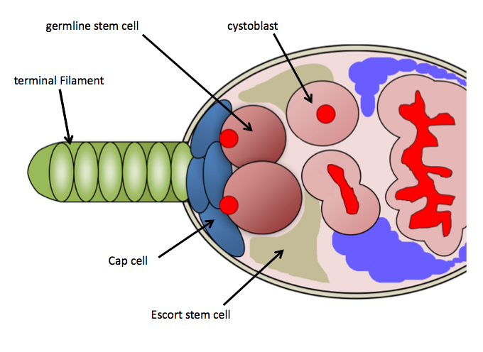
Stem-cell niche refers to a microenvironment, within the specific anatomic location where stem cells are found, which interacts with stem cells to regulate cell fate. The word 'niche' can be in reference to the ''in vivo'' or ''in vitro'' stem-cell microenvironment. During embryonic development, various niche factors act on embryonic stem cells to alter gene expression, and induce their proliferation or differentiation for the development of the fetus. Within the human body, stem-cell niches maintain adult stem cells in a quiescent state, but after tissue injury, the surrounding micro-environment actively signals to stem cells to promote either self-renewal or differentiation to form new tissues. Several factors are important to regulate stem-cell characteristics within the niche: cell–cell interactions between stem cells, as well as interactions between stem cells and neighbouring differentiated cells, interactions between stem cells and adhesion molecules, extracellular matrix co ... [...More Info...] [...Related Items...] OR: [Wikipedia] [Google] [Baidu] |
Precursor Cell
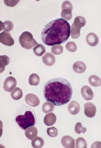
In cell biology, a precursor cell, also called a blast cell or simply blast, is a partially differentiated cell, usually referred to as a unipotent cell that has lost most of its stem cell properties. A precursor cell is also known as a progenitor cell but progenitor cells are multipotent. Precursor cells are known as the intermediate cell before they become differentiated after being a stem cell. Usually, a precursor cell is a stem cell with the capacity to differentiate into only one cell type. Sometimes, ''precursor cell'' is used as an alternative term for unipotent stem cells. In embryology, precursor cells are a group of cells that later differentiate into one organ. A blastoma is any cancer created by malignancies of precursor cells. Precursor cells, and progenitor cells, have many potential uses in medicine. , there is research being done to use these cells to build heart valves, blood vessels and other tissues, by using blood and muscle precursor, or progenitor c ... [...More Info...] [...Related Items...] OR: [Wikipedia] [Google] [Baidu] |
Kinase Insert Domain Receptor
Kinase insert domain receptor (KDR, a type IV receptor tyrosine kinase) also known as vascular endothelial growth factor receptor 2 (VEGFR-2) is a VEGF receptor. ''KDR'' is the human gene encoding it. KDR has also been designated as CD309 (cluster of differentiation 309). KDR is also known as Flk1 (Fetal Liver Kinase 1). The Q472H germline ''KDR'' genetic variant affects VEGFR-2 phosphorylation and has been found to associate with microvessel density in NSCLC. Interactions Kinase insert domain receptor has been shown to interact with SHC2, Annexin A5 and SHC1. See also * Cluster of differentiation * VEGF receptors VEGF receptors are receptors for vascular endothelial growth factor (VEGF). There are three main subtypes of VEGFR, numbered 1, 2 and 3. Also, they may be membrane-bound (mbVEGFR) or soluble (sVEGFR), depending on alternative splicing. Inh ... References Further reading * * * * * * * * External links * * Clusters of diff ... [...More Info...] [...Related Items...] OR: [Wikipedia] [Google] [Baidu] |
Knockout Mice
A knockout mouse, or knock-out mouse, is a genetically modified mouse (''Mus musculus'') in which researchers have inactivated, or "knocked out", an existing gene by replacing it or disrupting it with an artificial piece of DNA. They are important animal models for studying the role of genes which have been sequenced but whose functions have not been determined. By causing a specific gene to be inactive in the mouse, and observing any differences from normal behaviour or physiology, researchers can infer its probable function. Mice are currently the laboratory animal species most closely related to humans for which the knockout technique can easily be applied. They are widely used in knockout experiments, especially those investigating genetic questions that relate to human physiology. Gene knockout in rats is much harder and has only been possible since 2003. The first recorded knockout mouse was created by Mario R. Capecchi, Martin Evans, and Oliver Smithies in 1989, for whi ... [...More Info...] [...Related Items...] OR: [Wikipedia] [Google] [Baidu] |
Hemangioblast
Hemangioblasts are the multipotent precursor cells that can differentiate into both hematopoietic stem cells, hematopoietic and endothelial cells. In the mouse embryo, the emergence of blood islands in the yolk sac at embryonic day 7 marks the onset of hematopoiesis. From these blood islands, the hematopoietic cells and vasculature are formed shortly after. Hemangioblasts are the progenitors that form the blood islands. To date, the hemangioblast has been identified in human, mouse and zebrafish embryos. Hemangioblasts have been first extracted from embryonic cultures and manipulated by cytokines to differentiate along either hematopoietic or endothelial route. It has been shown that these pre-endothelial/pre-hematopoietic cells in the embryo arise out of a phenotype CD34 population. It was then found that hemangioblasts are also present in the tissue of post-natal individuals, such as in newborn infants and adults. Adult hemangioblast There is now emerging evidence of hemangiobla ... [...More Info...] [...Related Items...] OR: [Wikipedia] [Google] [Baidu] |
Endothelium
The endothelium is a single layer of squamous endothelial cells that line the interior surface of blood vessels and lymphatic vessels. The endothelium forms an interface between circulating blood or lymph in the lumen and the rest of the vessel wall. Endothelial cells form the barrier between vessels and tissue and control the flow of substances and fluid into and out of a tissue. Endothelial cells in direct contact with blood are called vascular endothelial cells whereas those in direct contact with lymph are known as lymphatic endothelial cells. Vascular endothelial cells line the entire circulatory system, from the heart to the smallest capillaries. These cells have unique functions that include fluid filtration, such as in the glomerulus of the kidney, blood vessel tone, hemostasis, neutrophil recruitment, and hormone trafficking. Endothelium of the interior surfaces of the heart chambers is called endocardium. An impaired function can lead to serious health issues throug ... [...More Info...] [...Related Items...] OR: [Wikipedia] [Google] [Baidu] |
Mitosis
In cell biology, mitosis () is a part of the cell cycle in which replicated chromosomes are separated into two new nuclei. Cell division by mitosis gives rise to genetically identical cells in which the total number of chromosomes is maintained. Therefore, mitosis is also known as equational division. In general, mitosis is preceded by S phase of interphase (during which DNA replication occurs) and is often followed by telophase and cytokinesis; which divides the cytoplasm, organelles and cell membrane of one cell into two new cells containing roughly equal shares of these cellular components. The different stages of mitosis altogether define the mitotic (M) phase of an animal cell cycle—the division of the mother cell into two daughter cells genetically identical to each other. The process of mitosis is divided into stages corresponding to the completion of one set of activities and the start of the next. These stages are preprophase (specific to plant cells), prophase ... [...More Info...] [...Related Items...] OR: [Wikipedia] [Google] [Baidu] |
Oxygen Saturation (medicine)
Oxygen saturation is the fraction of oxygen-saturated hemoglobin relative to total hemoglobin (unsaturated + saturated) in the blood. The human body requires and regulates a very precise and specific balance of oxygen in the blood. Normal arterial blood oxygen saturation levels in humans are 97–100 percent. If the level is below 90 percent, it is considered low and called hypoxemia. Arterial blood oxygen levels below 80 percent may compromise organ function, such as the brain and heart, and should be promptly addressed. Continued low oxygen levels may lead to respiratory or cardiac arrest. Oxygen therapy may be used to assist in raising blood oxygen levels. Oxygenation occurs when oxygen molecules () enter the tissues of the body. For example, blood is oxygenated in the lungs, where oxygen molecules travel from the air and into the blood. Oxygenation is commonly used to refer to medical oxygen saturation. Definition In medicine, oxygen saturation, commonly referred to as "sa ... [...More Info...] [...Related Items...] OR: [Wikipedia] [Google] [Baidu] |
Umbilical Vesicle
The yolk sac is a membranous sac attached to an embryo, formed by cells of the hypoblast layer of the bilaminar embryonic disc. This is alternatively called the umbilical vesicle by the Terminologia Embryologica (TE), though ''yolk sac'' is far more widely used. In humans, the yolk sac is important in early embryonic blood supply, and much of it is incorporated into the primordial gut during the fourth week of embryonic development. In humans The yolk sac is the first element seen within the gestational sac during pregnancy, usually at 3 days gestation. The yolk sac is situated on the front (ventral) part of the embryo; it is lined by extra-embryonic endoderm, outside of which is a layer of extra-embryonic mesenchyme, derived from the epiblast. Blood is conveyed to the wall of the yolk sac by the primitive aorta and after circulating through a wide-meshed capillary plexus, is returned by the vitelline veins to the tubular heart of the embryo. This constitutes the vitelline ... [...More Info...] [...Related Items...] OR: [Wikipedia] [Google] [Baidu] |
Blood Islands
Blood islands are structures around the developing embryo which lead to many different parts of the circulatory system. Blood islands arise external to the developing embryo on the umbilical vesicle, allantois, connecting stalk and chorion. They are also known as Pander's islands or Wolff's islands, after Heinz Christian Pander or Caspar Friedrich Wolff. Development In humans, the formation of extraembryonic blood vessels starts at the beginning of the third week after fertilization. Vasculogenesis begins as mesodermal cells differentiate into hemangioblasts, which in turn differentiate into angioblasts. Clusters of angioblasts make up the blood islands. Within the blood islands, lumens begin to appear by the growth of intercellular clefts. The flattened cells at the periphery form the endothelium. Mesenchymal cells exterior to this form the muscular and connective tissue components of blood vessels. Roughly 3 weeks after fertilization, red blood cells, still with a nucleus ... [...More Info...] [...Related Items...] OR: [Wikipedia] [Google] [Baidu] |
Bone Cell
An osteocyte, an oblate shaped type of bone cell with dendritic processes, is the most commonly found cell in mature bone. It can live as long as the organism itself. The adult human body has about 42 billion of them. Osteocytes do not divide and have an average half life of 25 years. They are derived from osteoprogenitor cells, some of which differentiate into active osteoblasts (which may further differentiate to osteocytes). Osteoblasts/osteocytes develop in mesenchyme. In mature bones, osteocytes and their processes reside inside spaces called lacunae (Latin for a ''pit'') and canaliculi, respectively. Osteocytes are simply osteoblasts trapped in the matrix that they secrete. They are networked to each other via long cytoplasmic extensions that occupy tiny canals called canaliculi, which are used for exchange of nutrients and waste through gap junctions. Although osteocytes have reduced synthetic activity and (like osteoblasts) are not capable of mitotic division, they are ... [...More Info...] [...Related Items...] OR: [Wikipedia] [Google] [Baidu] |


