|
Umbilical Vesicle
The yolk sac is a membranous sac attached to an embryo, formed by cells of the hypoblast layer of the bilaminar embryonic disc. This is alternatively called the umbilical vesicle by the Terminologia Embryologica (TE), though ''yolk sac'' is far more widely used. In humans, the yolk sac is important in early embryonic blood supply, and much of it is incorporated into the primordial gut during the fourth week of embryonic development. In humans The yolk sac is the first element seen within the gestational sac during pregnancy, usually at 3 days gestation. The yolk sac is situated on the front (ventral) part of the embryo; it is lined by extra-embryonic endoderm, outside of which is a layer of extra-embryonic mesenchyme, derived from the epiblast. Blood is conveyed to the wall of the yolk sac by the primitive aorta and after circulating through a wide-meshed capillary plexus, is returned by the vitelline veins to the tubular heart of the embryo. This constitutes the vitelline ... [...More Info...] [...Related Items...] OR: [Wikipedia] [Google] [Baidu] |
Embryo
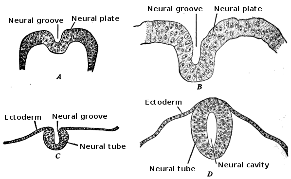
An embryo is an initial stage of development of a multicellular organism. In organisms that reproduce sexually, embryonic development is the part of the life cycle that begins just after fertilization of the female egg cell by the male sperm cell. The resulting fusion of these two cells produces a single-celled zygote that undergoes many cell divisions that produce cells known as blastomeres. The blastomeres are arranged as a solid ball that when reaching a certain size, called a morula, takes in fluid to create a cavity called a blastocoel. The structure is then termed a blastula, or a blastocyst in mammals. The mammalian blastocyst hatches before implantating into the endometrial lining of the womb. Once implanted the embryo will continue its development through the next stages of gastrulation, neurulation, and organogenesis. Gastrulation is the formation of the three germ layers that will form all of the different parts of the body. Neurulation forms the nervous ... [...More Info...] [...Related Items...] OR: [Wikipedia] [Google] [Baidu] |
Haematopoiesis
Haematopoiesis (, from Greek , 'blood' and 'to make'; also hematopoiesis in American English; sometimes also h(a)emopoiesis) is the formation of blood cellular components. All cellular blood components are derived from haematopoietic stem cells. In a healthy adult person, approximately – new blood cells are produced daily in order to maintain steady state levels in the peripheral circulation.Semester 4 medical lectures at Uppsala University 2008 by Leif Jansson Process Haematopoietic stem cells (HSCs) Haematopoietic stem cells (HSCs) reside in the medulla of the bone (bone marrow) and have the unique ability to give rise to all of the different mature blood cell types and tissues. HSCs are self-renewing cells: when they differentiate, at least some of their daughter cells remain as HSCs so the pool of stem cells is not depleted. This phenomenon is called asymmetric division. The other daughters of HSCs ( myeloid and lymphoid progenitor cells) can follow any of the other ... [...More Info...] [...Related Items...] OR: [Wikipedia] [Google] [Baidu] |
Organogenesis
Organogenesis is the phase of embryonic development that starts at the end of gastrulation and continues until birth. During organogenesis, the three germ layers formed from gastrulation (the ectoderm, endoderm, and mesoderm) form the internal organs of the organism. The cells of each of the three germ layers undergo differentiation, a process where less-specialized cells become more-specialized through the expression of a specific set of genes. Cell differentiation is driven by cell signaling cascades. Differentiation is influenced by extracellular signals such as growth factors that are exchanged to adjacent cells which is called juxtracrine signaling or to neighboring cells over short distances which is called paracrine signaling. Intracellular signals consist of a cell signaling itself (autocrine signaling), also play a role in organ formation. These signaling pathways allow for cell rearrangement and ensure that organs form at specific sites within the organism. The organoge ... [...More Info...] [...Related Items...] OR: [Wikipedia] [Google] [Baidu] |
Gestational Sac
The gestational sac is the large cavity of fluid surrounding the embryo. During early embryogenesis it consists of the extraembryonic coelom, also called the chorionic cavity. The gestational sac is normally contained within the uterus. It is the only available structure that can be used to determine if an intrauterine pregnancy exists until the embryo can be identified. On obstetric ultrasound, the gestational sac is a dark (anechoic) space surrounded by a white (hyperechoic) rim. Structure The gestational sac is spherical in shape, and is usually located in the upper part (fundus) of the uterus. By approximately nine weeks of gestational age, due to folding of the trilaminar germ disc, the amniotic sac expands and occupy the majority of the volume of the gestational sac, eventually reducing the extraembryonic coelom (the gestational sac or the chorionic cavity) to a thin layer between the parietal somatopleuric and visceral splanchnopleuric layer of extraembryonic mesoderm. Deve ... [...More Info...] [...Related Items...] OR: [Wikipedia] [Google] [Baidu] |
Mesoderm
The mesoderm is the middle layer of the three germ layers that develops during gastrulation in the very early development of the embryo of most animals. The outer layer is the ectoderm, and the inner layer is the endoderm.Langman's Medical Embryology, 11th edition. 2010. The mesoderm forms mesenchyme, mesothelium, non-epithelial blood cells and coelomocytes. Mesothelium lines coeloms. Mesoderm forms the muscles in a process known as myogenesis, septa (cross-wise partitions) and mesenteries (length-wise partitions); and forms part of the gonads (the rest being the gametes). Myogenesis is specifically a function of mesenchyme. The mesoderm differentiates from the rest of the embryo through intercellular signaling, after which the mesoderm is polarized by an organizing center. The position of the organizing center is in turn determined by the regions in which beta-catenin is protected from degradation by GSK-3. Beta-catenin acts as a co-factor that alters the activity of ... [...More Info...] [...Related Items...] OR: [Wikipedia] [Google] [Baidu] |
Heuser's Membrane
Heuser's membrane (or the exocoelomic membrane) is a short lived combination of hypoblast cells and extracellular matrix In biology, the extracellular matrix (ECM), also called intercellular matrix, is a three-dimensional network consisting of extracellular macromolecules and minerals, such as collagen, enzymes, glycoproteins and hydroxyapatite that provide stru .... At day 9-10 of embryonic development, cells from the hypoblast begin to migrate to the embryonic pole, forming a layer of cells just beneath the cytotrophoblast, called Heuser's membrane. It surrounds the exocoelomic cavity (primary yolk sac), i.e. it lines the inner surface of the cytotrophoblast. At this point, the exocoelomic cavity replaces the blastocyst cavity. At days 11 to 12, there is further delineation of the trophoblastic cells giving rise to a layer of loosely arranged cells that inserts between Heuser's membrane and both syncytiotrophoblast and cytotrophoblast. References Embryology {{ ... [...More Info...] [...Related Items...] OR: [Wikipedia] [Google] [Baidu] |
Ileum
The ileum () is the final section of the small intestine in most higher vertebrates, including mammals, reptiles, and birds. In fish, the divisions of the small intestine are not as clear and the terms posterior intestine or distal intestine may be used instead of ileum. Its main function is to absorb vitamin B12, bile salts, and whatever products of digestion that were not absorbed by the jejunum. The ileum follows the duodenum and jejunum and is separated from the cecum by the ileocecal valve (ICV). In humans, the ileum is about 2–4 m long, and the pH is usually between 7 and 8 (neutral or slightly basic). ''Ileum ''is derived from the Greek word ''eilein'', meaning "to twist up tightly". Structure The ileum is the third and final part of the small intestine. It follows the jejunum and ends at the ileocecal junction, where the terminal ileum communicates with the cecum of the large intestine through the ileocecal valve. The ileum, along with the jejunum, is suspended ... [...More Info...] [...Related Items...] OR: [Wikipedia] [Google] [Baidu] |
Navel
The navel (clinically known as the umbilicus, commonly known as the belly button or tummy button) is a protruding, flat, or hollowed area on the abdomen at the attachment site of the umbilical cord. All placental mammals have a navel, although it is generally more conspicuous in humans. Structure The umbilicus is used to visually separate the abdomen into quadrants. The umbilicus is a prominent scar on the abdomen, with its position being relatively consistent among humans. The skin around the waist at the level of the umbilicus is supplied by the tenth thoracic spinal nerve (T10 dermatome). The umbilicus itself typically lies at a vertical level corresponding to the junction between the L3 and L4 vertebrae, with a normal variation among people between the L3 and L5 vertebrae. Parts of the adult navel include the "umbilical cord remnant" or "umbilical tip", which is the often protruding scar left by the detachment of the umbilical cord. This is located in the center of the ... [...More Info...] [...Related Items...] OR: [Wikipedia] [Google] [Baidu] |
Ileocecal Valve
The ileocecal valve (ileal papilla, ileocaecal valve, Tulp's valve, Tulpius valve, Bauhin's valve, ileocecal eminence, valve of Varolius or colic valve) is a sphincter muscle valve that separates the small intestine and the large intestine. Its critical function is to limit the reflux of colonic contents into the ileum. Approximately two liters of fluid enters the colon daily through the ileocecal valve. Microanatomy The histology of the ileocecal valve shows an abrupt change from a villous mucosa pattern of the ileum to a more colonic mucosa. A thickening of the muscularis mucosa, which is the smooth muscle tissue found beneath the mucosal layer of the digestive tract. A thickening of the muscularis externa is also noted. There is also a variable amount of lymphatic tissue found at the valve. The ileocecal valve has a papillose structure. Clinical significance Colonoscopy During colonoscopy, the ileocecal valve is used, along with the appendiceal orifice, in the identifi ... [...More Info...] [...Related Items...] OR: [Wikipedia] [Google] [Baidu] |
Meckel's Diverticulum
A Meckel's diverticulum, a true congenital diverticulum, is a slight bulge in the small intestine present at birth and a vestigial remnant of the omphalomesenteric duct (also called the vitelline duct or yolk stalk). It is the most common malformation of the Human gastrointestinal tract, gastrointestinal tract and is present in approximately 2% of the population, with males more frequently experiencing symptoms. Meckel's diverticulum was first explained by Fabricius Hildanus in the sixteenth century and later named after Johann Friedrich Meckel, who described the embryological origin of this type of diverticulum in 1809. Signs and symptoms The majority of people with a Meckel's diverticulum are asymptomatic. An asymptomatic Meckel's diverticulum is called a ''silent'' Meckel's diverticulum. If symptoms do occur, they typically appear before the age of two years. The most common presenting symptom is painless rectal bleeding such as melaena-like black offensive stools, followed by i ... [...More Info...] [...Related Items...] OR: [Wikipedia] [Google] [Baidu] |
Placenta
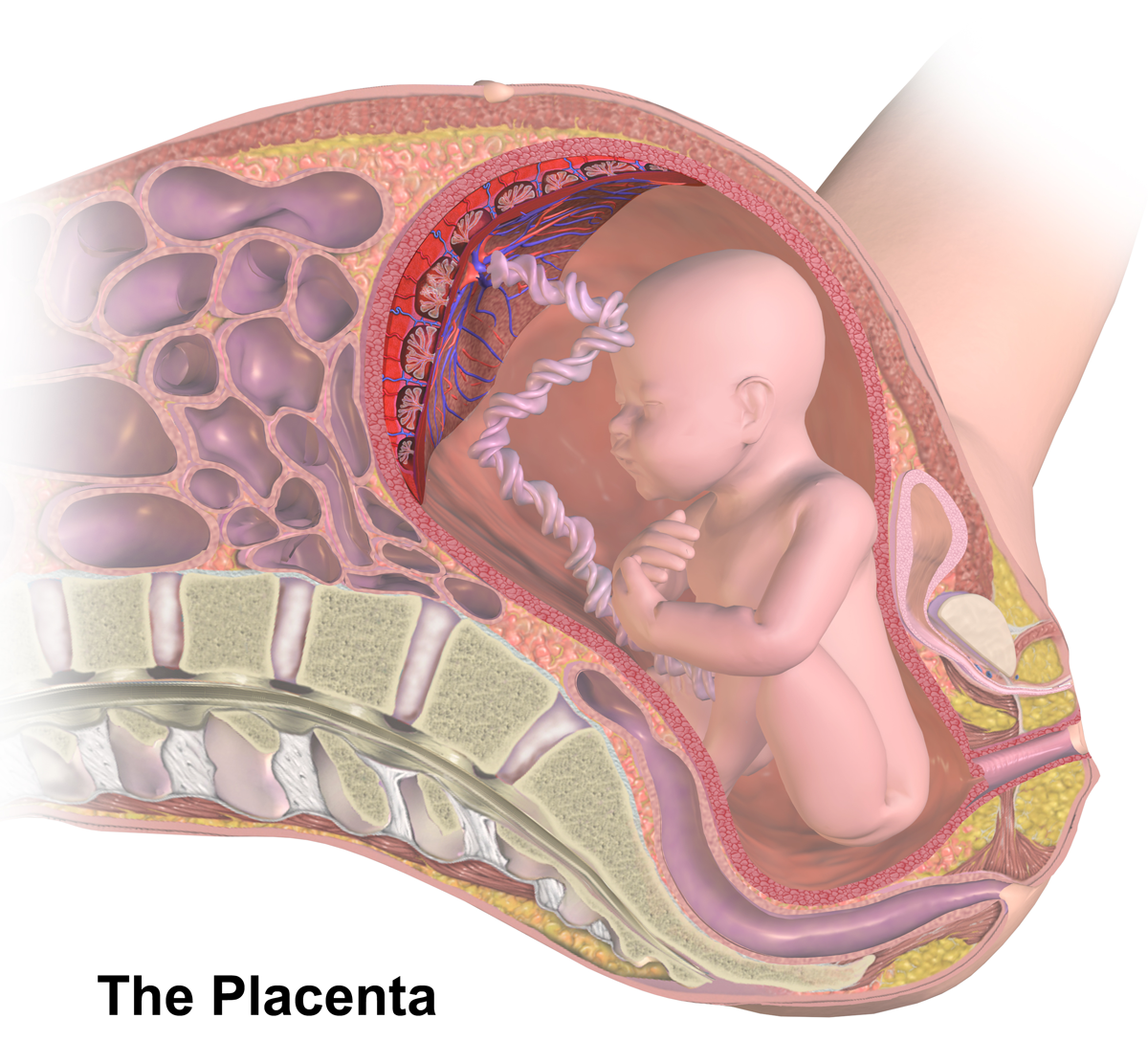
The placenta is a temporary embryonic and later fetal organ that begins developing from the blastocyst shortly after implantation. It plays critical roles in facilitating nutrient, gas and waste exchange between the physically separate maternal and fetal circulations, and is an important endocrine organ, producing hormones that regulate both maternal and fetal physiology during pregnancy. The placenta connects to the fetus via the umbilical cord, and on the opposite aspect to the maternal uterus in a species-dependent manner. In humans, a thin layer of maternal decidual (endometrial) tissue comes away with the placenta when it is expelled from the uterus following birth (sometimes incorrectly referred to as the 'maternal part' of the placenta). Placentas are a defining characteristic of placental mammals, but are also found in marsupials and some non-mammals with varying levels of development. Mammalian placentas probably first evolved about 150 million to 200 million years ... [...More Info...] [...Related Items...] OR: [Wikipedia] [Google] [Baidu] |
Chorion
The chorion is the outermost fetal membrane around the embryo in mammals, birds and reptiles (amniotes). It develops from an outer fold on the surface of the yolk sac, which lies outside the zona pellucida (in mammals), known as the vitelline membrane in other animals. In insects it is developed by the follicle cells while the egg is in the ovary.Chapman, R.F. (1998) "The insects: structure and function", Section ''The egg and embryology''. Previewed in Google Bookon 26 Sep 2009. Structure In humans and other mammals (excluding monotremes), the chorion is one of the fetal membranes that exist during pregnancy between the developing fetus and mother. The chorion and the amnion together form the amniotic sac. In humans it is formed by extraembryonic mesoderm and the two layers of trophoblast that surround the embryo and other membranes; the chorionic villi emerge from the chorion, invade the endometrium, and allow the transfer of nutrients from maternal blood to fetal blood. ... [...More Info...] [...Related Items...] OR: [Wikipedia] [Google] [Baidu] |


