|
H Reflex
The H-reflex (or Hoffmann's reflex) is a reflectory reaction of muscles after electrical stimulation of sensory fibers (Ia afferents stemming from muscle spindles) in their innervating nerves (for example, those located behind the knee). The H-reflex test is performed using an electric stimulator, which gives usually a square-wave current of short duration and small amplitude (higher stimulations might involve alpha fibers, causing an F-wave, compromising the results), and an EMG set, to record the muscle response. That response is usually a clear wave, called H-wave, 28-35 ms after the stimulus, not to be confused with an F-wave. An M-wave, an early response, occurs 3-6 ms after the onset of stimulation. The H and F-waves are later responses. As the stimulus increases, the amplitude of the F-wave increases only slightly, and the H-wave decreases, and at supramaximal stimulus, the H-wave will disappear. The M-wave does the opposite of the H-wave. As the stimulus increases the ... [...More Info...] [...Related Items...] OR: [Wikipedia] [Google] [Baidu] |
Hoffmann's Reflex
Hoffmann's reflex (Hoffmann's sign, sometimes simply "''Hoffmann's''", also finger flexor reflex) is a neurological examination finding elicited by a reflex test which can help verify the presence or absence of issues arising from the corticospinal tract. It is named after neurologist Johann Hoffmann (neurologist), Johann Hoffmann. Usually considered a pathological reflex in a clinical setting, the Hoffmann's reflex has also been used as a measure of spinal reflex processing (adaptation) in response to Physical exercise, exercise training. Procedure The Hoffmann's reflex test itself involves loosely holding the middle finger and flicking the fingernail downward, allowing the middle finger to flick upward reflexively. A positive response is seen when there is flexion and adduction of the thumb on the same hand. ... [...More Info...] [...Related Items...] OR: [Wikipedia] [Google] [Baidu] |
Reflectory Reaction
In biology, a reflex, or reflex action, is an involuntary, unplanned sequence or action and nearly instantaneous response to a Stimulus (physiology), stimulus. Reflexes are found with varying levels of complexity in organisms with a nervous system. A reflex occurs via Neural pathway, neural pathways in the nervous system called Reflex arc, reflex arcs. A stimulus initiates a neural signal, which is carried to a synapse. The signal is then transferred across the synapse to a motor neuron which evokes a target response. These neural signals do not always travel to the brain, so many reflexes are an automatic response to a stimulus that does not receive or need conscious thought. Many reflexes are fine-tuned to increase organism survival and self-defense. This is observed in reflexes such as the Startle response, startle reflex, which provides an automatic response to an unexpected stimuli, and the feline righting reflex, which reorients a cat's body when falling to ensure safe l ... [...More Info...] [...Related Items...] OR: [Wikipedia] [Google] [Baidu] |
Electrical
Electricity is the set of physical phenomena associated with the presence and motion of matter that has a property of electric charge. Electricity is related to magnetism, both being part of the phenomenon of electromagnetism, as described by Maxwell's equations. Various common phenomena are related to electricity, including lightning, static electricity, electric heating, electric discharges and many others. The presence of an electric charge, which can be either positive or negative, produces an electric field. The movement of electric charges is an electric current and produces a magnetic field. When a charge is placed in a location with a non-zero electric field, a force will act on it. The magnitude of this force is given by Coulomb's law. If the charge moves, the electric field would be doing work on the electric charge. Thus we can speak of electric potential at a certain point in space, which is equal to the work done by an external agent in carrying a unit of positiv ... [...More Info...] [...Related Items...] OR: [Wikipedia] [Google] [Baidu] |
Ia Afferent
A type Ia sensory fiber, or a primary afferent fiber is a type of afferent nerve fiber. It is the sensory fiber of a stretch receptor called the muscle spindle found in muscles, which constantly monitors the rate at which a muscle stretch changes. The information carried by type Ia fibers contributes to the sense of proprioception. Function of muscle spindles For the body to keep moving properly and with finesse, the nervous system has to have a constant input of sensory data coming from areas such as the muscles and joints. In order to receive a continuous stream of sensory data, the body has developed special sensory receptors called proprioceptors. Muscle spindles are a type of proprioceptor, and they are found inside the muscle itself. They lie parallel with the contractile fibers. This gives them the ability to monitor muscle length with precision. Types of sensory fibers This change in length of the spindle is transduced (transformed into electric membrane potential ... [...More Info...] [...Related Items...] OR: [Wikipedia] [Google] [Baidu] |
Muscle Spindles
Muscle spindles are stretch receptors within the body of a skeletal muscle that primarily detect changes in the length of the muscle. They convey length information to the central nervous system via afferent nerve fibers. This information can be processed by the brain as proprioception. The responses of muscle spindles to changes in length also play an important role in regulating the contraction of muscles, for example, by activating motor neurons via the stretch reflex to resist muscle stretch. The muscle spindle has both sensory and motor components. * Sensory information conveyed by primary type Ia sensory fibers which spiral around muscle fibres within the spindle, and secondary type II sensory fibers * Activation of muscle fibres within the spindle by up to a dozen gamma motor neurons and to a lesser extent by one or two beta motor neurons Structure Muscle spindles are found within the belly of a skeletal muscle. Muscle spindles are fusiform (spindle-shaped), and the speci ... [...More Info...] [...Related Items...] OR: [Wikipedia] [Google] [Baidu] |
Knee
In humans and other primates, the knee joins the thigh with the leg and consists of two joints: one between the femur and tibia (tibiofemoral joint), and one between the femur and patella (patellofemoral joint). It is the largest joint in the human body. The knee is a modified hinge joint, which permits flexion and extension as well as slight internal and external rotation. The knee is vulnerable to injury and to the development of osteoarthritis. It is often termed a ''compound joint'' having tibiofemoral and patellofemoral components. (The fibular collateral ligament is often considered with tibiofemoral components.) Structure The knee is a modified hinge joint, a type of synovial joint, which is composed of three functional compartments: the patellofemoral articulation, consisting of the patella, or "kneecap", and the patellar groove on the front of the femur through which it slides; and the medial and lateral tibiofemoral articulations linking the femur, or thigh bone ... [...More Info...] [...Related Items...] OR: [Wikipedia] [Google] [Baidu] |
Alpha Motor Neuron
Alpha (α) motor neurons (also called alpha motoneurons), are large, multipolar lower motor neurons of the brainstem and spinal cord. They innervate extrafusal muscle fibers of skeletal muscle and are directly responsible for initiating their contraction. Alpha motor neurons are distinct from gamma motor neurons, which innervate intrafusal muscle fibers of muscle spindles. While their cell bodies are found in the central nervous system (CNS), α motor neurons are also considered part of the somatic nervous system—a branch of the peripheral nervous system (PNS)—because their axons extend into the periphery to innervate skeletal muscles. An alpha motor neuron and the muscle fibers it innervates is a motor unit. A motor neuron pool contains the cell bodies of all the alpha motor neurons involved in contracting a single muscle. Location Alpha motor neurons (α-MNs) innervating the head and neck are found in the brainstem; the remaining α-MNs innervate the rest of the body and ... [...More Info...] [...Related Items...] OR: [Wikipedia] [Google] [Baidu] |
F Wave
In neuroscience, an F wave is one of several motor responses which may follow the Muscle contraction, direct motor response (M) evoked by electrical stimulation of peripheral motor or mixed (sensory and motor) nerves. F-waves are the second of two late voltage changes observed after stimulation is applied to the skin surface above the Anatomical terms of location#Proximal and distal, distal region of a nerve, in addition to the H-reflex (Hoffman's Reflex) which is a muscle reaction in response to electrical stimulation of innervating sensory fibers. Traversal of F-waves along the entire length of peripheral nerves between the spinal cord and muscle, allows for assessment of motor nerve conduction between distal stimulation sites in the arm and leg, and related motoneurons (MN's) in the cervical and lumbosacral cord. F-waves are able to assess both Afferent nerve fiber, afferent and Efferent nerve fiber, efferent loops of the alpha motor neuron in its entirety. As such, various proper ... [...More Info...] [...Related Items...] OR: [Wikipedia] [Google] [Baidu] |
Electromyography
Electromyography (EMG) is a technique for evaluating and recording the electrical activity produced by skeletal muscles. EMG is performed using an instrument called an electromyograph to produce a record called an electromyogram. An electromyograph detects the electric potential generated by muscle cells when these cells are electrically or neurologically activated. The signals can be analyzed to detect abnormalities, activation level, or recruitment order, or to analyze the biomechanics of human or animal movement. Needle EMG is an electrodiagnostic medicine technique commonly used by neurologists. Surface EMG is a non-medical procedure used to assess muscle activation by several professionals, including physiotherapists, kinesiologists and biomedical engineers. In Computer Science, EMG is also used as middleware in gesture recognition towards allowing the input of physical action to a computer as a form of human-computer interaction. Clinical uses EMG testing has a variety of ... [...More Info...] [...Related Items...] OR: [Wikipedia] [Google] [Baidu] |
Stretch Reflex
The stretch reflex (myotatic reflex), or more accurately "muscle stretch reflex", is a muscle contraction in response to stretching within the muscle. The reflex functions to maintain the muscle at a constant length. The term deep tendon reflex is often used by many health workers and students to refer to this reflex. "Tendons have little to do with the response, other than being responsible for mechanically transmitting the sudden stretch from the reflex hammer to the muscle spindle. In addition, some muscles with stretch reflexes have no tendons (e.g., "jaw jerk" of the masseter muscle)". As an example of a spinal reflex, it results in a fast response that involves an afferent signal into the spinal cord and an efferent signal out to the muscle. The stretch reflex can be a monosynaptic reflex which provides automatic regulation of skeletal muscle length, whereby the signal entering the spinal cord arises from a change in muscle length or velocity. It can also include a polysyna ... [...More Info...] [...Related Items...] OR: [Wikipedia] [Google] [Baidu] |
Knee Jerk Reflex
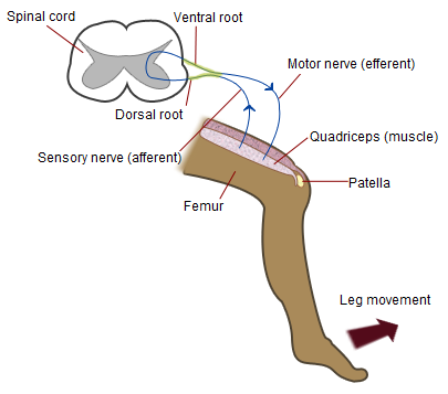
The patellar reflex, also called the knee reflex or knee-jerk, is a stretch reflex which tests the L2, L3, and L4 segments of the spinal cord. Mechanism Striking of the patellar tendon with a reflex hammer just below the patella stretches the muscle spindle in the quadriceps muscle. This produces a signal which travels back to the spinal cord and synapses (without interneurons) at the level of L3 or L4 in the spinal cord, completely independent of higher centres. From there, an alpha motor neuron conducts an efferent impulse back to the quadriceps femoris muscle, triggering contraction. This contraction, coordinated with the relaxation of the antagonistic flexor hamstring muscle causes the leg to kick. There is a latency of around 18 ms between stretch of the patellar tendon and the beginning of contraction of the quadriceps femoris muscle. This is a reflex of proprioception which helps maintain posture and balance, allowing to keep one's balance with little effort or conscious th ... [...More Info...] [...Related Items...] OR: [Wikipedia] [Google] [Baidu] |
Zero Gravity
Weightlessness is the complete or near-complete absence of the sensation of weight. It is also termed zero gravity, zero G-force, or zero-G. Weight is a measurement of the force on an object at rest in a relatively strong gravitational field (such as on the surface of the Earth). These weight-sensations originate from contact with supporting floors, seats, beds, scales, and the like. A sensation of weight is also produced, even when the gravitational field is zero, when contact forces act upon and overcome a body's inertia by mechanical, non-gravitational forces- such as in a centrifuge, a rotating space station, or within an accelerating vehicle. When the gravitational field is non-uniform, a body in free fall experiences tidal effects and is not stress-free. Near a black hole, such tidal effects can be very strong. In the case of the Earth, the effects are minor, especially on objects of relatively small dimensions (such as the human body or a spacecraft) and the overall ... [...More Info...] [...Related Items...] OR: [Wikipedia] [Google] [Baidu] |


