|
Granzyme
Granzymes are serine proteases released by cytoplasmic granules within cytotoxic T cells and natural killer (NK) cells. They induce programmed cell death (apoptosis) in the target cell, thus eliminating cells that have become cancerous or are infected with viruses or bacteria. Granzymes also kill bacteria and inhibit viral replication. In NK cells and T cells, granzymes are packaged in cytotoxic granules along with perforin. Granzymes can also be detected in the rough endoplasmic reticulum, golgi complex, and the trans-golgi reticulum. The contents of the cytotoxic granules function to permit entry of the granzymes into the target cell cytosol. The granules are released into an immune synapse formed with a target cell, where perforin mediates the delivery of the granzymes into endosomes in the target cell, and finally into the target cell cytosol. Granzymes are part of the serine esterase family. They are closely related to other immune serine proteases expressed by innate immune cel ... [...More Info...] [...Related Items...] OR: [Wikipedia] [Google] [Baidu] |
Granzyme B
Granzyme B (GrB) is one of the serine protease granzymes most commonly found in the granules of natural killer cells (NK cells) and cytotoxic T cells. It is secreted by these cells along with the pore forming protein perforin to mediate apoptosis in target cells. Granzyme B has also been found to be produced by a wide range of non-cytotoxic cells ranging from basophils and mast cells to smooth muscle cells. The secondary functions of granzyme B are also numerous. Granzyme B has shown to be involved in inducing inflammation by stimulating cytokine release and is also involved in extracellular matrix remodelling. Elevated levels of granzyme B are also implicated in a number of autoimmune diseases, several skin diseases, and type 1 diabetes. Structure In humans, granzyme B is encoded by ''GZMB'' on chromosome 14q.11.2, which is 3.2kb long and consists of 5 exons. It is one of the most abundant granzymes of which there are 5 in humans and 10 in mice. Granzyme B is thought to hav ... [...More Info...] [...Related Items...] OR: [Wikipedia] [Google] [Baidu] |
GZMK
Granzyme K (GrK) is a protein that is encoded by the ''GZMK'' gene on chromosome 5 in humans. Granzymes are a family of serine proteases which have various intracellular and extracellular roles. GrK is found in granules of natural killer (NK) cells and cytotoxic T lymphocytes (CTLs), and is traditionally described as cytotoxic towards targeted foreign, infected, or cancerous cells. NK cells and CTLs can induce apoptosis through the granule secretory pathway, which involves the secretion of granzymes along with perforin at immunological synapse In immunology, an immunological synapse (or immune synapse) is the interface between an antigen-presenting cell or target cell and a lymphocyte such as a T/B cell or Natural Killer cell. The interface was originally named after the neuronal syn ...s. Intracellularly, GrK may cleave a variety of substrates, such as the nucleosome assembly protein (NAP), HMG2, and Ape1 in the ER-associated SET complex, along with other targets that have dow ... [...More Info...] [...Related Items...] OR: [Wikipedia] [Google] [Baidu] |
GZMA
Granzyme A (, ''CTLA3'', ''HuTPS'', ''T-cell associated protease 1'', ''cytotoxic T lymphocyte serine protease'', ''TSP-1'', ''T-cell derived serine proteinase'') is an enzyme. that in humans is encoded by the GZMA gene, and is one of the five granzymes encoded in the human genome . This enzyme is present in cytotoxic T lymphocyte granules. Cytolytic T lymphocytes (CTL) and natural killer (NK) cells share the remarkable ability to recognize, bind, and lyse specific target cells. They are thought to protect their host by lysing cells bearing on their surface 'nonself' antigens, usually peptides or proteins resulting from infection by intracellular pathogens. The protein described here is a T cell- and natural killer cell-specific serine protease that may function as a common component necessary for lysis of target cells by cytotoxic T lymphocytes and natural killer cells. This enzyme catalyses the following chemical reaction: :Hydrolysis of proteins, including fibronectin, type ... [...More Info...] [...Related Items...] OR: [Wikipedia] [Google] [Baidu] |
GZMH
Granzyme H is a protein that in humans is encoded by the ''GZMH'' gene In biology, the word gene (from , ; "...Wilhelm Johannsen coined the word gene to describe the Mendelian units of heredity..." meaning ''generation'' or ''birth'' or ''gender'') can have several different meanings. The Mendelian gene is a ba .... References Further reading * * * * * * * * * * * * * * {{gene-14-stub ... [...More Info...] [...Related Items...] OR: [Wikipedia] [Google] [Baidu] |
Perforin
Perforin-1 is a protein that in humans is encoded by the ''PRF1'' gene and the ''Prf1'' gene in mice. Function Perforin is a pore forming cytolytic protein found in the granules of cytotoxic T lymphocytes (CTLs) and natural killer cells (NK cells). Upon degranulation, perforin molecules translocate to the target cell with the help of calreticulin, which works as a chaperone protein to prevent perforin from degrading. Perforin then binds to the target cell's plasma membrane via membrane phospholipids while phosphatidylcholine binds calcium ions to increase perforin's affinity to the membrane. Perforin oligomerises in a Ca2+ dependent manner to form pores on the target cell. The pore formed allows for the passive diffusion of a family of pro-apoptotic proteases, known as the granzymes, into the target cell. The lytic membrane-inserting part of perforin is the MACPF domain. This region shares homology with cholesterol-dependent cytolysins from Gram-positive bacteria. Perforin has ... [...More Info...] [...Related Items...] OR: [Wikipedia] [Google] [Baidu] |
Natural Killer Cell
Natural killer cells, also known as NK cells or large granular lymphocytes (LGL), are a type of cytotoxic lymphocyte critical to the innate immune system that belong to the rapidly expanding family of known innate lymphoid cells (ILC) and represent 5–20% of all circulating lymphocytes in humans. The role of NK cells is analogous to that of cytotoxic T cells in the vertebrate adaptive immune response. NK cells provide rapid responses to virus-infected cell and other intracellular pathogens acting at around 3 days after infection, and respond to tumor formation. Typically, immune cells detect the major histocompatibility complex (MHC) presented on infected cell surfaces, triggering cytokine release, causing the death of the infected cell by lysis or apoptosis. NK cells are unique, however, as they have the ability to recognize and kill stressed cells in the absence of antibodies and MHC, allowing for a much faster immune reaction. They were named "natural killers" because ... [...More Info...] [...Related Items...] OR: [Wikipedia] [Google] [Baidu] |
Inflammation
Inflammation (from la, wikt:en:inflammatio#Latin, inflammatio) is part of the complex biological response of body tissues to harmful stimuli, such as pathogens, damaged cells, or Irritation, irritants, and is a protective response involving immune cells, blood vessels, and molecular mediators. The function of inflammation is to eliminate the initial cause of cell injury, clear out necrotic cells and tissues damaged from the original insult and the inflammatory process, and initiate tissue repair. The five cardinal signs are heat, pain, redness, swelling, and Functio laesa, loss of function (Latin ''calor'', ''dolor'', ''rubor'', ''tumor'', and ''functio laesa''). Inflammation is a generic response, and therefore it is considered as a mechanism of innate immune system, innate immunity, as compared to adaptive immune system, adaptive immunity, which is specific for each pathogen. Too little inflammation could lead to progressive tissue destruction by the harmful stimulus (e.g. b ... [...More Info...] [...Related Items...] OR: [Wikipedia] [Google] [Baidu] |
ELISPOT
The enzyme-linked immune absorbent spot (ELISpot) is a type of assay that focuses on quantitatively measuring the frequency of cytokine secretion for a single cell. The ELISpot Assay is also a form of immunostaining since it is classified as a technique that uses antibodies to detect a protein analyte, with the word analyte referring to any biological or chemical substance being identified or measured. The FluoroSpot Assay is a variation of the ELISpot assay. The FluoroSpot Assay uses fluorescence in order to analyze multiple analytes, meaning it can detect the secretion of more than one type of protein. History Cecil Czerkinsky first described ELISpot in 1983 as a new way to quantify the production of an antigen-specific immunoglobulin by hybridoma cells. In 1988, Czerkinsky developed an ELISA spot assay that quantified the secretion of a lymphokine by T cells. In the same year, dual-color ELISpot was combined with computer imaging for the first time, which allowed for the enu ... [...More Info...] [...Related Items...] OR: [Wikipedia] [Google] [Baidu] |
Flow Cytometry
Flow cytometry (FC) is a technique used to detect and measure physical and chemical characteristics of a population of cells or particles. In this process, a sample containing cells or particles is suspended in a fluid and injected into the flow cytometer instrument. The sample is focused to ideally flow one cell at a time through a laser beam, where the light scattered is characteristic to the cells and their components. Cells are often labeled with fluorescent markers so light is absorbed and then emitted in a band of wavelengths. Tens of thousands of cells can be quickly examined and the data gathered are processed by a computer. Flow cytometry is routinely used in basic research, clinical practice, and clinical trials. Uses for flow cytometry include: * Cell counting * Cell sorting * Determining cell characteristics and function * Detecting microorganisms * Biomarker detection * Protein engineering detection * Diagnosis of health disorders such as blood cancers * Measuring ... [...More Info...] [...Related Items...] OR: [Wikipedia] [Google] [Baidu] |
Bcl-2 Homologous Antagonist Killer
Bcl-2 homologous antagonist/killer is a protein that in humans is encoded by the ''BAK1'' gene on chromosome 6. The protein encoded by this gene belongs to the BCL2 protein family. BCL2 family members form oligomers or heterodimers and act as anti- or pro-apoptotic regulators that are involved in a wide variety of cellular activities. This protein localizes to mitochondria, and functions to induce apoptosis. It interacts with and accelerates the opening of the mitochondrial voltage-dependent anion channel, which leads to a loss in membrane potential and the release of cytochrome c. This protein also interacts with the tumor suppressor P53 after exposure to cell stress. Structure BAK1 is a pro-apoptotic Bcl-2 protein containing four Bcl-2 homology (BH) domains: BH1, BH2, BH3, and BH4. These domains are composed of nine α-helices, with a hydrophobic α-helix core surrounded by amphipathic helices and a transmembrane C-terminal α-helix anchored to the mitochondrial outer membran ... [...More Info...] [...Related Items...] OR: [Wikipedia] [Google] [Baidu] |
ELISA
The enzyme-linked immunosorbent assay (ELISA) (, ) is a commonly used analytical biochemistry assay, first described by Eva Engvall and Peter Perlmann in 1971. The assay uses a solid-phase type of enzyme immunoassay (EIA) to detect the presence of a ligand (commonly a protein) in a liquid sample using antibodies directed against the protein to be measured. ELISA has been used as a diagnostic tool in medicine, plant pathology, and biotechnology, as well as a quality control check in various industries. In the most simple form of an ELISA, antigens from the sample to be tested are attached to a surface. Then, a matching antibody is applied over the surface so it can bind the antigen. This antibody is linked to an enzyme and then any unbound antibodies are removed. In the final step, a substance containing the enzyme's substrate is added. If there was binding, the subsequent reaction produces a detectable signal, most commonly a color change. Performing an ELISA involves at least ... [...More Info...] [...Related Items...] OR: [Wikipedia] [Google] [Baidu] |
Western Blot
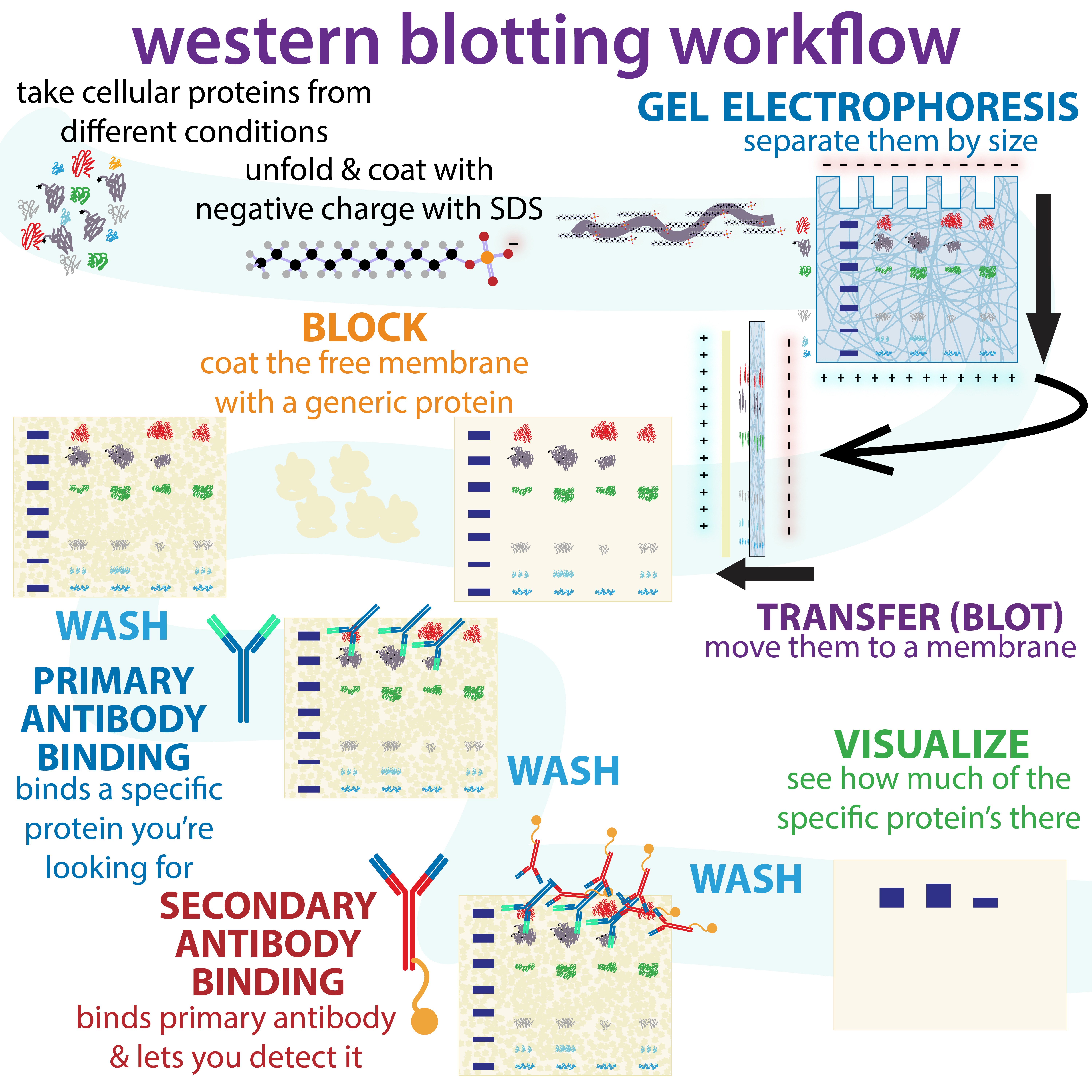
The western blot (sometimes called the protein immunoblot), or western blotting, is a widely used analytical technique in molecular biology and immunogenetics to detect specific proteins in a sample of tissue homogenate or extract. Besides detecting the proteins, this technique is also utilized to visualize, distinguish, and quantify the different proteins in a complicated protein combination. Western blot technique uses three elements to achieve its task of separating a specific protein from a complex: separation by size, transfer of protein to a solid support, and marking target protein using a primary and secondary antibody to visualize. A synthetic or animal-derived antibody (known as the primary antibody) is created that recognizes and binds to a specific target protein. The electrophoresis membrane is washed in a solution containing the primary antibody, before excess antibody is washed off. A secondary antibody is added which recognizes and binds to the primary antibody ... [...More Info...] [...Related Items...] OR: [Wikipedia] [Google] [Baidu] |


